Lab 4: Brain Stem 2
Lab Summary
During this lab, you will locate each cranial nerve nucleus, using the myelin stained brain stem series that you oriented on the brainstem drawing in lab 3. As you identify each nucleus, be sure to recall its function, its associated cranial nerve(s), the exit or entrance point of the nerve from the brainstem, and the blood supply to that region of the brain stem.
SECTION 1: Cranial Nerve Nuclei Introduction
SECTION 2: Sensory Nuclei
SECTION 3: Motor Nuclei
SECTION 4: Blood Supply and Ascending/Descending Pathways
SECTION 5: Review
Cranial Nerve Nuclei Introduction
Objectives
- Identify the cranial nerves having purely sensory function, motor function and those nerves having both sensory and motor functions.
- Describe the central structures (nuclei and tracts) that are involved in mediating the sensory and motor functions or each cranial nerve and identify where in the brain stem these structures can be found.
The study of the brain stem is a particularly important part of the neurobiology course since proper neurological diagnosis depends heavily on an understanding of the cranial nerves, their nuclei and the organization of ascending and descending sensory and motor pathways within the brainstem. Before identifying the nuclei, be sure that you have determined the location and orientation of the 14 brain stem slides in the myelin stained brainstem series from lab 3. As you examine each slide in this lab, also use the fixed brain preparations to identify any superficial evidence of the cranial nerves, their nuclei or tracts. For example, in addition to identifying the brainstem entrance and exit points for all of the cranial nerves except the olfactory (CN1) and optic (CN2) (these are more related to the diencephalon), you should be able to locate the facial colliculus on the floor of the 4th ventricle representing the area where facial nerve fibers wrap around the abducens nucleus dorsally before exiting from the ventral surface at the ponto-medullary junction. Other superficial landmarks related to underlying cranial nerve nuclei include the hypoglossal and vagal trigones (4th ventricle), gracile and cuneate tubercles (caudal medulla), and vestibular area (4th ventricle).
Before examining each of the 14 sections in the myelin stained series for the cranial nerve nuclei and their axon pathways study the Cranial Nerve Table 4.1 identifying where the nuclei related to each cranial nerve can be found (medulla, pons and/or midbrain) and what axons pathways or tracts ascend or descend from them. Test yourself using the linked table in the end of chapter review.
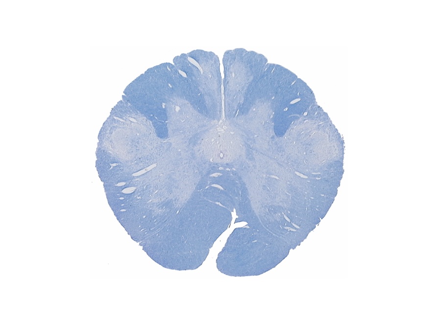
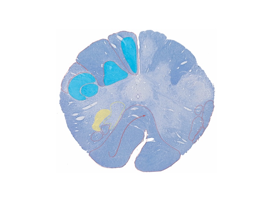
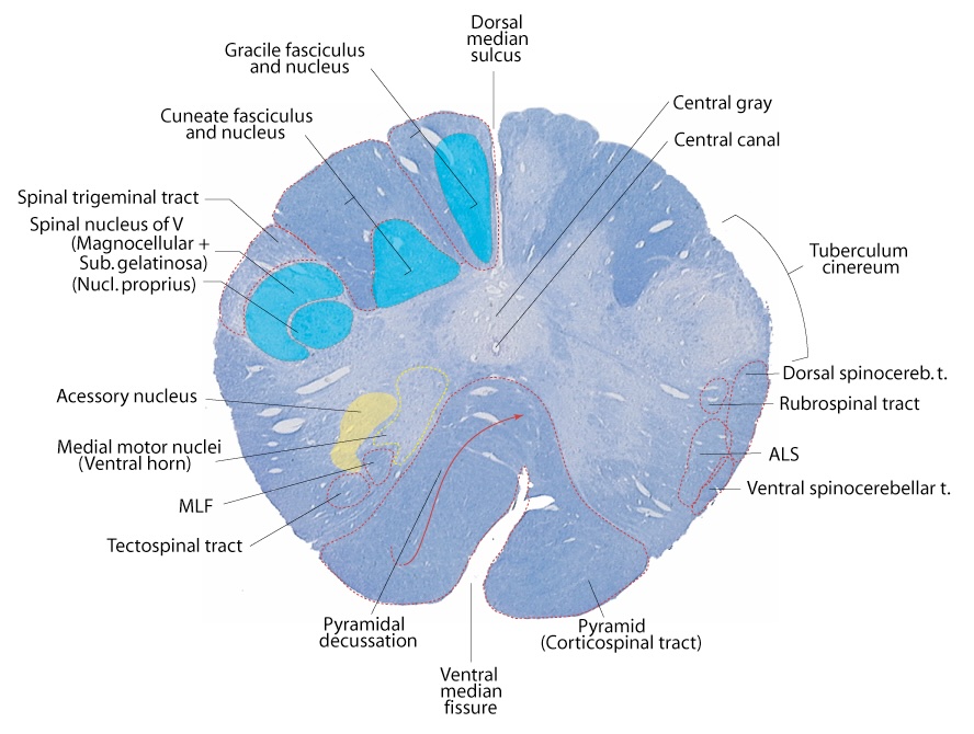
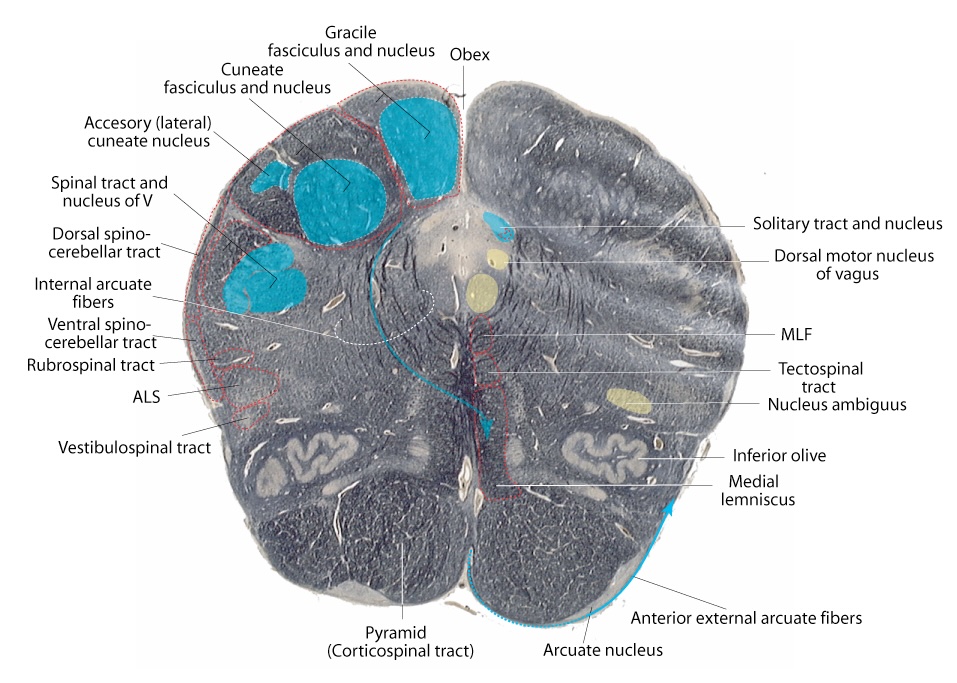
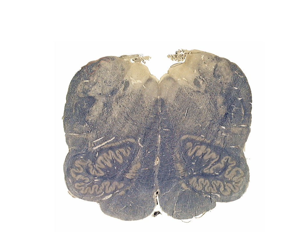
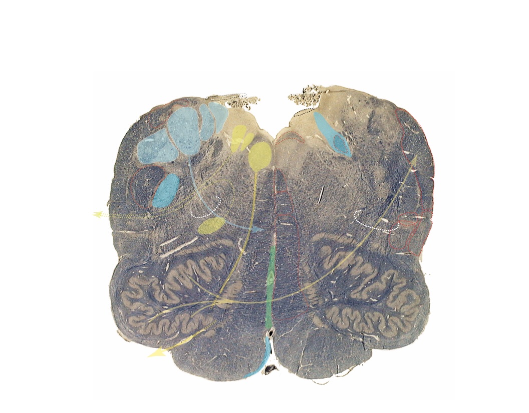
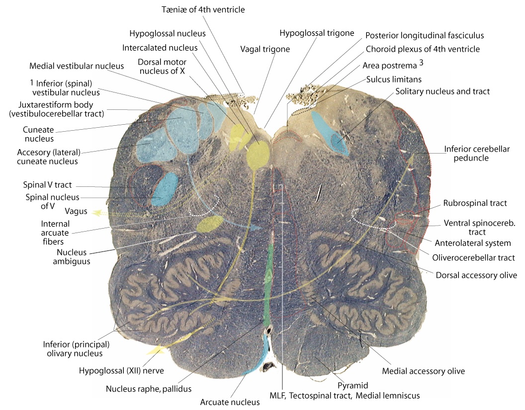
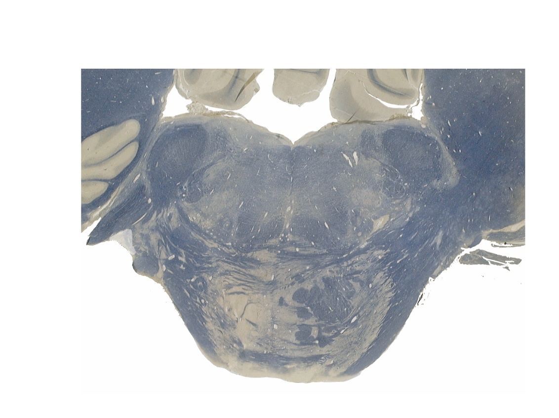
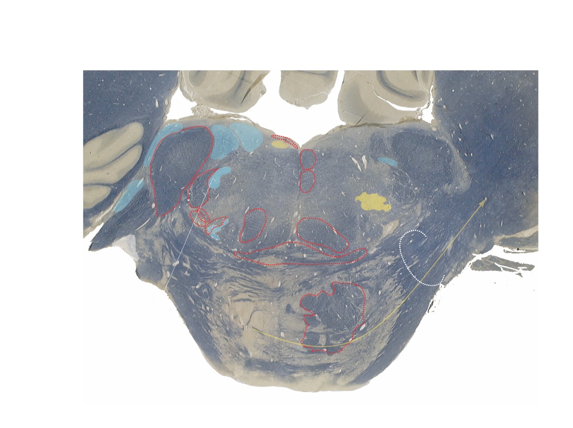
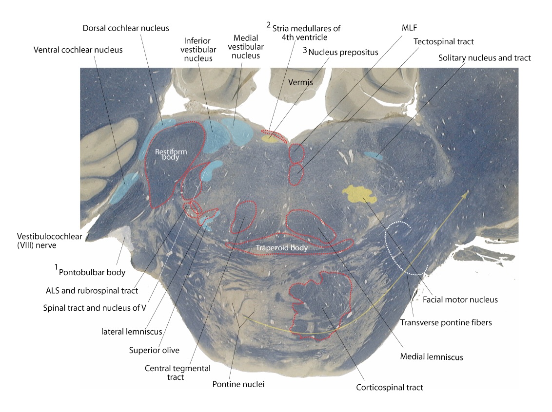
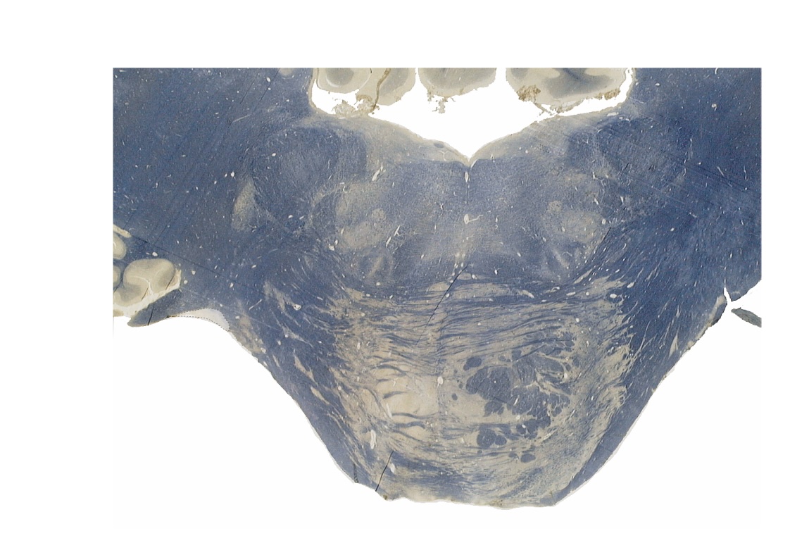
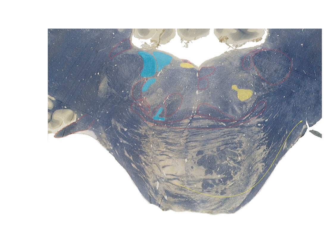
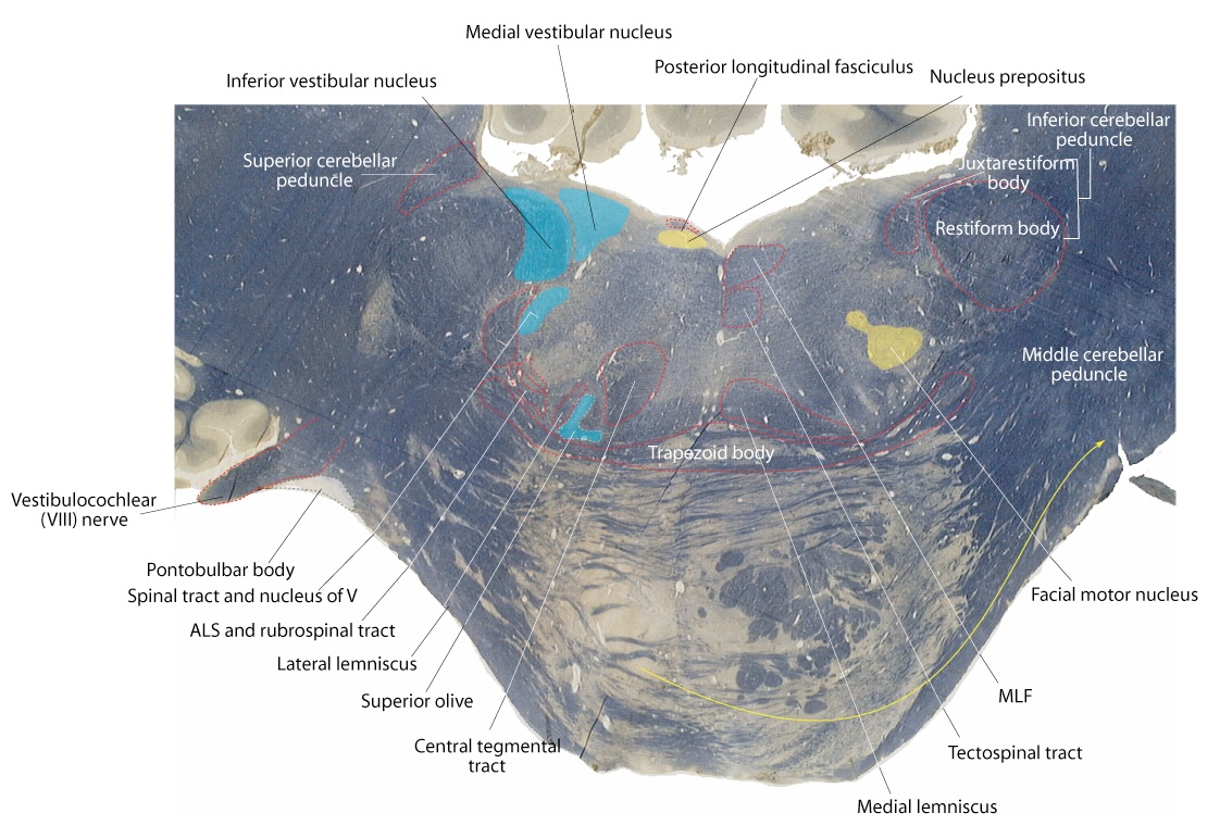
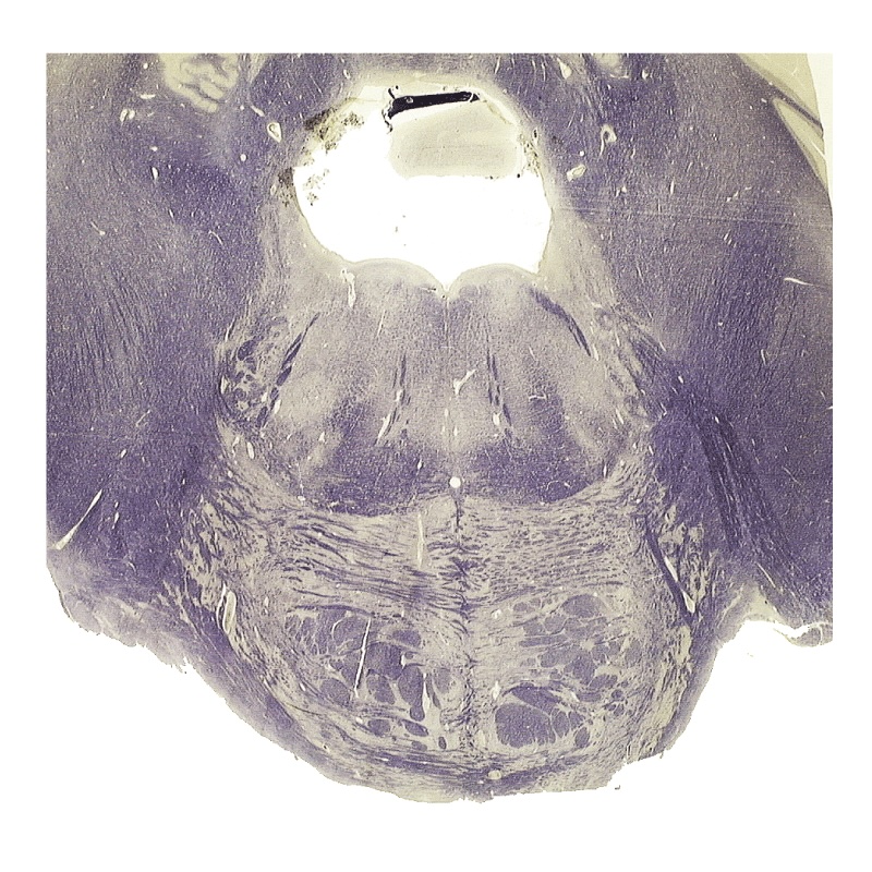
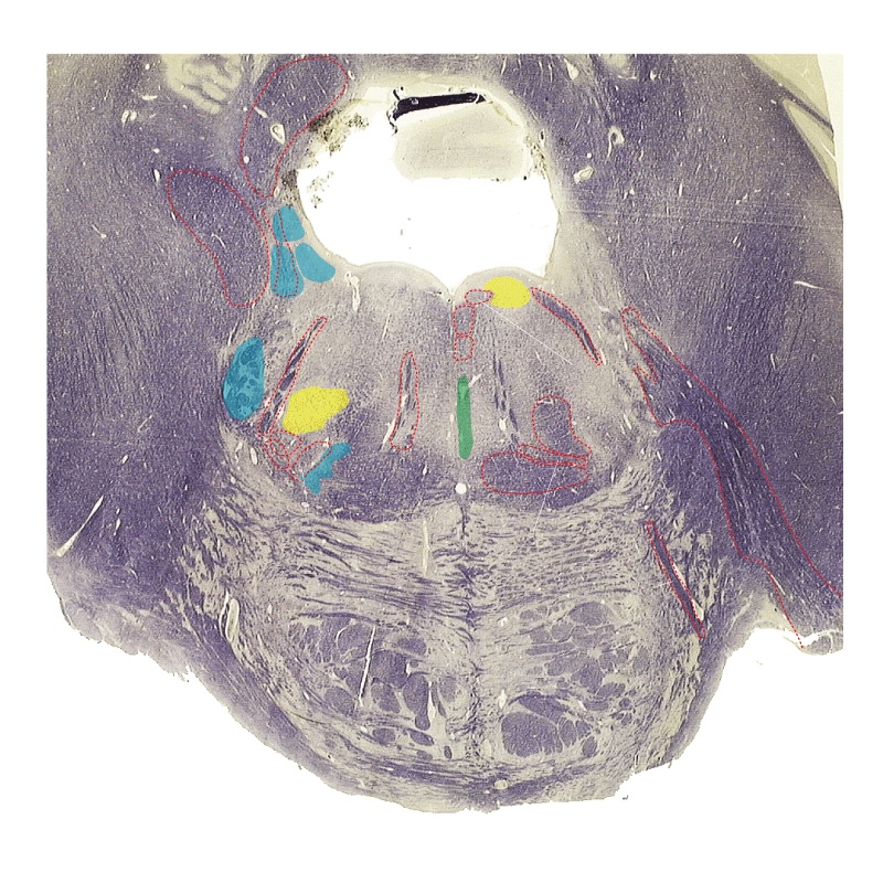
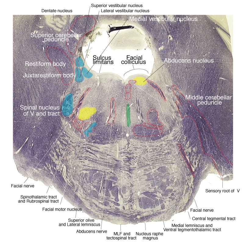
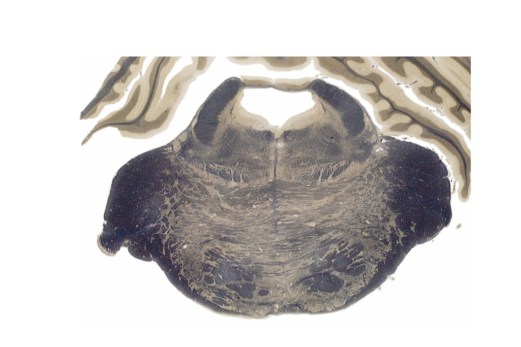
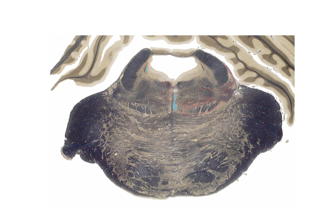
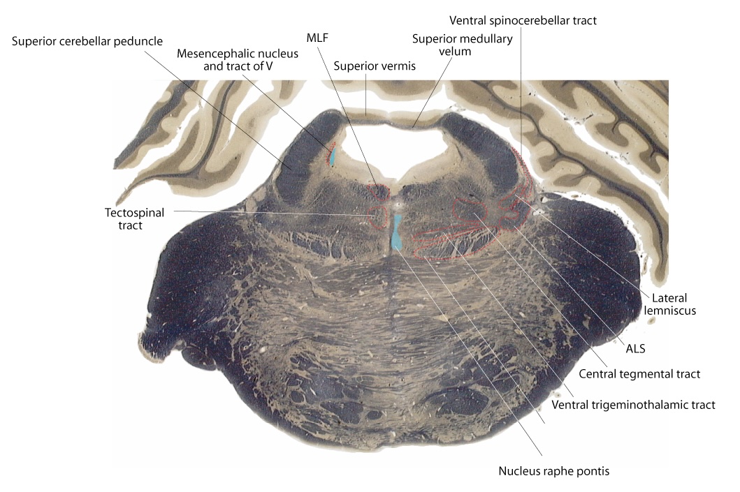
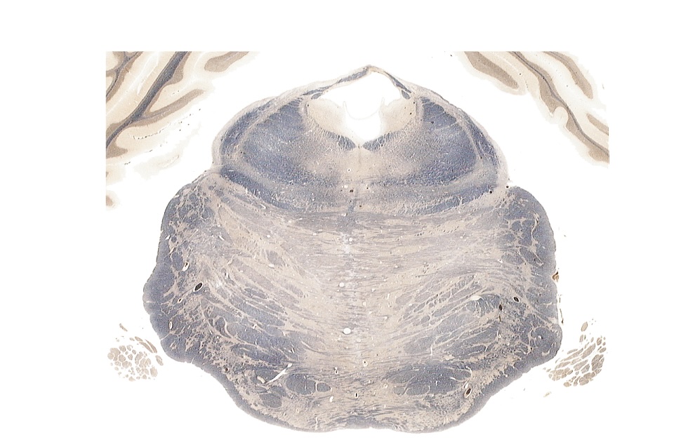
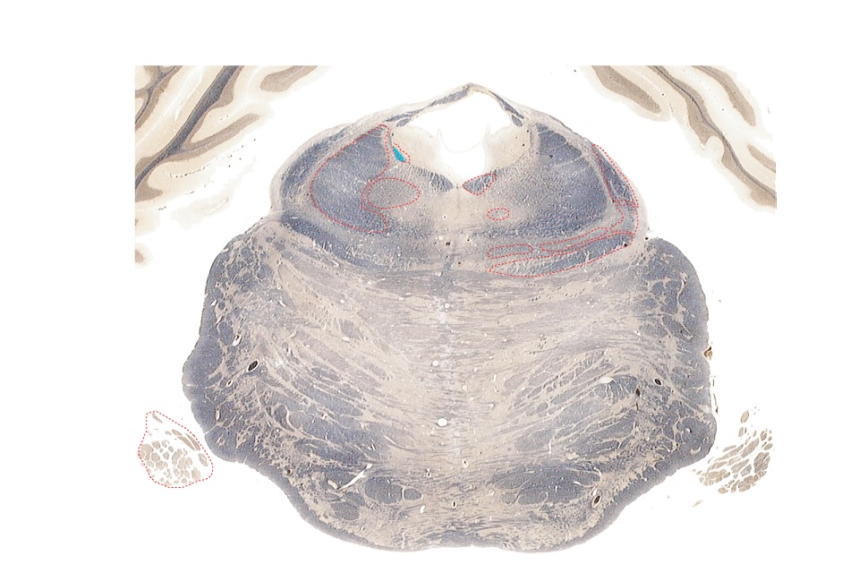
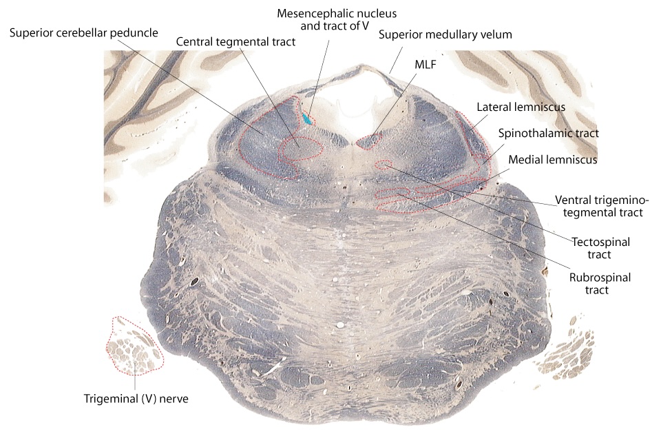
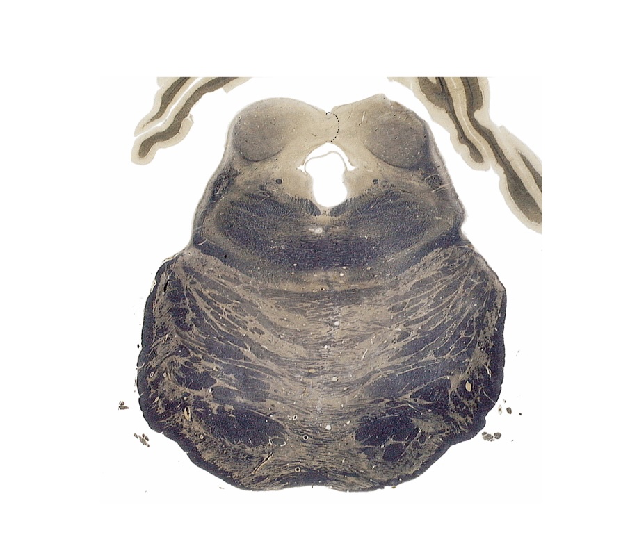
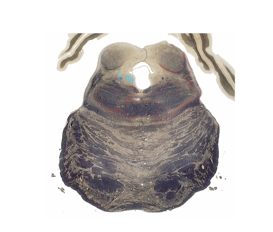
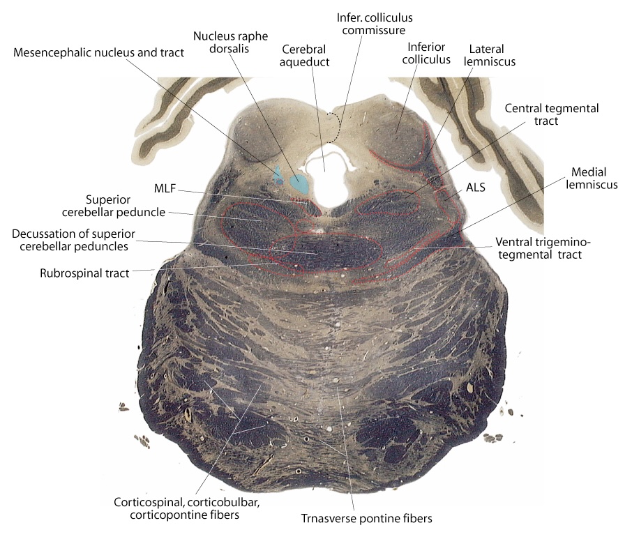
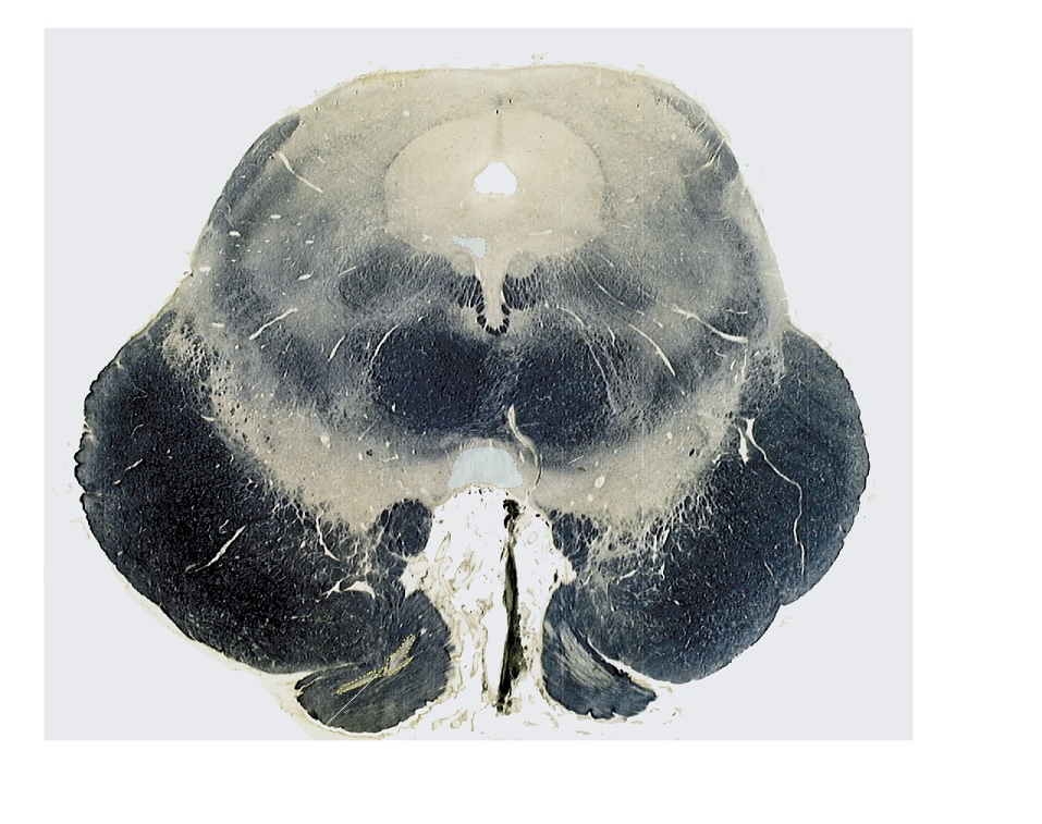
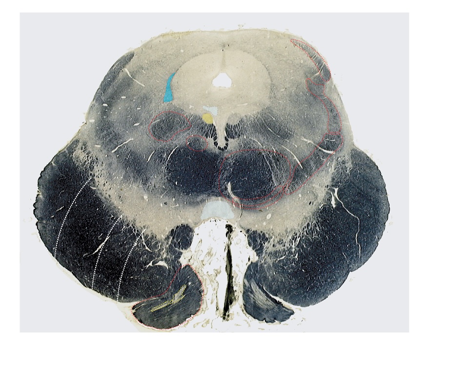
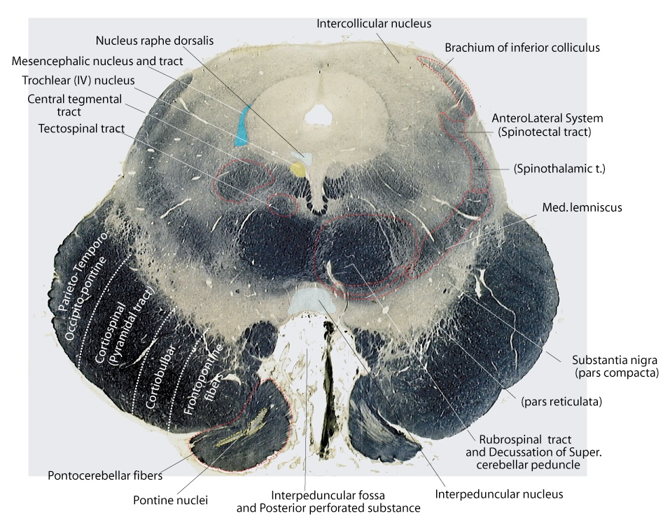
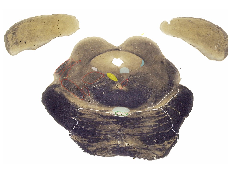
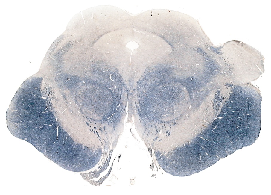
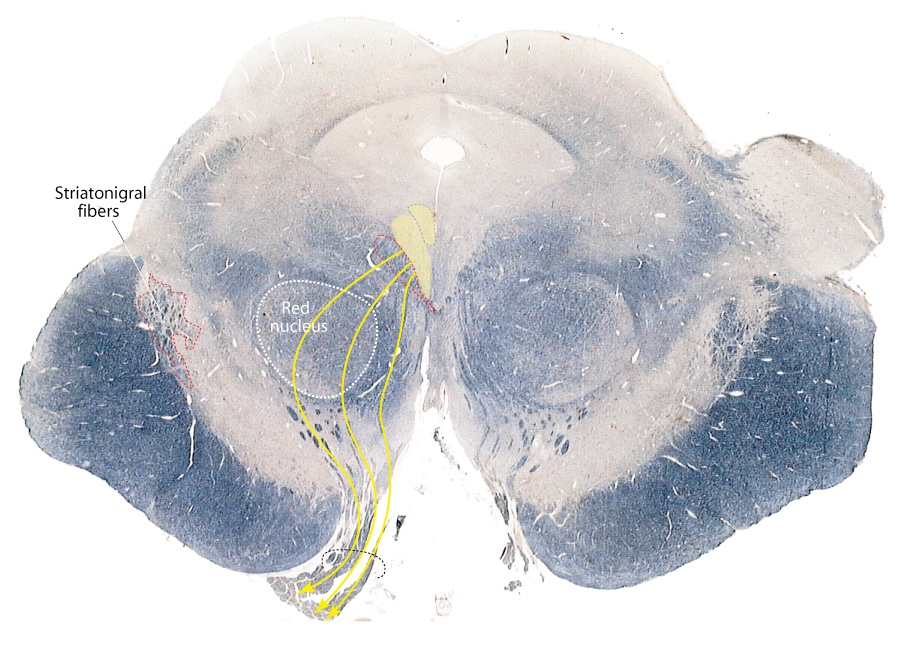
Now using the information in the table as a guide, begin to examine each section and determine the location of the nuclei, the ‘neighborhood’ (other nearby nuclei and tracts) that they and their afferent and efferent axon tracts live in. Remember to use your understanding of the embryology of the brainstem from the Brainstem lectures to help you locate the cranial nerve sensory and motor nuclei. In Figure 4.1you will find each unlabeled section from the myelin stained brainstem series paired with a section in which the tracts and nuclei are labeled. Try to identify the cranial nerve nuclei and the ascending and descending tracts in each unlabeled section and then check your answers using the labeled section. Systematically work your way through the Medullary, Pontine and the Midbrain sections (Figure 4.1). As you identify the cranial nerve nuclei and tracts draw them on the brainstem outline (Interactive 4.1).

After you have successfully located all of the cranial nerve nuclei and tracts in the myelin stained brainstem series, examine the sections in the Medullary, Pontine, Midbrain and Sagittal Tablas in lab. These are additional unlabeled sections through the brainstem to give you practice at identifying the cranial nerve nuclei and brainstem structures in a variety of sections and planes of orientation. Work with your lab instructors as you go through these sections and think about the functional role of each structure.
Sensory Nuclei
Objectives
- Identify the cranial nerves having purely sensory function, motor function and those nerves having both sensory and motor functions.
- Describe the central structures (nuclei and tracts) that are involved in mediating the sensory and motor functions or each cranial nerve and identify where in the brain stem these structures can be found.
Somatic

Trigeminal Nuclei – Function: Faciocranial sensation. In a section of the lower medulla, at the level of the decussation of the pyramids, locate the spinal nucleus of V in the same position as the superficial dorsal horn of the cervical cord. Lateral to the spinal nucleus of V, in a position similar to that of Lissauer’s tract to the dorsal horn of the spinal cord, is the spinal tract of V. Recall that the Vth nerve enters the brain stem at the mid-pontine level. The primary afferent axons carrying sensory information for the face arise from cells of the semilunar (trigeminal) ganglion. These axons contribute to the spinal tract of V and descend adjacent to the spinal nucleus of V from mid-pontine to caudal medullary levels terminating in the nucleus in a somatotopic distribution (Figure 4.2). Trace the spinal nucleus through each brain stem slide, up to the mid-pontine level, just above the facial colliculus. At this level the spinal nucleus and tract end, and the main (principal) nucleus of V can be found instead. Dorsal and slightly medial to the main nucleus of V, the mesencephalic nucleus of V emerges. Initially it occupies a position just ventral to the superior cerebellar peduncle, but as it extends into the midbrain it comes to occupy a position bordering the lateral edge of the periaqueductal gray. Cells of the mesencephalic nucleus are the only primary sensory neurons within the brain. The neurons in this nucleus convey proprioceptive and pressure information from the teeth, periodontium, hard palate and joint capsules. Where are the primary sensory neurons found for other sensory pathways of the brain and spinal cord?
Visceral
Nucleus Solitarius – Function: Taste; Visceral afferents. Like the trigeminal spinal nucleus, the solitarius is a long cell column extending the length of the medulla. The primary afferent axons of ganglion cells form a tract that is surrounded by the cells of the nucleus. You will find this tract dorsal and medial to the spinal nucleus of V.
Special

Vestibular Nucleus – Function: Position sense. The complex of four vestibular nuclei extends from the mid-medulla to mid-pons. Figure 4.3 gives the levels of each nucleus. The boundaries of the inferior nucleus can be distinguished by its characteristic content of myelinated axons, which are mostly primary afferents from the vestibular ganglion descending through the medulla from their entry at the medulla-pons junction.
Locate the medial longitudinal fasciculus (MLF) at the midline, just ventral to the nuclei bordering the ventricle. It maintains this position throughout the brain stem from the oculomotor nucleus to the decussation of the pyramids and then shifts to a ventromedial position in the cervical cord. The ascending MLF interconnects the vestibular, oculomotor, trochlear and abducens nuclei. The descending MLF mainly arises from the medial vestibular nucleus and descends as the medial vestibulospinal tract in the spinal cord. What other descending tract arises from one of the vestibular nuclei?
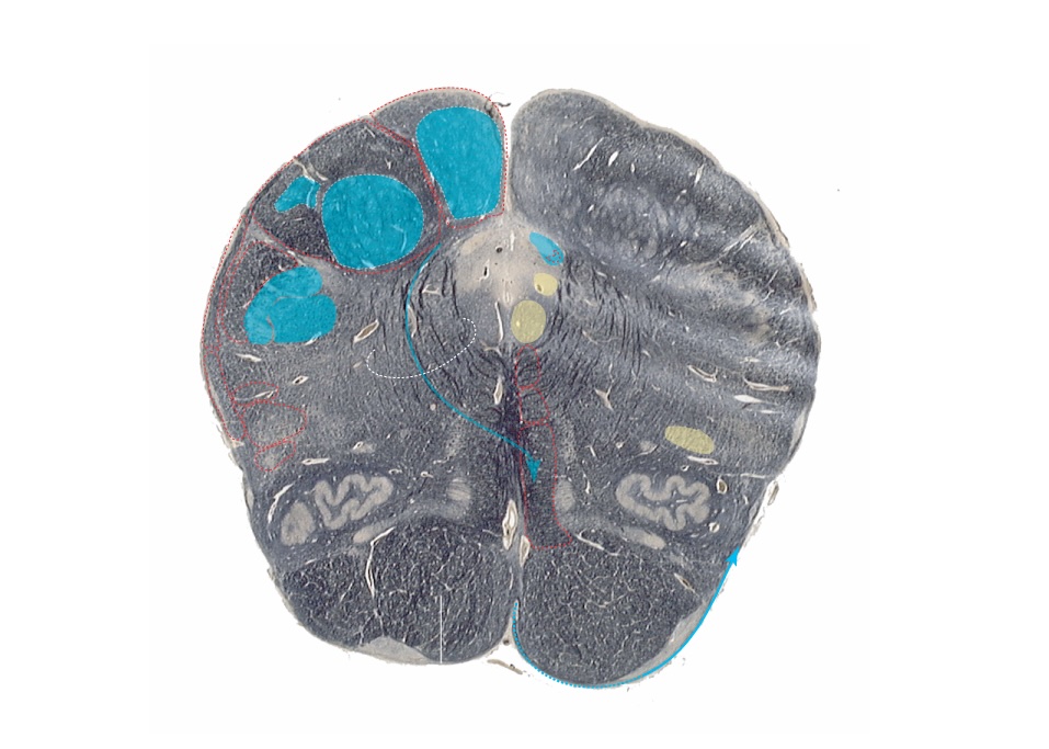
Review the positions of the sensory nuclei in Figure 4.4.

Motor Nuclei
Objectives
- Identify the cranial nerves having purely sensory function, motor function and those nerves having both sensory and motor functions.
- Describe the central structures (nuclei and tracts) that are involved in mediating the sensory and motor functions or each cranial nerve and identify where in the brain stem these structures can be found.
Somatic Motor
Oculomotor Nucleus – Function: Innervates 4 extraocular muscles and the levator palpebrae. On a section through the superior colliculus, you will find the oculomotor complex ventral to the periaqueductal gray. It consists of cell groups that supply striated muscles. Note the staining of the myelinated axons arising from these motor neurons. The MLF lies laterally, along the border of the nucleus.
Trochlear Nucleus – Function: Innervates the superior oblique. This nucleus can be found at the level of the inferior colliculus, but because the trochlear nucleus is small, some sections may not pass through the nucleus (Find a neighbor that has one!). Some sections may also have the decussation, which is located slightly caudal to the level of the nucleus. The MLF is directly ventral to the nucleus.
Abducens Nucleus – Function: Innervates the lateral rectus. Found in the mid-pons, lying on the medial floor of the fourth ventricle, the right and left nuclei form distinctive, paired medial bulges. The fascicles of the VIIth nerve arch over this nucleus and form these “bulges” on the floor of the fourth ventricle known as the facial colliculus. In sections through this area, the fibers of the facial nerve can often be seen medial and ventral to the abducens nucleus, as they course medial and dorsal from the facial nucleus and then after coursing over the abducens nucleus they angle ventral and lateral toward their point of exit at the ponto-medullary junction.
Hypoglossal Nucleus – Function: Innervates the muscles of the tongue. This nucleus extends from the obex almost to the lateral recess of the IVth ventricle. It maintains a midline position and has a characteristic myelinated appearance. The exit of the nerve can be found between the olive and pyramids on the lateral surface of the medulla.
Branchial Motor
Trigeminal Motor Nucleus – Function: Innervates the muscles of mastication. Note its position medial to the main sensory nucleus of V at mid-pontine levels.
Facial Nucleus – Function: Innervates the muscles of facial expression. This nucleus is located in the caudal pons, and lies ventral and medial to the trigeminal spinal nucleus. Efferent fibers exiting this nucleus to become the facial nerve travel rostral and dorsal to the ventricular surface where they arch over the abducens nucleus to form the facial colliculus on the floor of the IVth ventricle. After traveling over the abducens nucleus the axons of this nerve proceed in a ventral and lateral course to exit from the brainstem at the ponto-medullary junction.
Ambiguus Nucleus – Function: Innervates the muscles of the pharynx and larynx. These scattered cells are found throughout the length of the lower medulla. They are located dorsal to the inferior olive, lateral to the medial lemniscus, and medial and ventral to the spinal nucleus of V.
Spinal Accessory Nucleus – Function: Innervates trapezius and sternocleidomastoid muscles. This nucleus extends from C5 or C6 to the level of the decussation of the pyramids, and occupies the lateral region of the ventral horn in the spinal cord.
Visceral Motor
Edinger-Westphal Nucleus – Function: Innervates ganglia of the ciliary muscles and iris sphincter. This is a lighter staining area dorsal to the somatic motor region of the oculomotor complex.
Motor Nucleus of the Vagus – Function: Innervates ganglia to viscera of neck, thorax, and abdomen. This pale nucleus lies on the ventricular surface, dorsolateral to the hypoglossal nucleus and extends throughout the lower and middle medulla. What is the functional significance of the fact that the Edinger-Westphal and motor vagus nuclei are pale staining compared to the other cranial motor nuclei in myelin stained sections? After identifying these nuclei on the sections of this lab use the Kodachrome displays of horizontal, coronal and sagittal sections through the human brain to try and build a three dimensional perspective of the brainstem.
Review the positions of the cranial nerve nuclei in Figure 4.5A and then test yourself on the unlabeled figure in Figure 4.5B.
Blood Supply and Ascending/Descending Tracts
Objectives
- Identify the position of the major ascending and the descending pathways of the spinal cord as they traverse the brain stem. Describe how these spinal pathways are spatially related to the ascending and descending pathways of the cranial nerves. Similarly, identify the spatial relationship between the cranial nerve motor and sensory nuclei and these spinal pathways.
- Use the acquired knowledge of the relative spatial positioning of sensory and motor pathways and nuclear groups within the brainstem to anticipate the functional implications of various combinations of vascular and traumatic injury to areas of the brain steam and the exiting cranial nerves.
- Identify the source and distribution of the arterial supply to discrete regions of the medulla, pons and midbrain and identify the functional deficits that would occur following compromise to each.
Blood Supply
Use Figure 4.6 below of the blood supply for the medulla, pons and midbrain to become familiar with the vascular organization at each level of the brainstem. Different sources will have minor variations when discussing the blood supply of the brain since there can be substantial variation in the details from person to person. In this course we use the pattern described in Blumenfeld, Neuroanatomy Through Clinical Cases, Sinauer Associates, which is consistent with the majority of texts in the field.


On Figures 4.7A-C, name the blood vessel supplying the shaded region and the important cranial nerve nuclei found in that region. What functional deficits might be associated vascular compromise in each area?


Ascending and Descending Pathways
Use Figure 4.8A-B to identify the ascending and descending pathways carrying sensory and motor information through the brainstem. Pathways you should be able to identify include the: Dorsal Column/Medial Lemniscus, Spinothalamic, Spinocerebellar tracts, Trigeminothalamic, Corticospinal, Corticobulbar, Rubrospinal and the MLF.


Review
Question 1: Name three purely somatomotor cranial nerves and levels of their nuclei in brain stem.
• Trochlear Nerve – mesencephalon
• Abducens Nerve – pons
• Hypoglossal Nerve – medulla
Question 2: Name two cranial nerves that are responsible for the pupillary reflex.
Optic and Oculomotor nerve.
Questions 3-5
Question 6: Name the nucleus where sensory fibers that conduct pain and temperature from the face make their first synapse, i.e. second order neuron.
Spinal nucleus of trigeminal nerve.
Question 7: Identify the different arteries supplying the medulla in the illustration below:


Question 8: The figures below illustrate a common brain stem vascular infarct of the artery supplying the dorsal lateral area of the medulla. The pattern of deficits that follow is termed Wallenberg’s syndrome. Answer the following questions:

Question 8a: What artery supplies the area of damage?
PICA or the Vertebral Artery
Question 8b: What is the effect on pain and temperature sensation for the face and body (side and affected structure)?
Ipsilateral P&T loss for the face due to damage of the spinal n and tract of V and contralateral loss of P&T for the body due to damage of the spinothalamic tract
Question 8c: Patients with this syndrome typically complain of Ipsilateral ataxia, vertigo, nystagmus, nausea. This would be due to damage to what structure(s)?
Vestibular nuclei and inferior cerebellar peduncle (ICP)
Question 8d: Decreased ipsilateral taste and hoarsness (dysphasia) are present due to involvement of what nuclei?
Nucleus Solitarius and Ambiguus, respectively

