Proximal Lower Extremity
Lab Summary
The structures of the proximal lower extremity are considered from anterior, femoral triangle, medial, lateral and gluteal perspectives. The neurovascular structures of the femoral triangle are key in a vast array of clinical scenarios. The hip joint is discussed in terms of surrounding structures, anatomic and surgical approaches and its basic configuration. Vasculature and innervation of the lower extremity are taught with emphasis on their courses and functionality. Considerable time is spent on the gluteal region with its connections to pelvis and perineum.
Lab Objectives
- Describe the boundaries and contents of the femoral triangle.
- Describe the major muscle groups of the thigh and their innervation.
- Describe the vascular supply to the lower extremity from the inguinal ligament to popliteal fossa.
- Describe the superficial and deep venous drainages of the lower extremity.
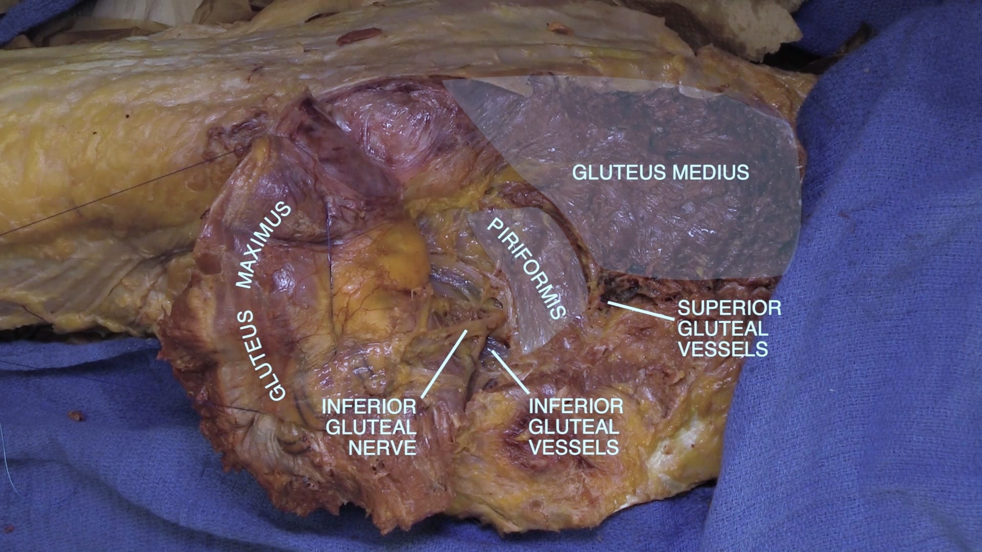
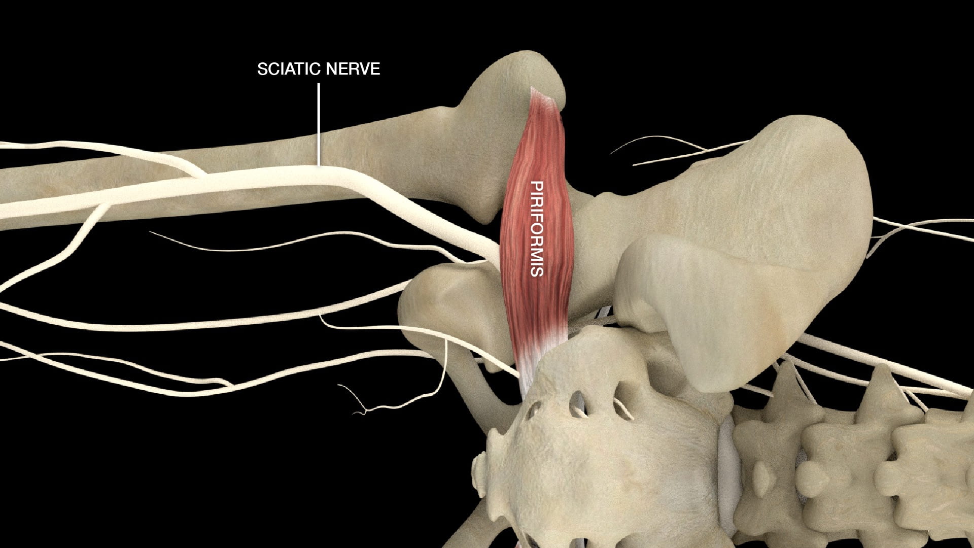
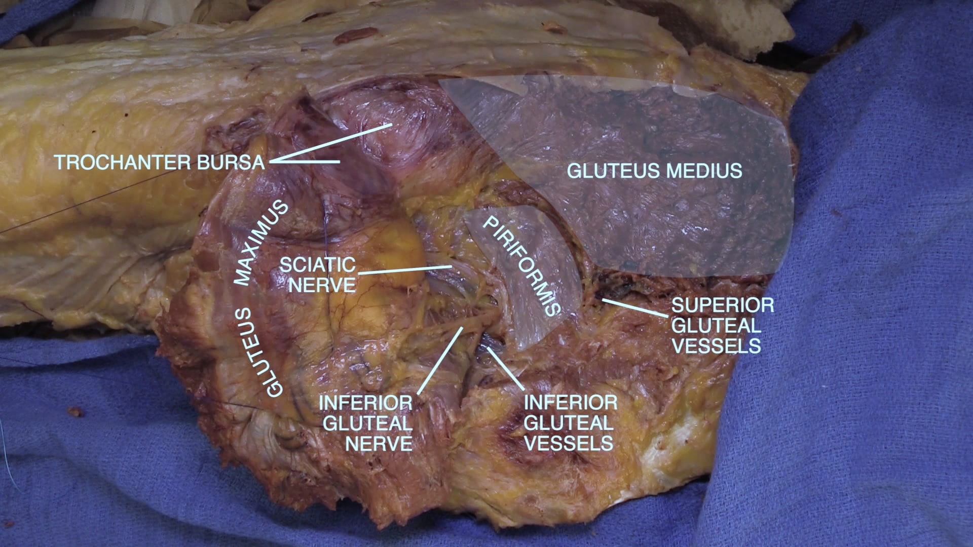
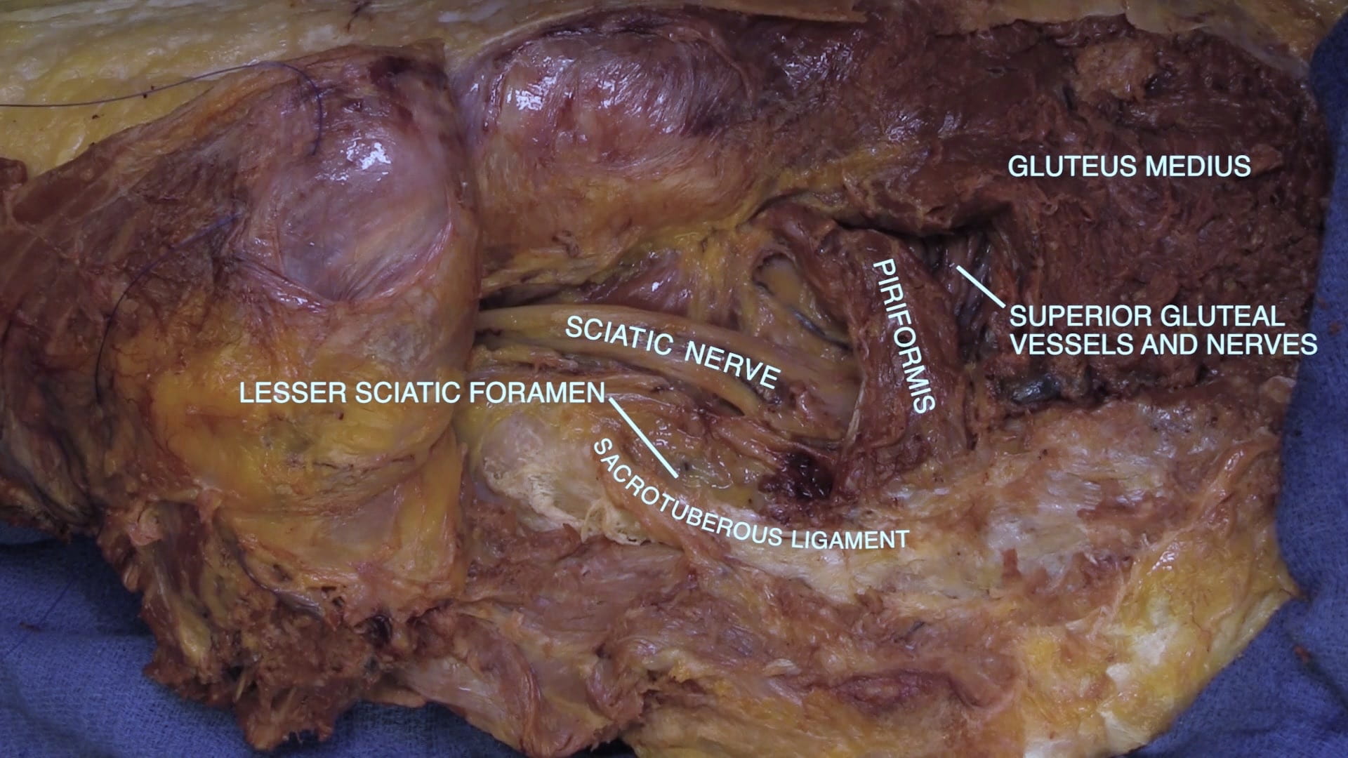
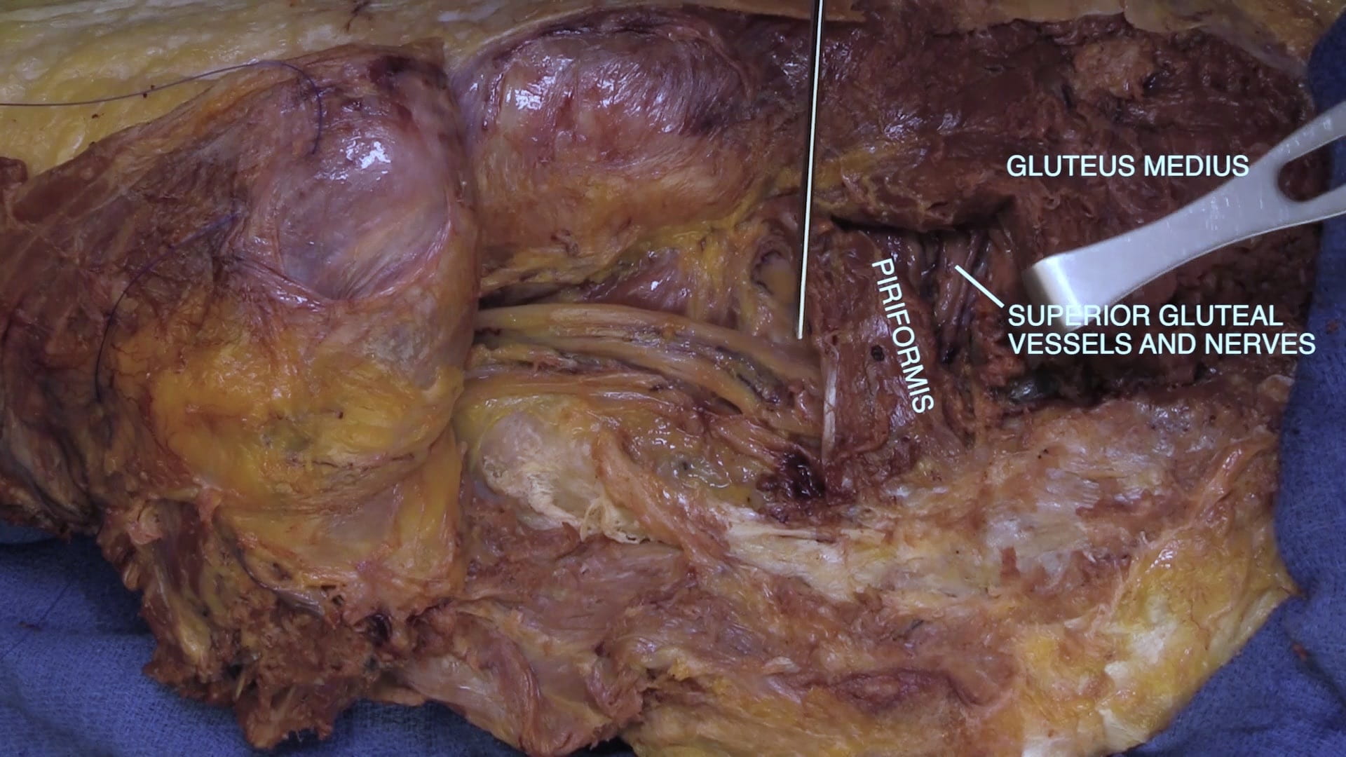
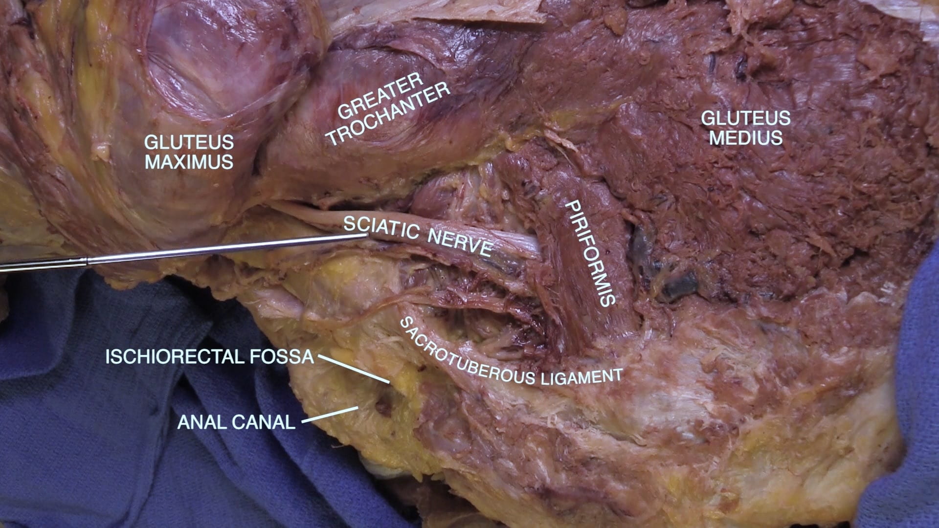
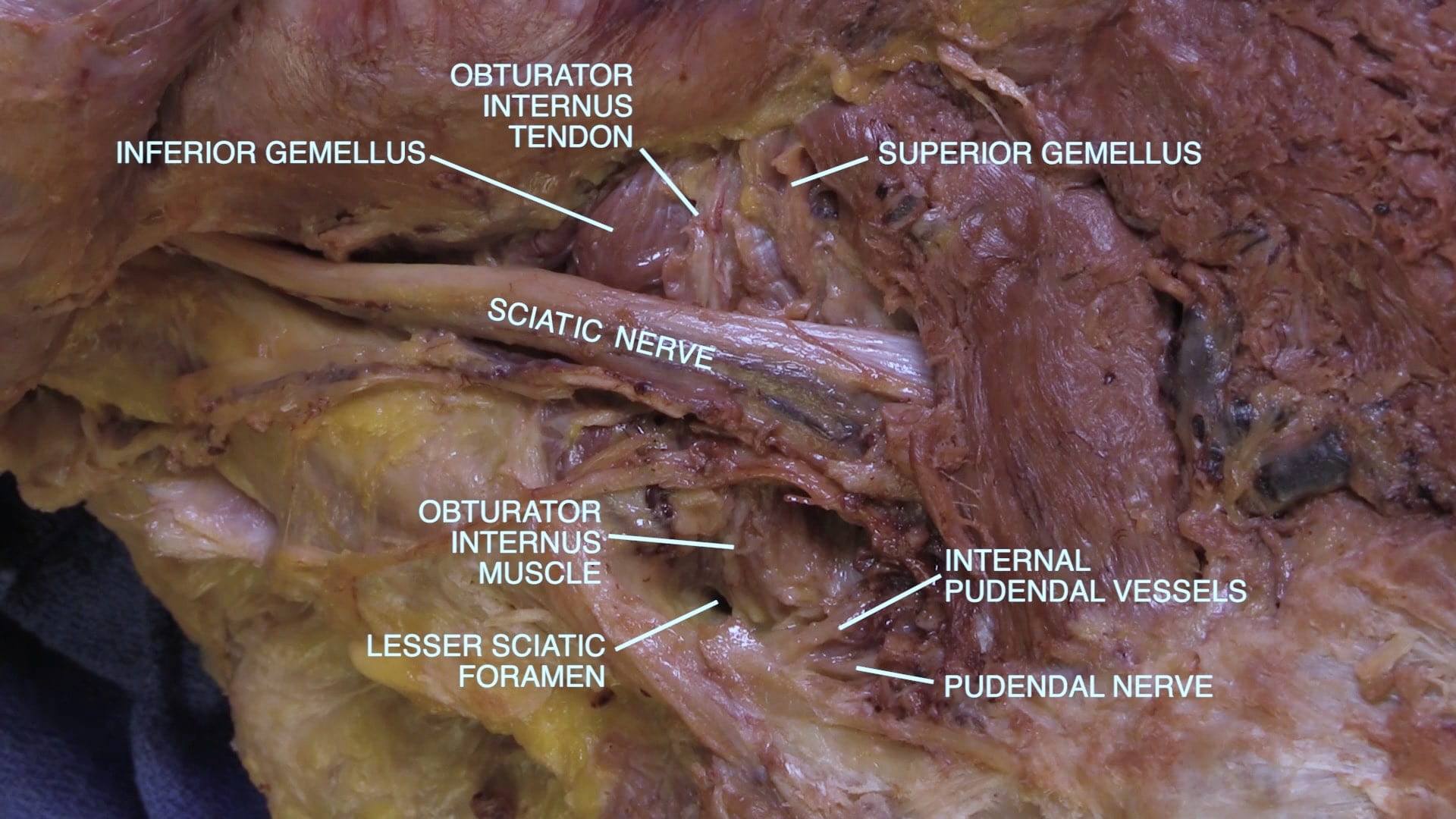
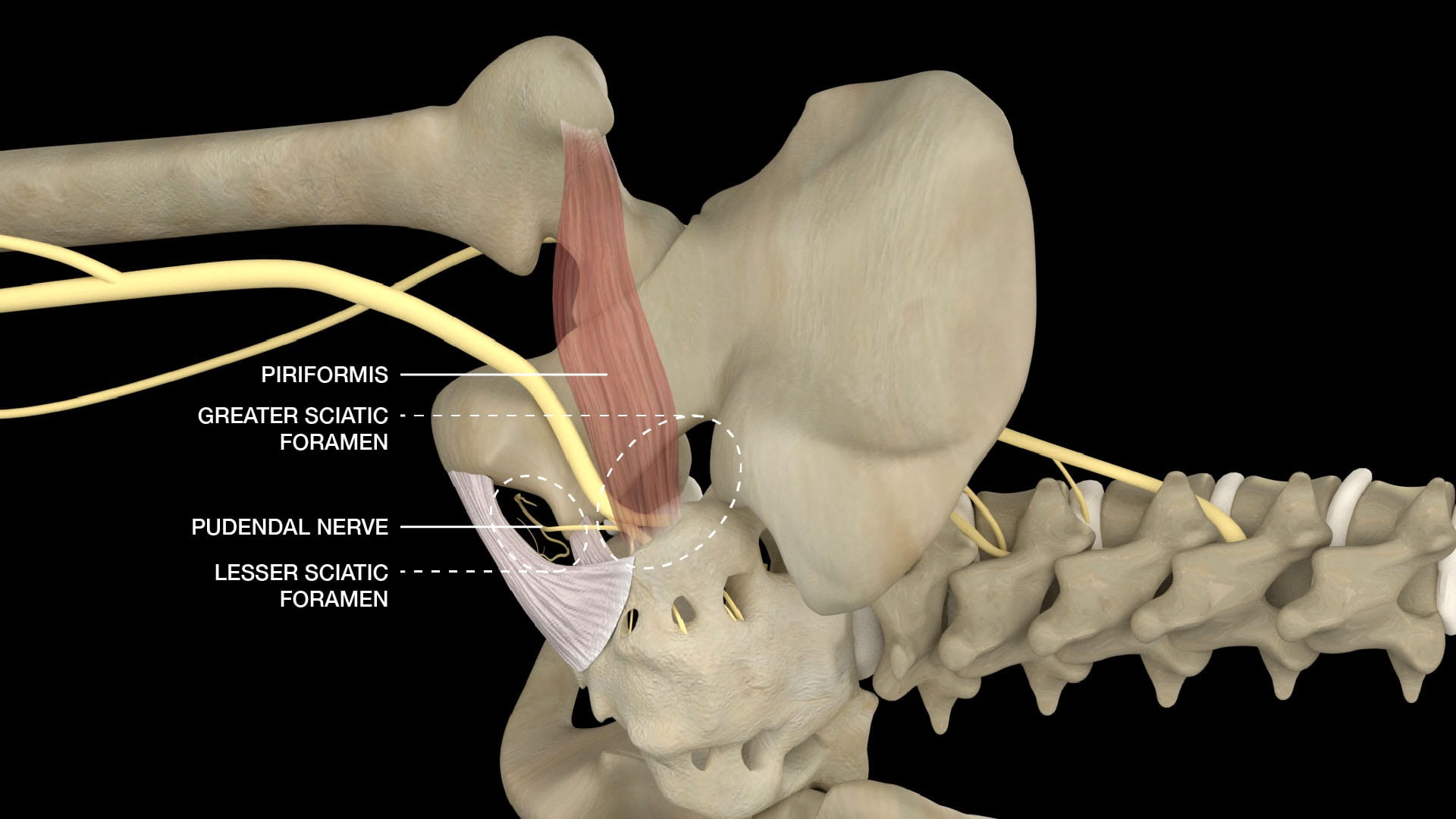
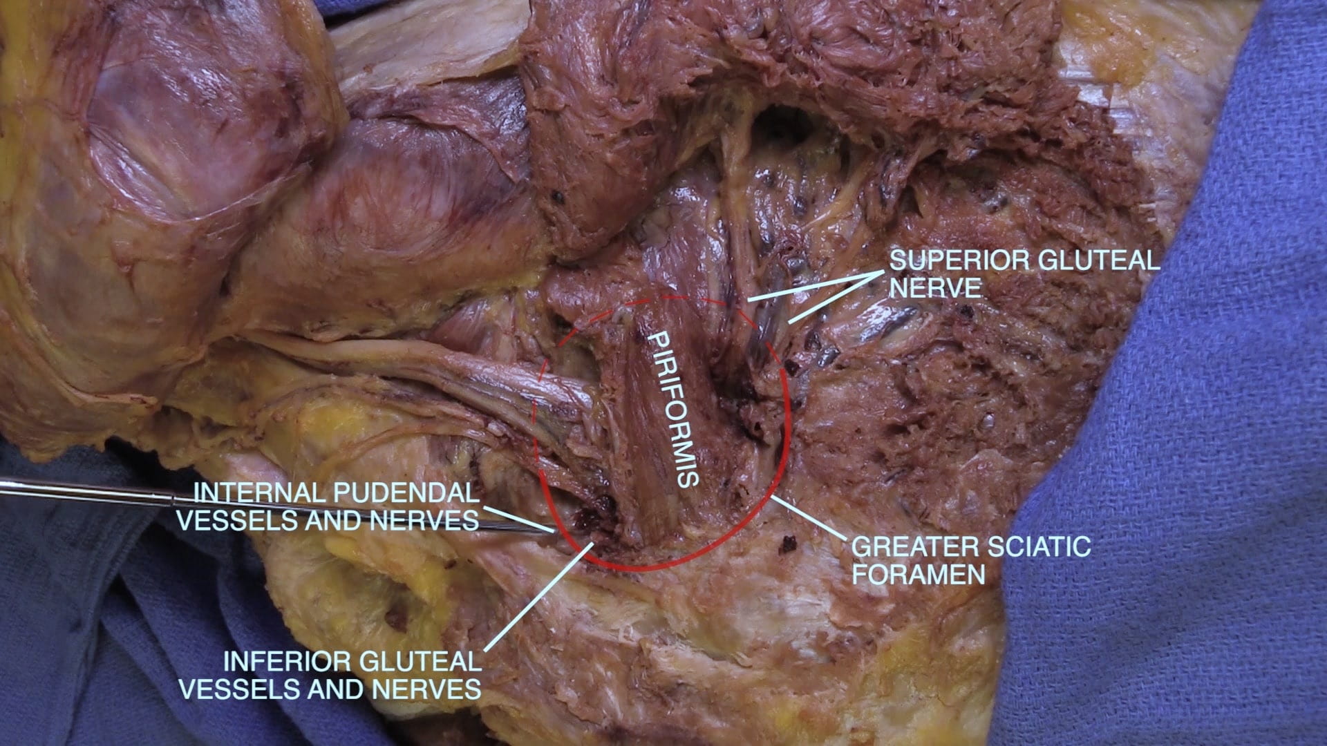
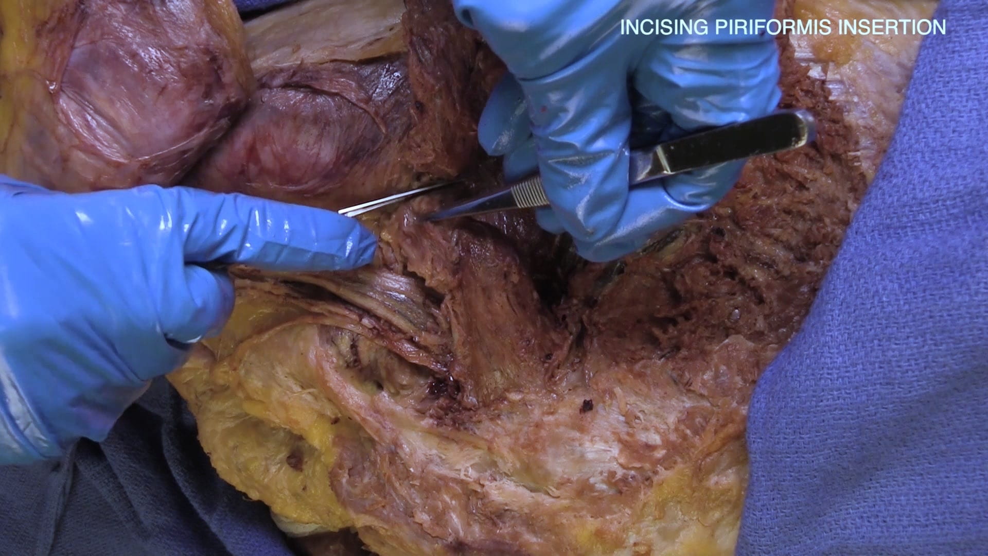
- Describe the relationship of the piriformis muscle to the sciatic nerve and the gluteal nerves and vessels.
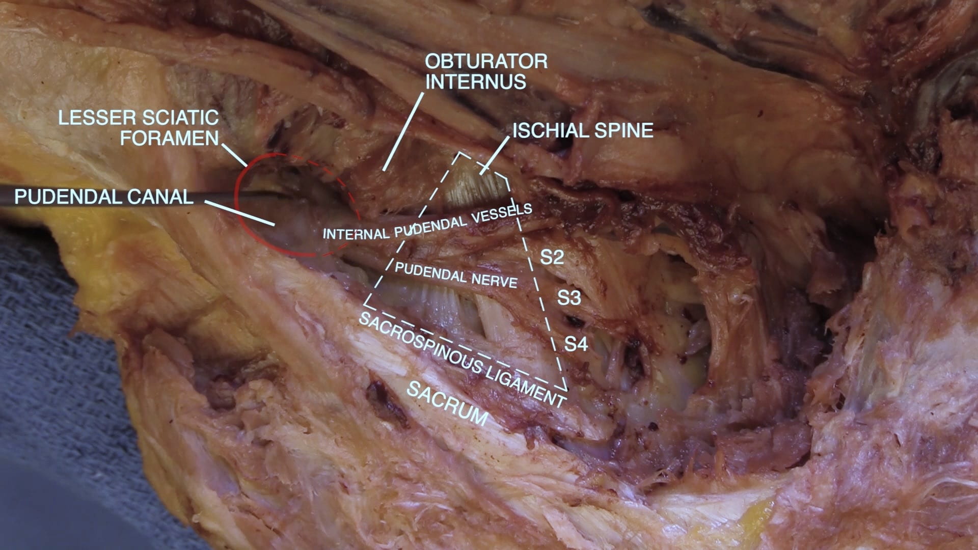
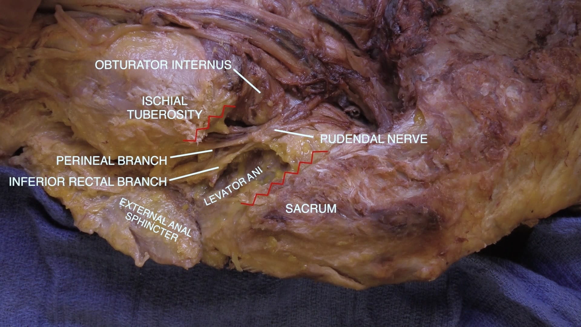
- Describe the course of the pudendal nerve from sacrum to perineum.
- Name the functions of the pudendal nerve.
Lecture List
Anterior Thigh, Femoral Triangle, Medial Thigh, Lateral Thigh, Gluteal Region Part I, Gluteal Region Part II
Anterior Thigh
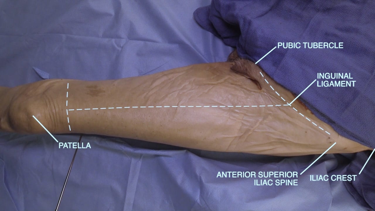
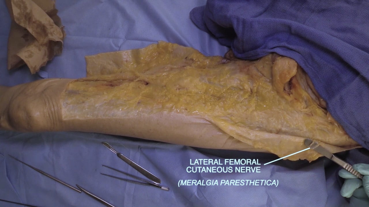
Exposing the Anterior Thigh
Superficial View Anterior Thigh
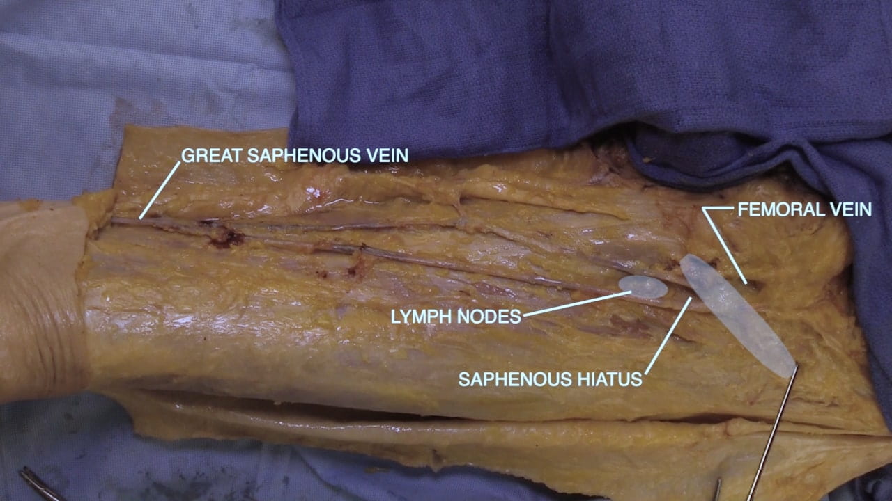
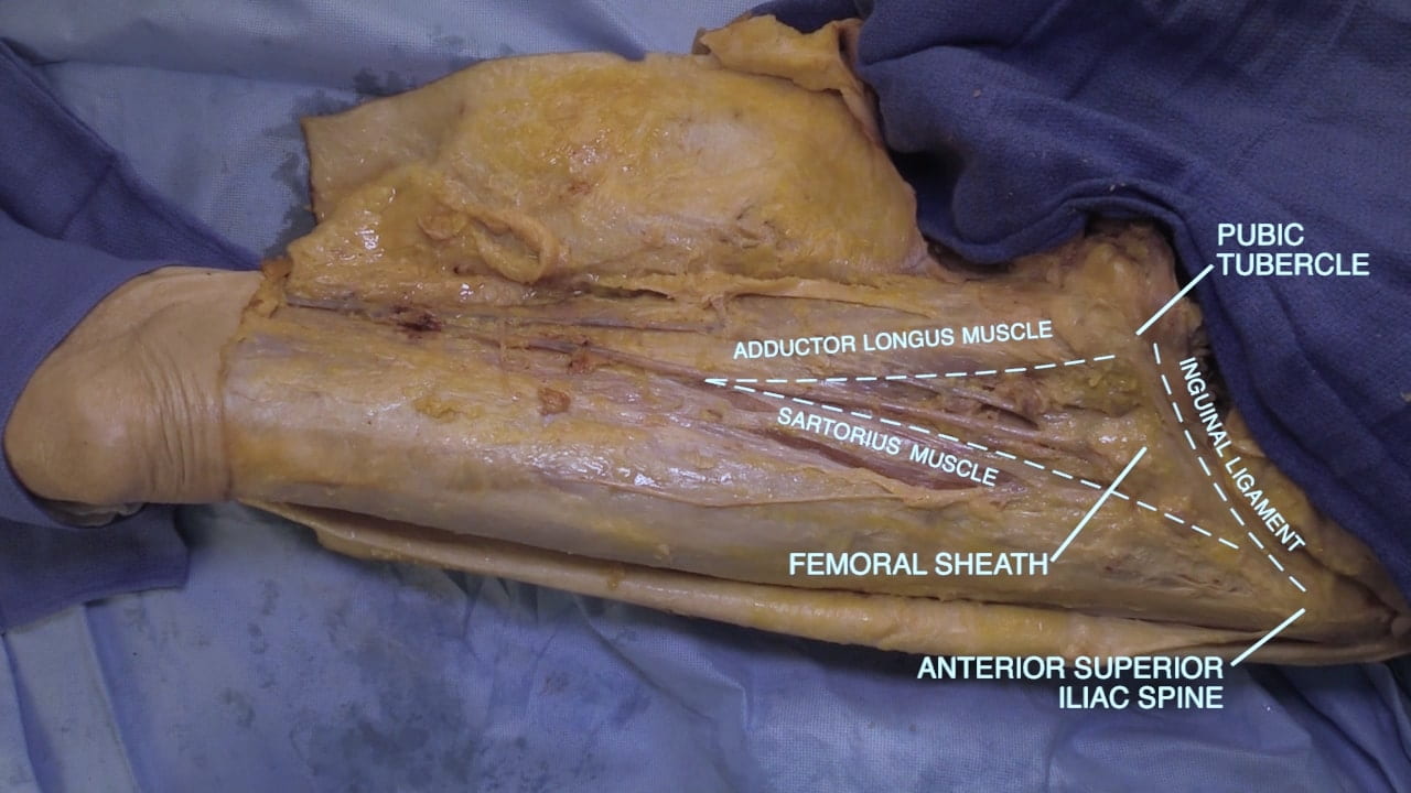
Dissect the superficial fascia on the medial thigh to expose the great saphenous vein from the lower thigh to the saphenous hiatus where it drains into the femoral vein.
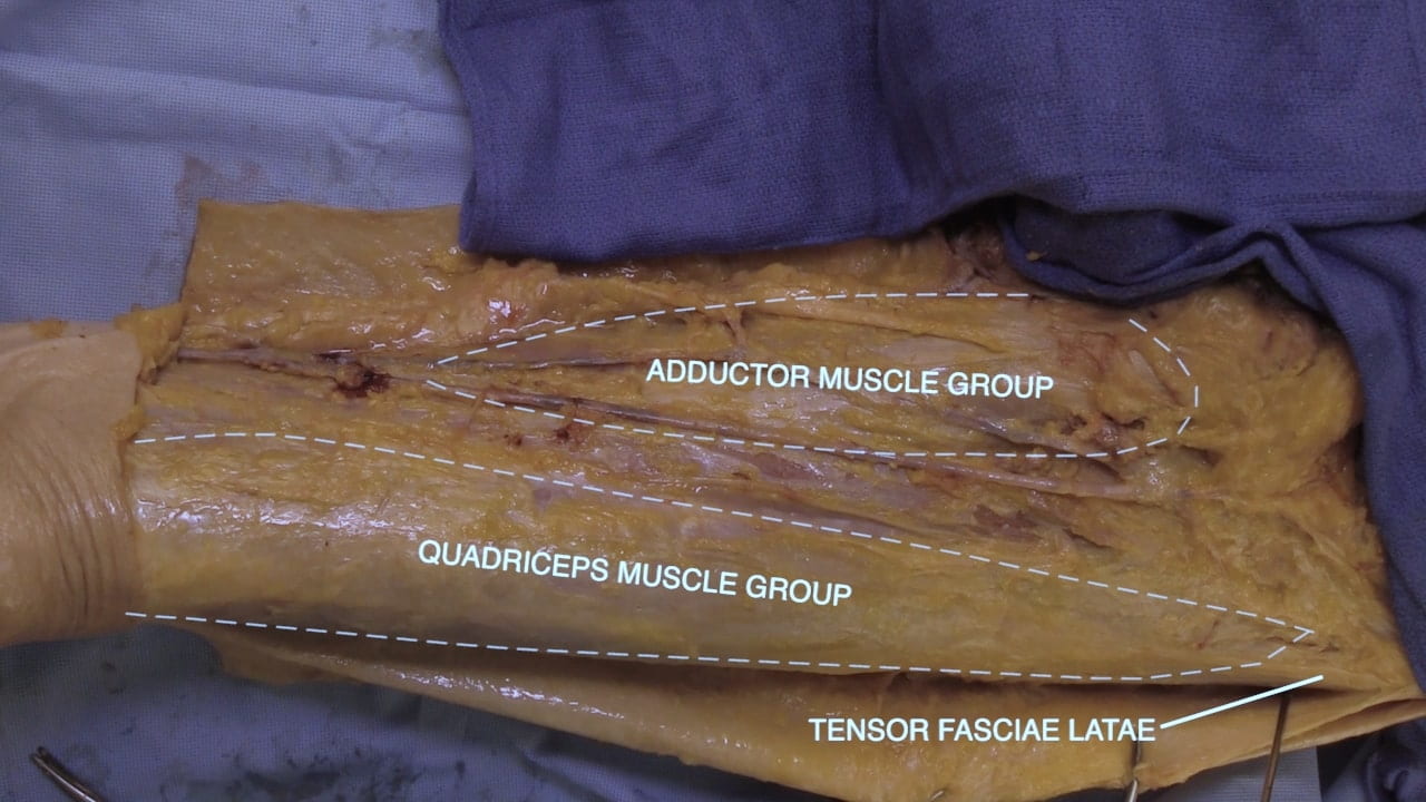
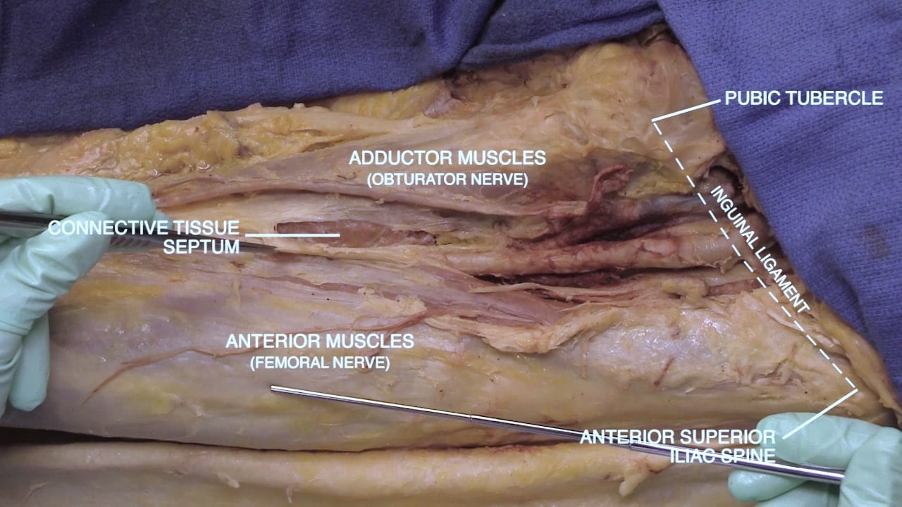
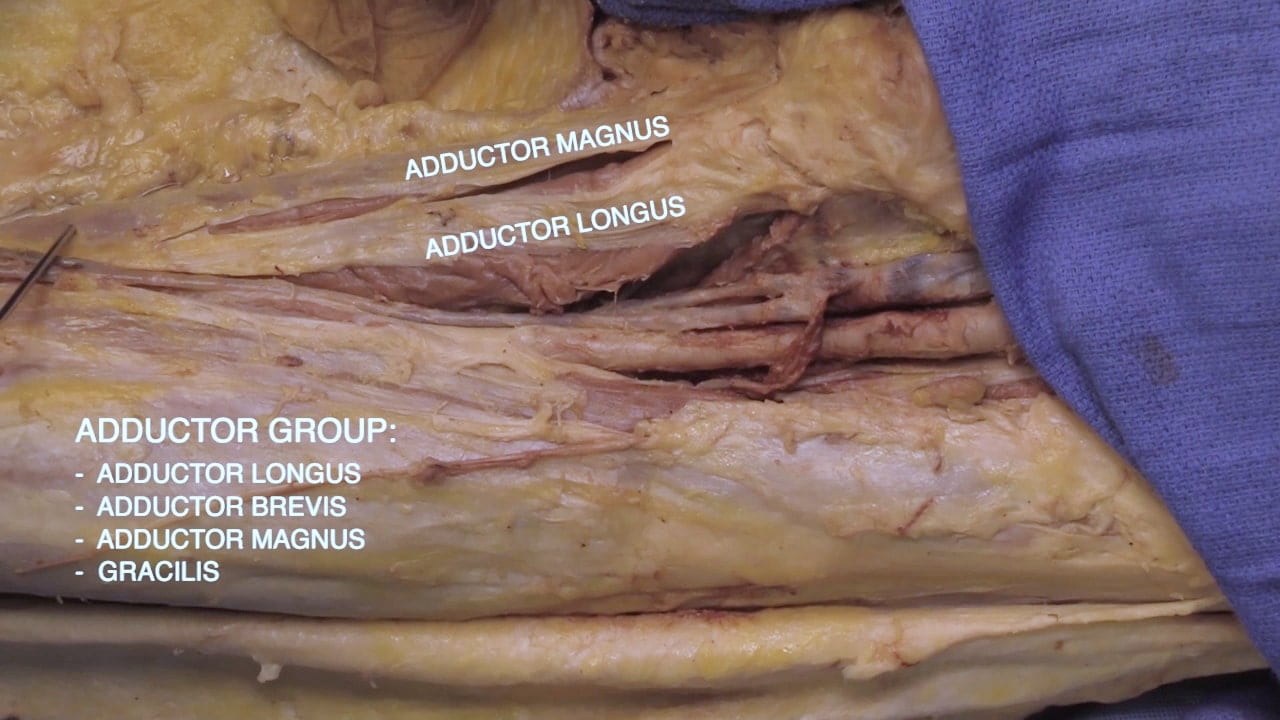
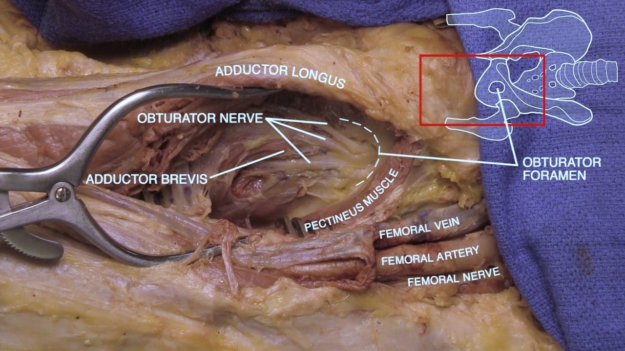
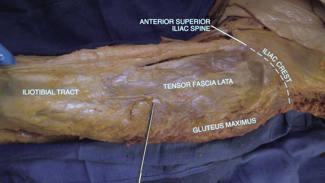
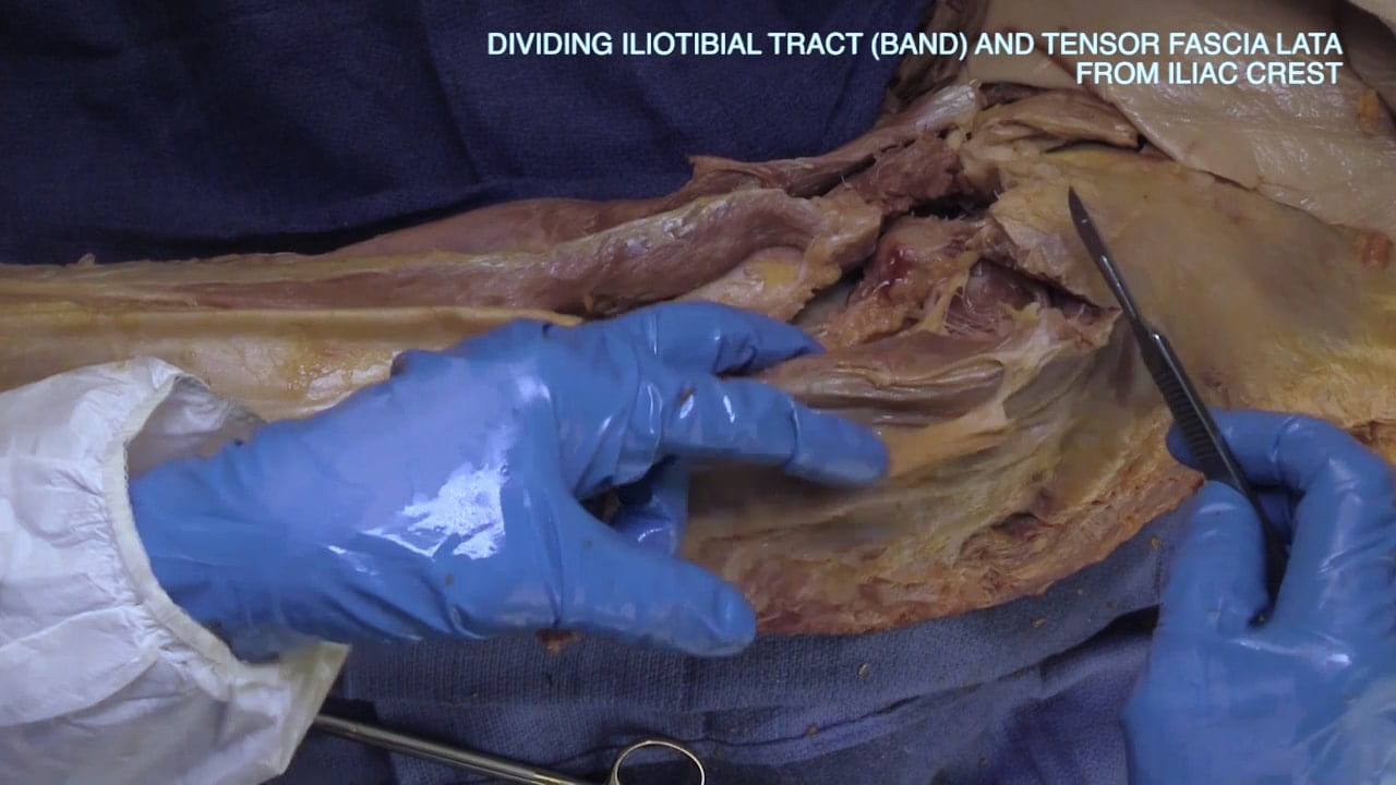
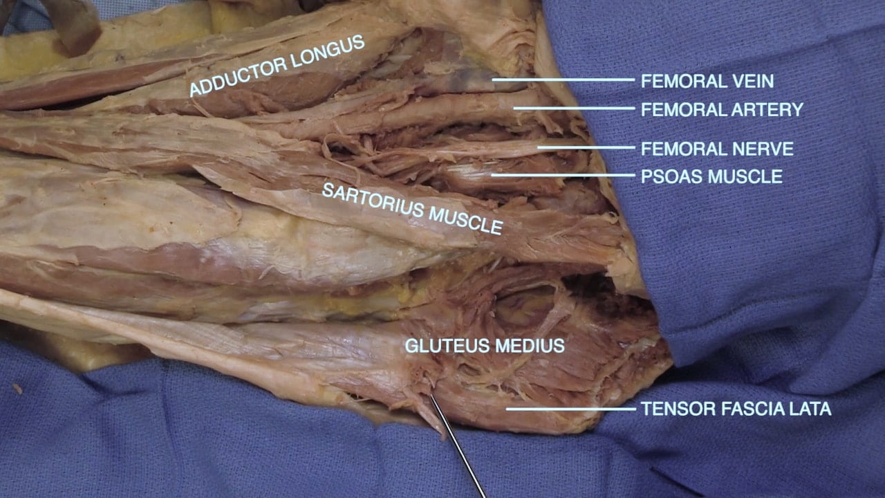
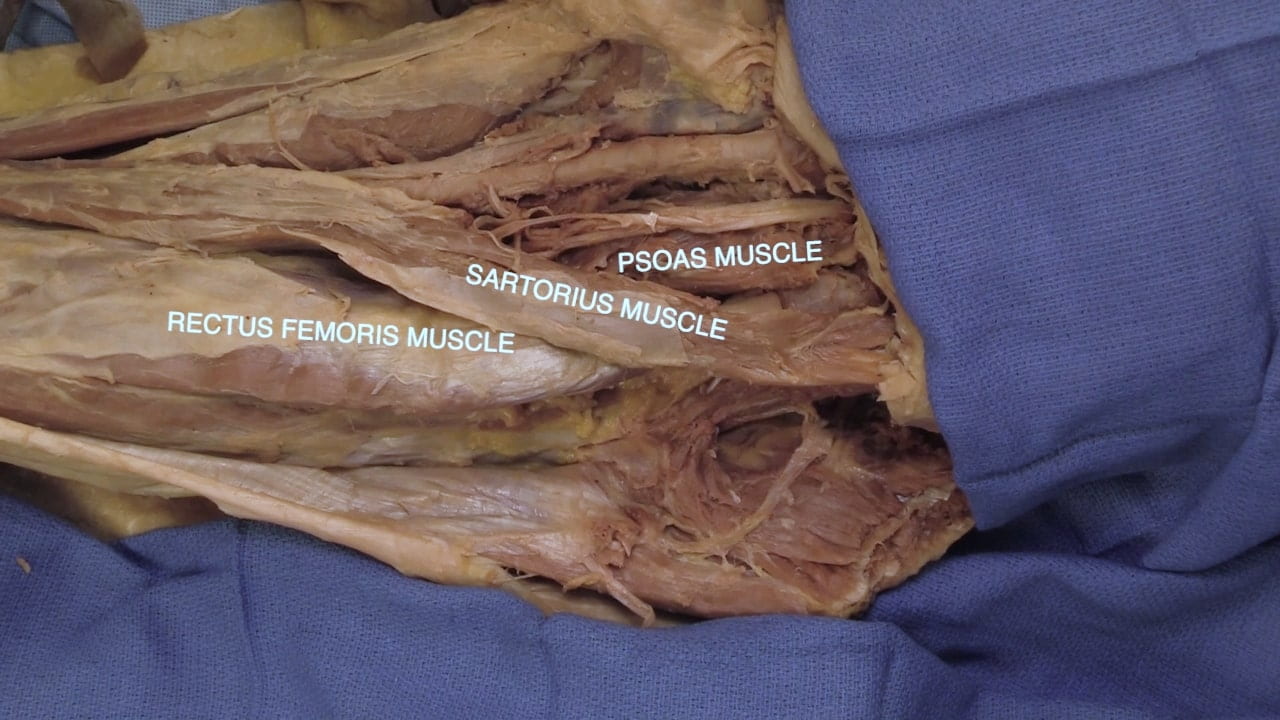
From medial to lateral, identify the adductor and quadriceps muscle groups as well as the tensor fascia latae.

