Midline Head and Neck

Lab Summary

In this lab the head and neck to upper thorax are taught. The approach is from a midline sagittal plane dissection of these structures. Skull base, nasal sinuses, nasopharynx, oral cavity, oropharynx and upper airway are emphasized.

Lab Objectives

  1. Describe structures of midline brain.
  2. Describe relationship of nasal and paranasal structures to base of brain.
  3. Describe the positions of the nasal cavity, nasopharynx, oral cavity, oropharynx, larynx and laryngopharynx.
  4. Describe the position of the paranasal sinuses.
  5. Describe the position and boundaries of the tonsillar fossa.
  6. Describe the vocal apparatus.
  7. Describe motor control of swallowing.

Lecture List

Sagittal Head and Neck Part I, II & III

Midline Head and Neck Part I

Sagittal Hemisection

Most donors will have had the brain removed by this point. The overall plan for this dissection is to make a midline sagittal hemisection of the head, neck and upper thorax. After completing this dissection, divide the brain sagittally in the midline and place it back in its anatomic position within the head.

Midline Head, Neck and Thorax

Make a midsagittal section through the skull, neck and upper thorax.

Divide the soft tissues along the midline with sharp dissection. Divide the bone with a coarse toothed hand saw.

Staying in the midline can be a challenge.

Extend the dissection to the upper thorax.

Upon completion of hemisection, place the brain back in its anatomic position within the head if previously removed.

Labels (left to right): Superior, cranial cavity, face, posterior, neck, thoracic cavity
1.2a) Midline head and neck

Midline Brain

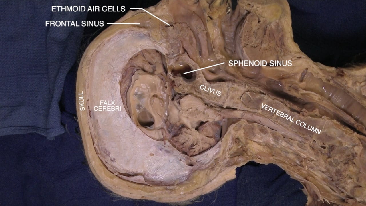
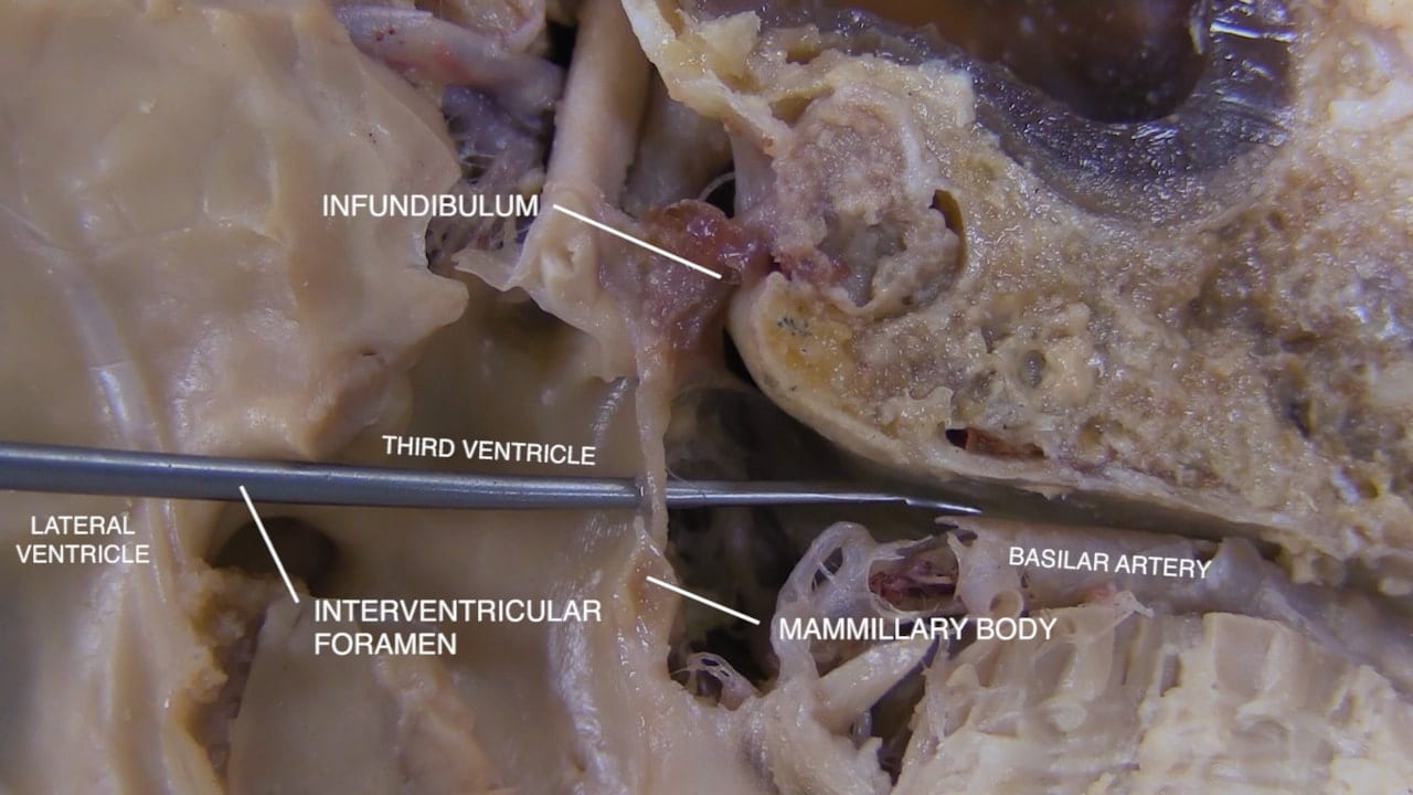
Orient by identifying the skull, frontal sinus, ethmoid air cells, sphenoid sinus, clivus and vertebral column.

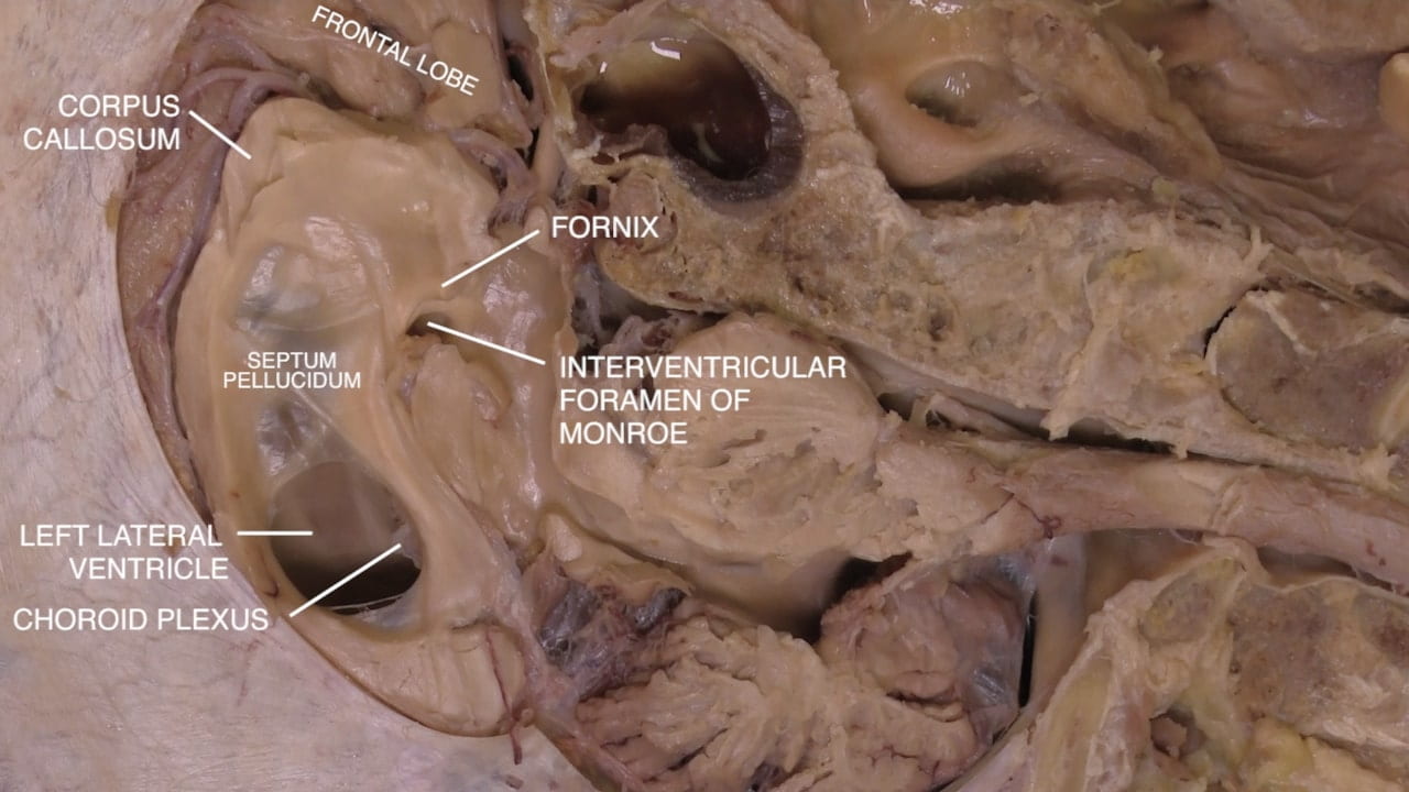
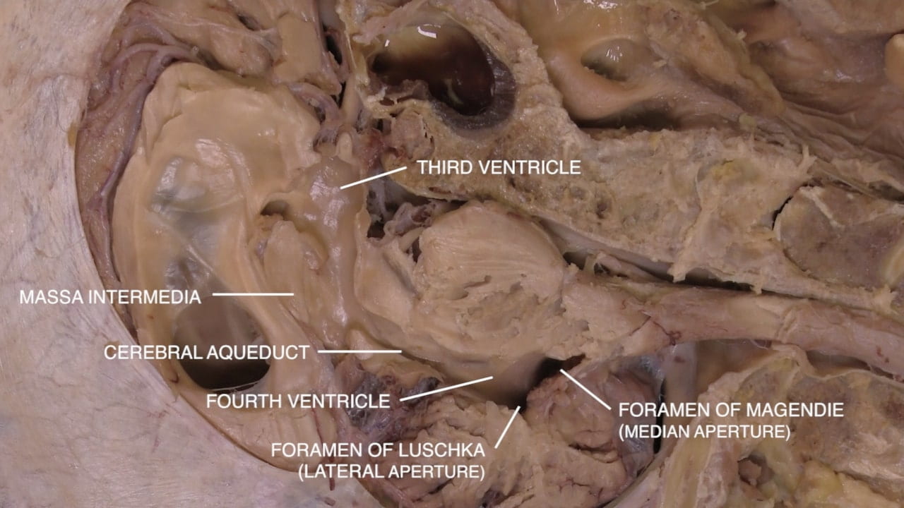
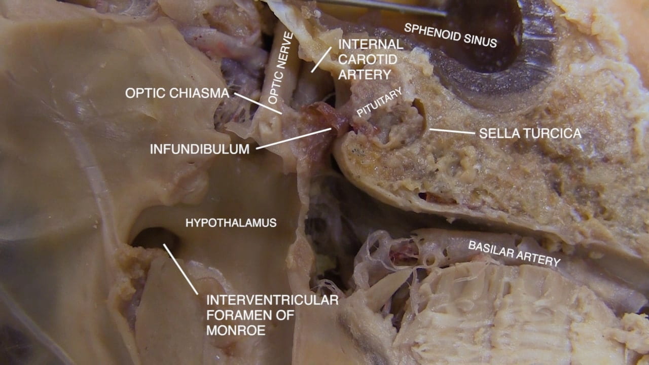
Identify structures of the midline brain:

  • Corpus callosum
  • Septum pellucidum
  • Fornix
  • Third ventricle
  • Hypothalamus
  • Foramen of Monro
  • Cerebral aqueduct
  • Fourth ventricle
  • Foramen of Luschka
  • Foramen of Magendie
  • Optic nerve and chiasm
  • Internal carotid artery
  • Infundibulum
  • Pituitary
  • Sella turcica
  • Basilar artery

Midline Head and Neck Part II

Nasal Cavity

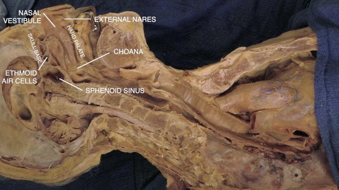
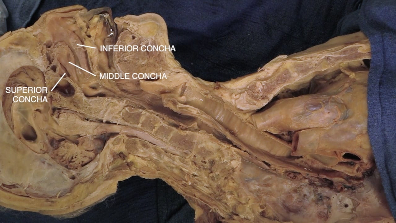
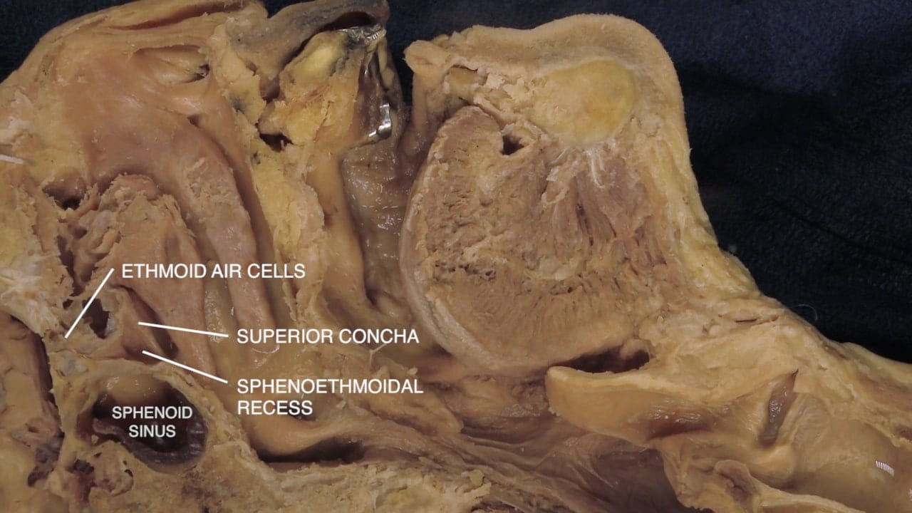
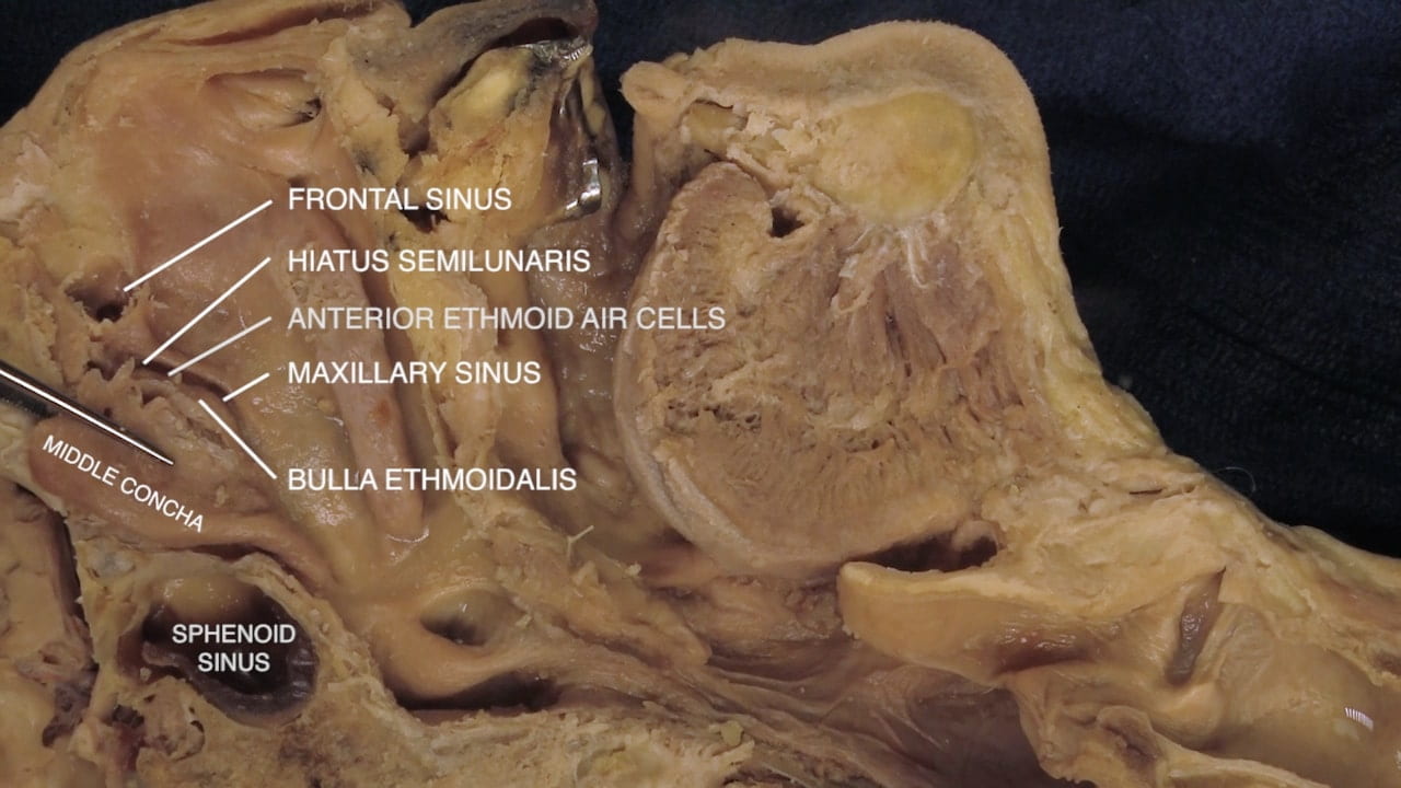
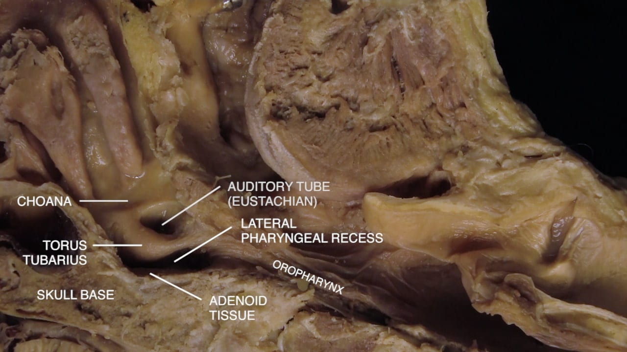
Identify structures of the nasal cavity and nasopharynx: skull base, hard palate, external nares, nasal vestibule, nasal cavity, choana, superior, middle and inferior concha, sphenoid sinus, ethmoid air cells.

Fold the middle conchus superiorly to reveal the hiatus semilunaris, which is the opening for the frontal and maxillary sinuses and middle ethmoid air cells. Explore dimensions of the maxillary sinus by placing a probe through this hiatus.

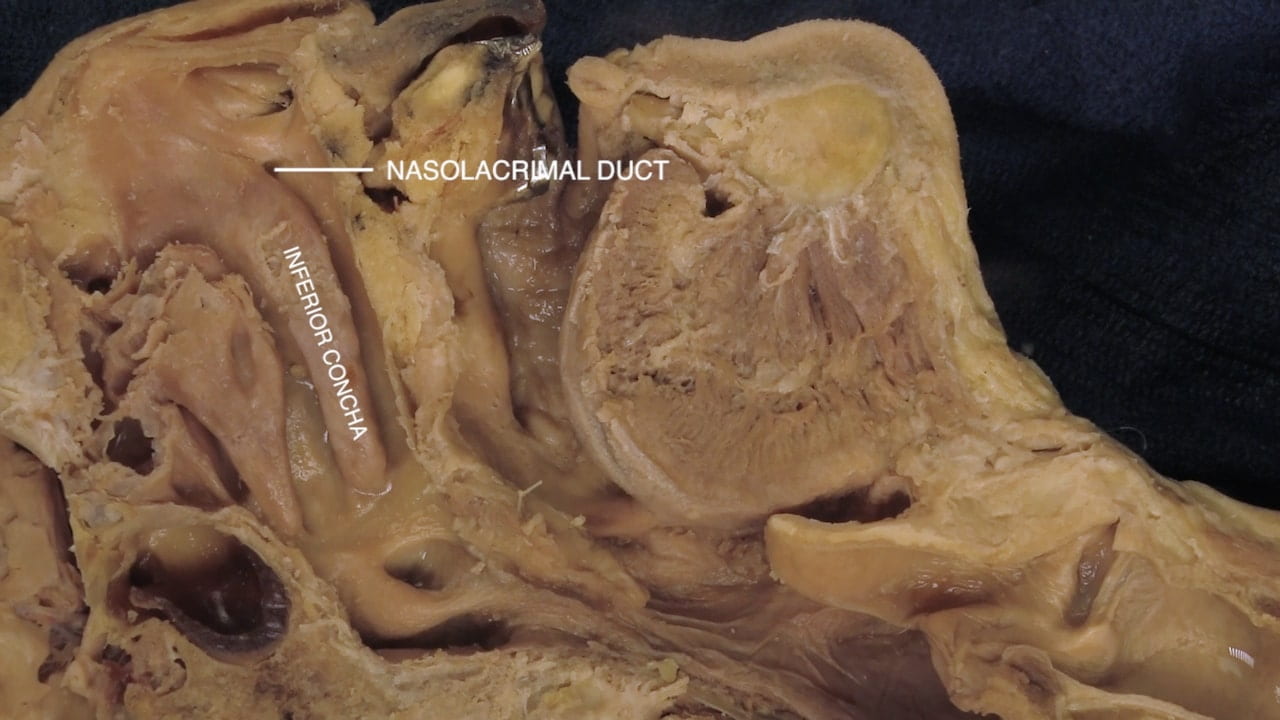
Lateral to the inferior conchus, locate the opening of the nasolacriminal duct. Posteriorly in the nasopharynx, locate the auditory tube at the posterior aspect of the conchae. Sensation to the nasopharynx is largely from the maxillary nerve (V2).

Midline Head and Neck Part III

Oral Cavity

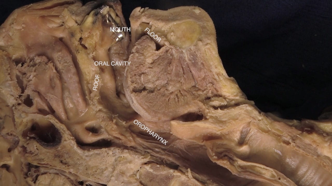
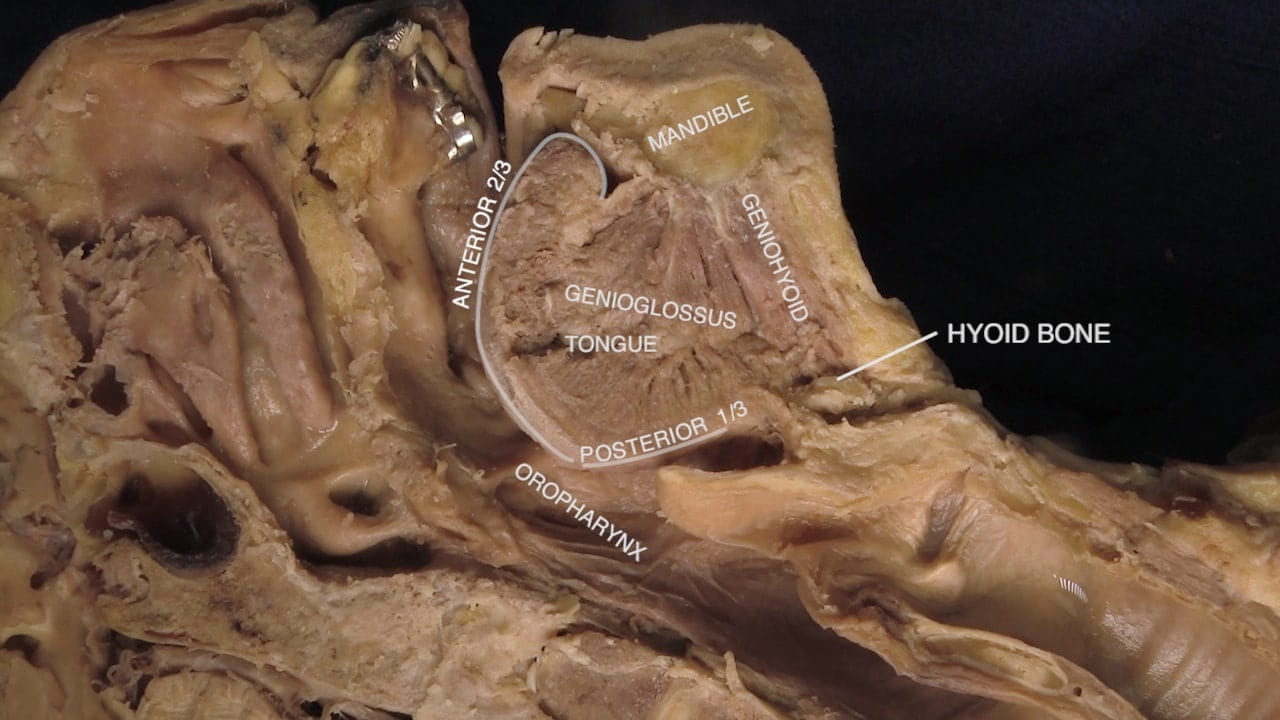
Orient by identifying structures of the oral cavity. The mouth is the entrance to the oral cavity. The hard and soft palate form the roof of the oral cavity. The floor of the mouth and tongue comprise the floor of the oral cavity. The oral cavity is continuous posteriorly with the oropharynx.

Identify the anterior 2/3 and posterior 1/3 of tongue.

  • Anterior 2/3 supplied by trigeminal nerve (V3) and facial nerve (VII).
  • Posterior 1/3 by glossopharyngeal nerve (IX).
  • Motor innervation to entire tongue by hypoglossal nerve (XII).

Pharynx

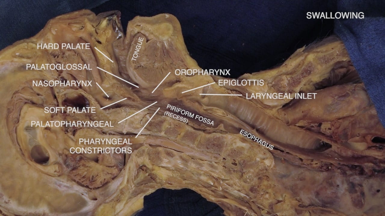
Examine the oropharynx as it extends from the palatoglossal fold to the laryngopharynx. It is bounded anteriorly to the posterior 1/3 of tongue, posteriorly to the pharyngeal wall and superiorly to the soft palate. Within the oropharnyx, identify the palatoglossal and palatopharyngeal folds forming the boundaries of the tonsillar fossa. Sensation is by the glossopharyngeal (IX) nerve; motor innervation by vagus (X) nerve.

Question 1: What vascular structure lies lateral to the tonsillar fossa?

Labels (left to right): Palatoglossal fold, nasopharynx, oropharynx, pharyngeal wall, tongue, palatopharyngeal fold, laryngopharynx
3.2a) Pharynx

Larynx

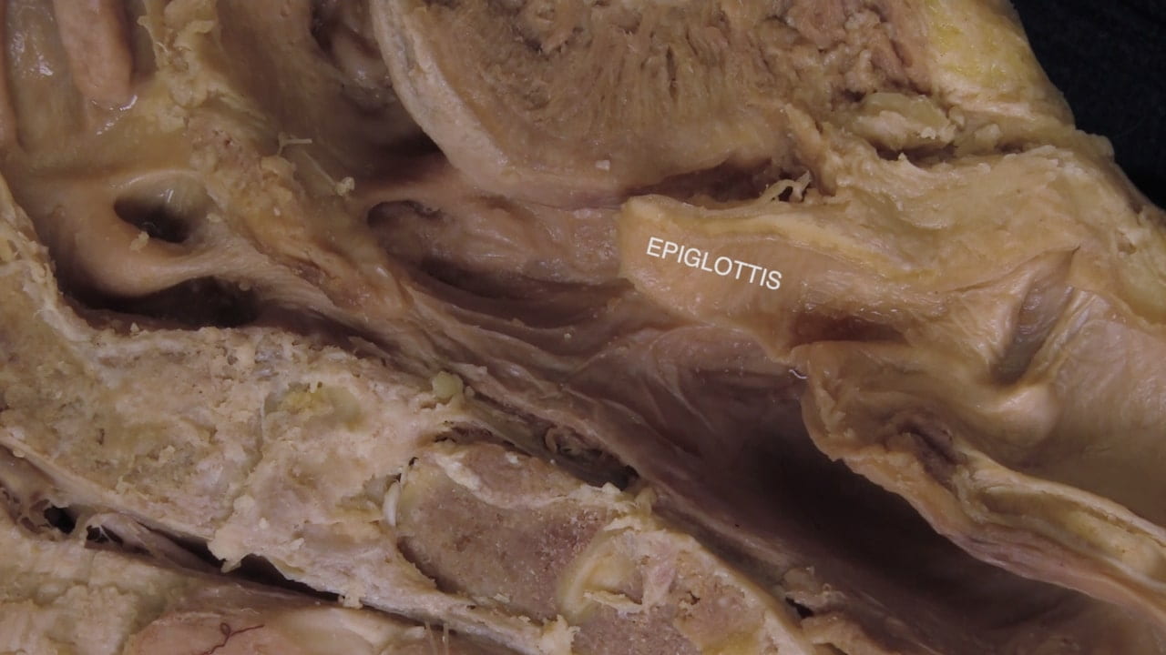
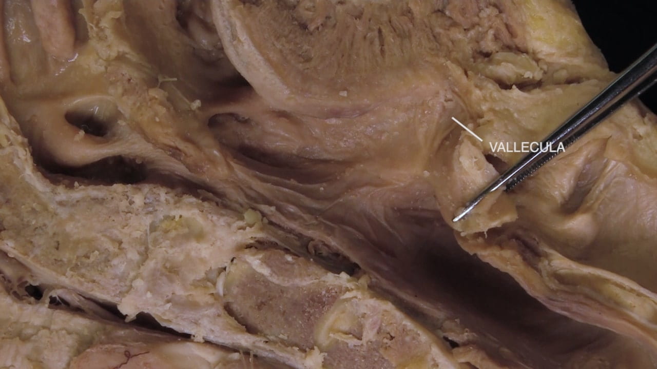
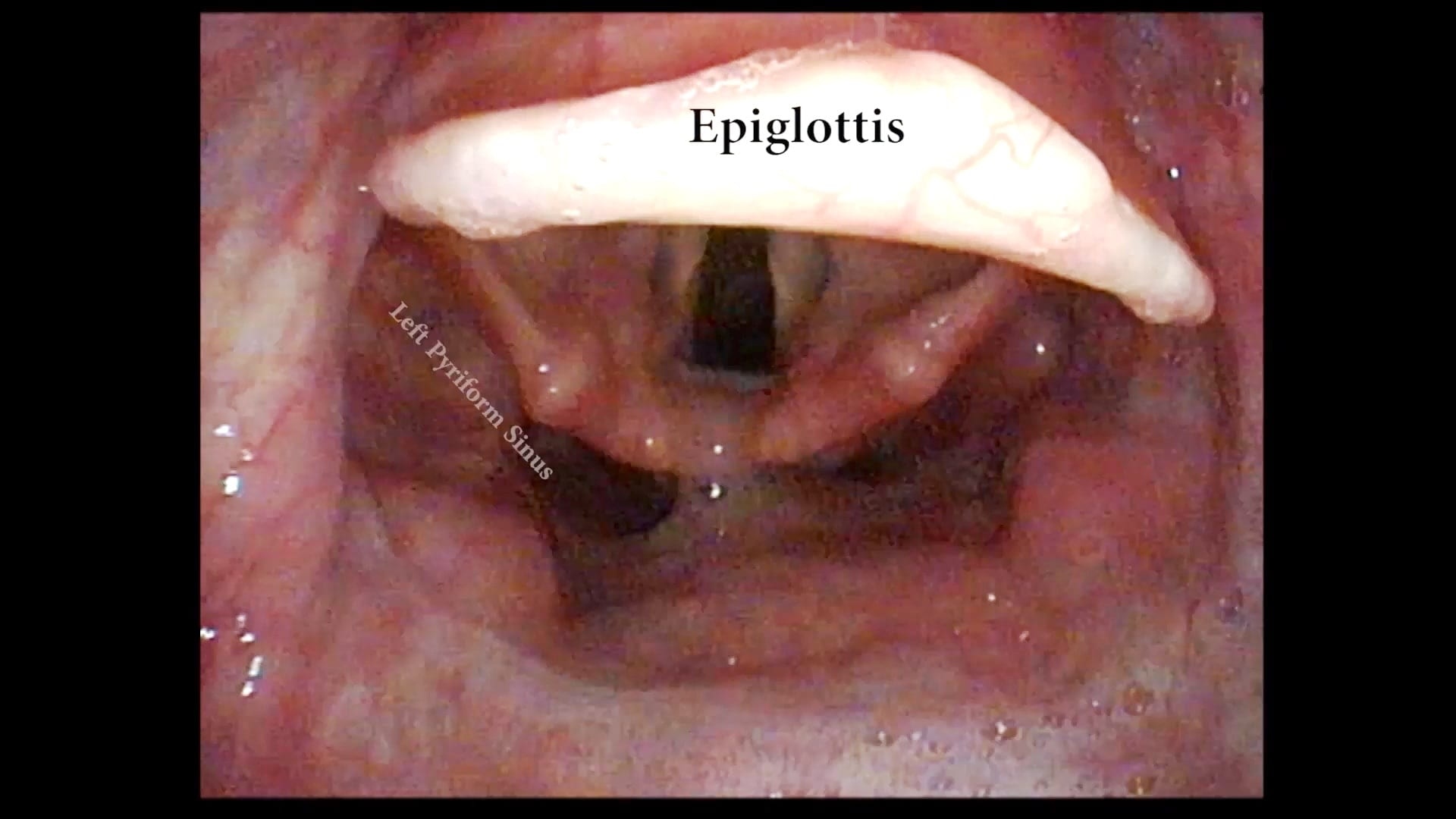
Identify the epiglottis. Retract the epiglottis posteriorly to identify the median and lateral glossoepiglottic folds forming the boundary of the vallecula. Anteriorly, the epiglottis protects the airway and directs food into the piriform recess.

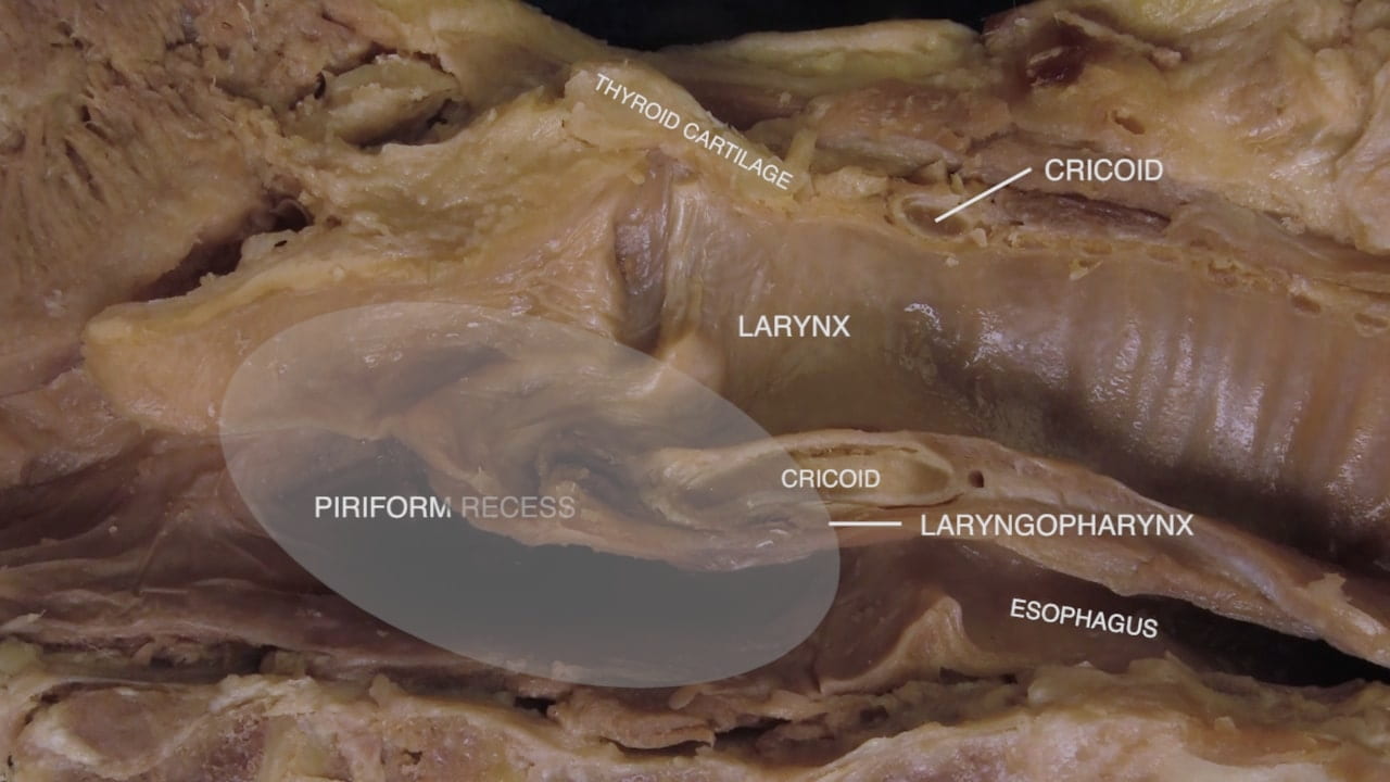
The laryngopharynx (piriform recess) is the portal from the oropharynx to esophagus. Inferiorly, the laryngopharynx is continuous with the esophagus. Neural control is by the vagus nerve (X).

Airway

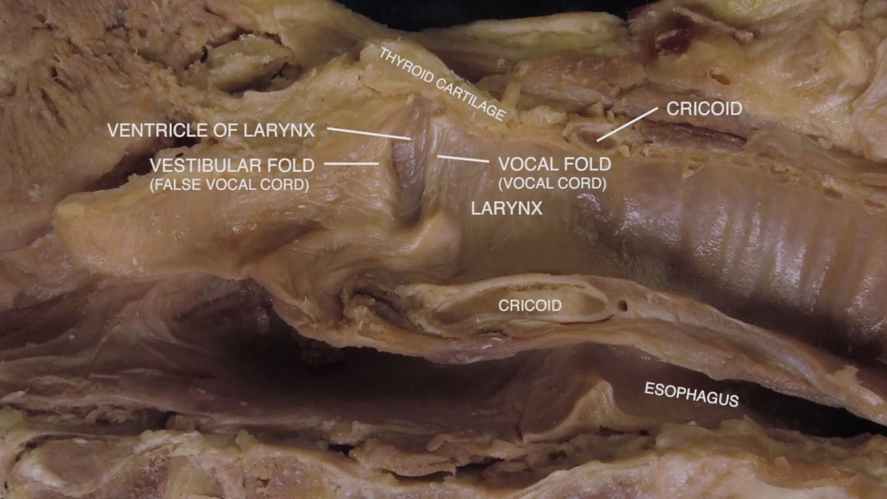
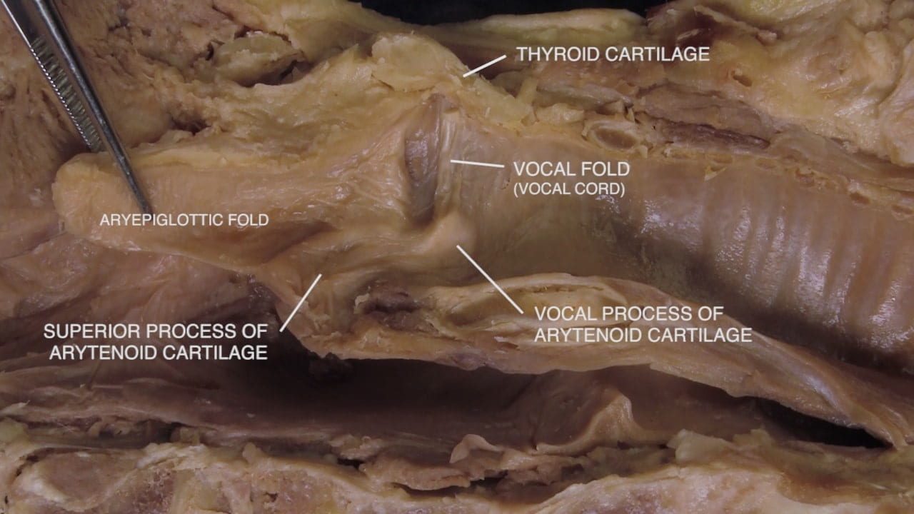
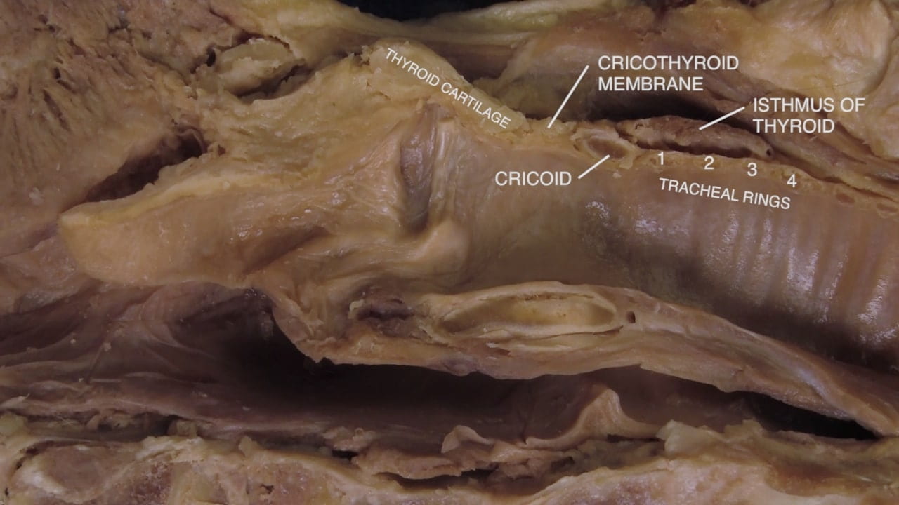
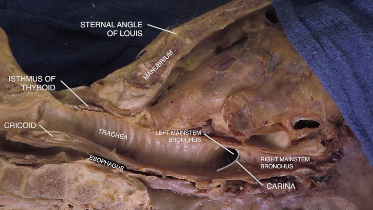
Anterior to the laryngopharynx and protected by the epiglottis, identify the larynx. Identify thyroid cartilage, anterior and posterior components of cricoid cartilage, vocal fold, vestibular fold, arytenoid cartilage, cricothyroid membrane, tracheal rings and isthmus of thyroid.

Question 2: What is clinical significance of the cricothyroid membrane?

Upper Chest

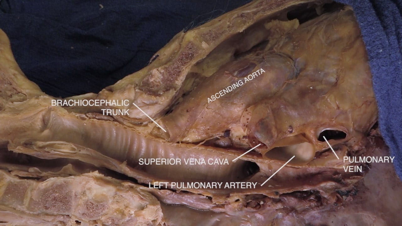
Follow the trachea inferiorly to the carina and mainstem bronchi. Note the relationships of surrounding structures: manubrium, ascending aorta and esophagus.

Swallowing

Consider the anatomic substrate of oral intake and swallowing.

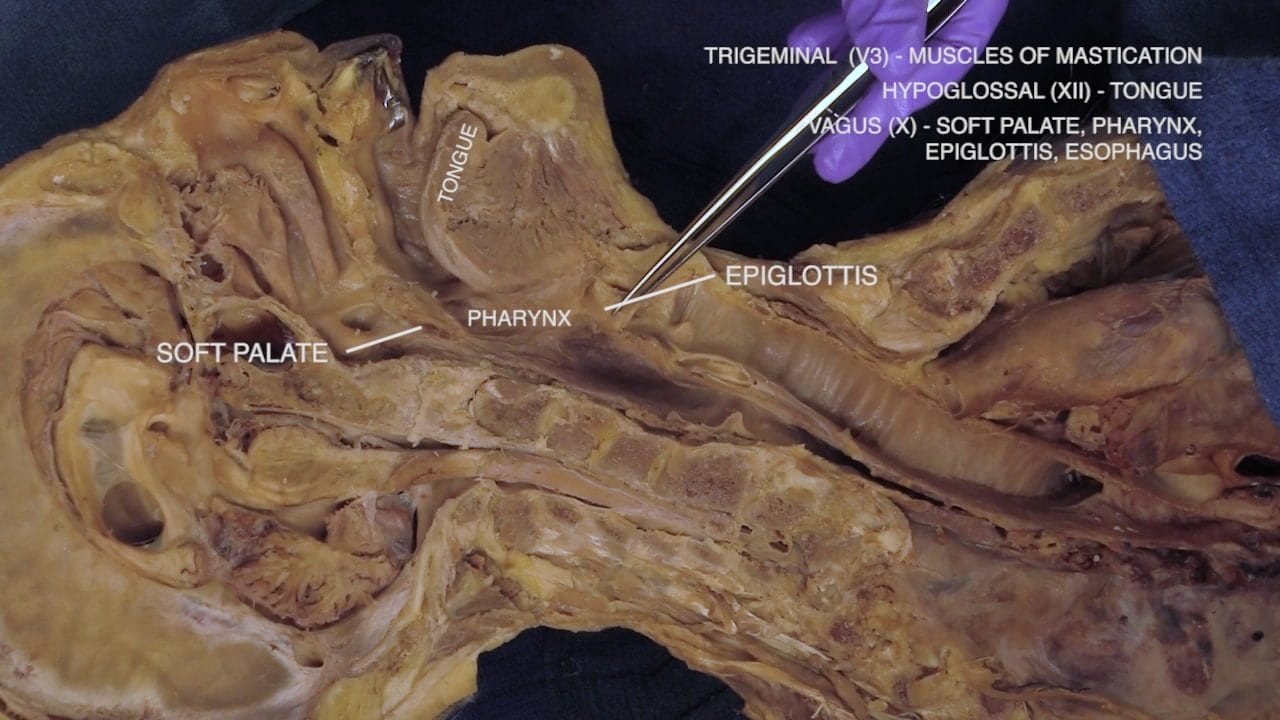
Motor Control:

  • Trigeminal (V3) – Muscles of mastication
  • Hypoglossal (XII) – Tongue
  • Vagus (X) – Soft palate, pharynx, epiglottis, esophagus

Upper Airway, William H. Rosenblatt

Review Quiz