Mediastinum

Lab Summary

Structures of the mediastinum from cervicothoracic junction to diaphragm are taught. The divisions of the mediastinum along with their components are included in this lab. Great vessels, nerves and their relationships to heart, chest wall and spine will be learned. There is detailed investigation of the apex or cupola of the chest cavity.

Lab Objectives

  1. Name the great vessels and their order of origin from the arch of the aorta.
  2. Describe the relationship of the subclavian vessels to the anterior scalene muscle.
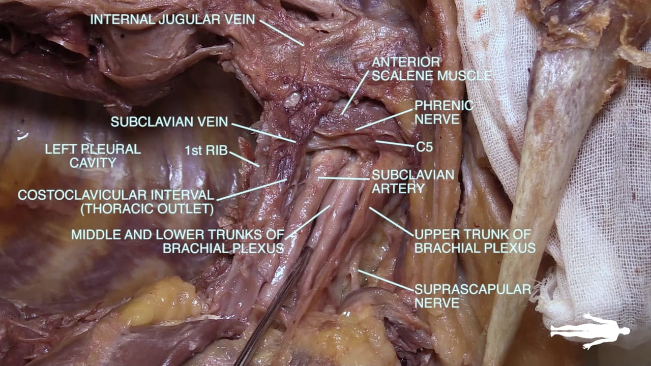
  3. Name the main structures in the thoracic outlet (costoclavicular interval).
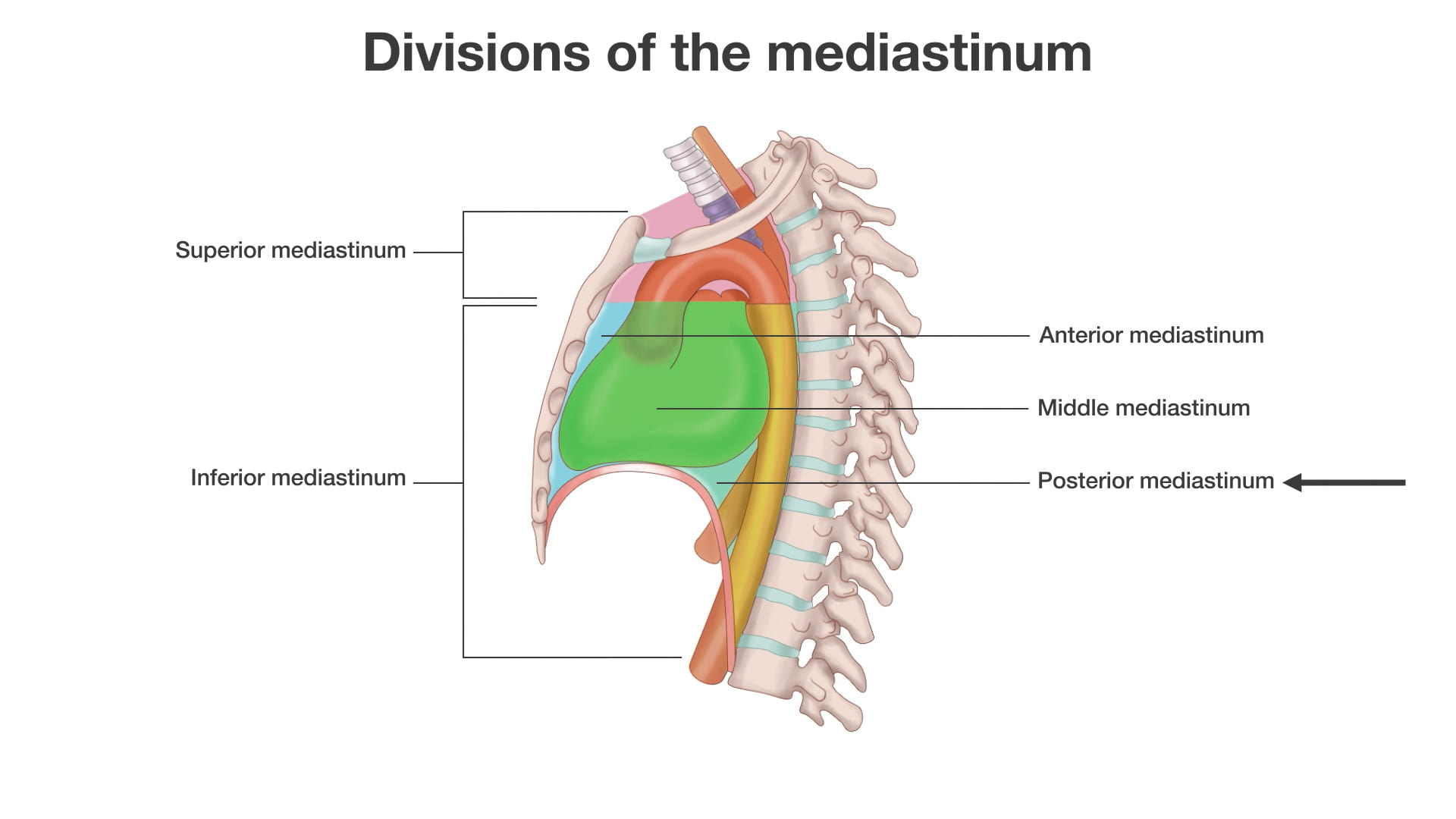
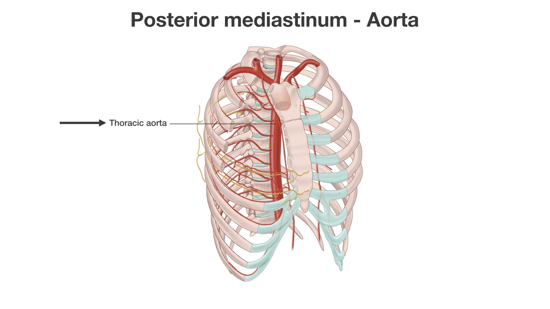
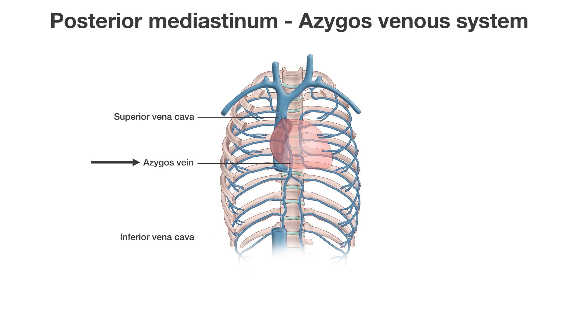
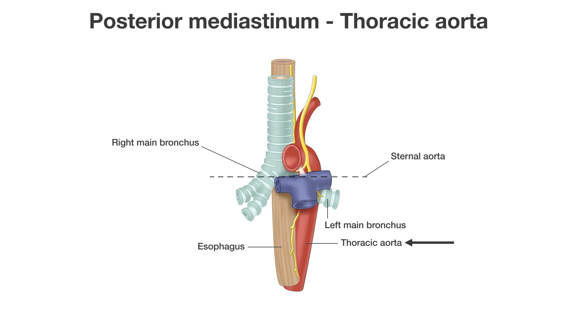
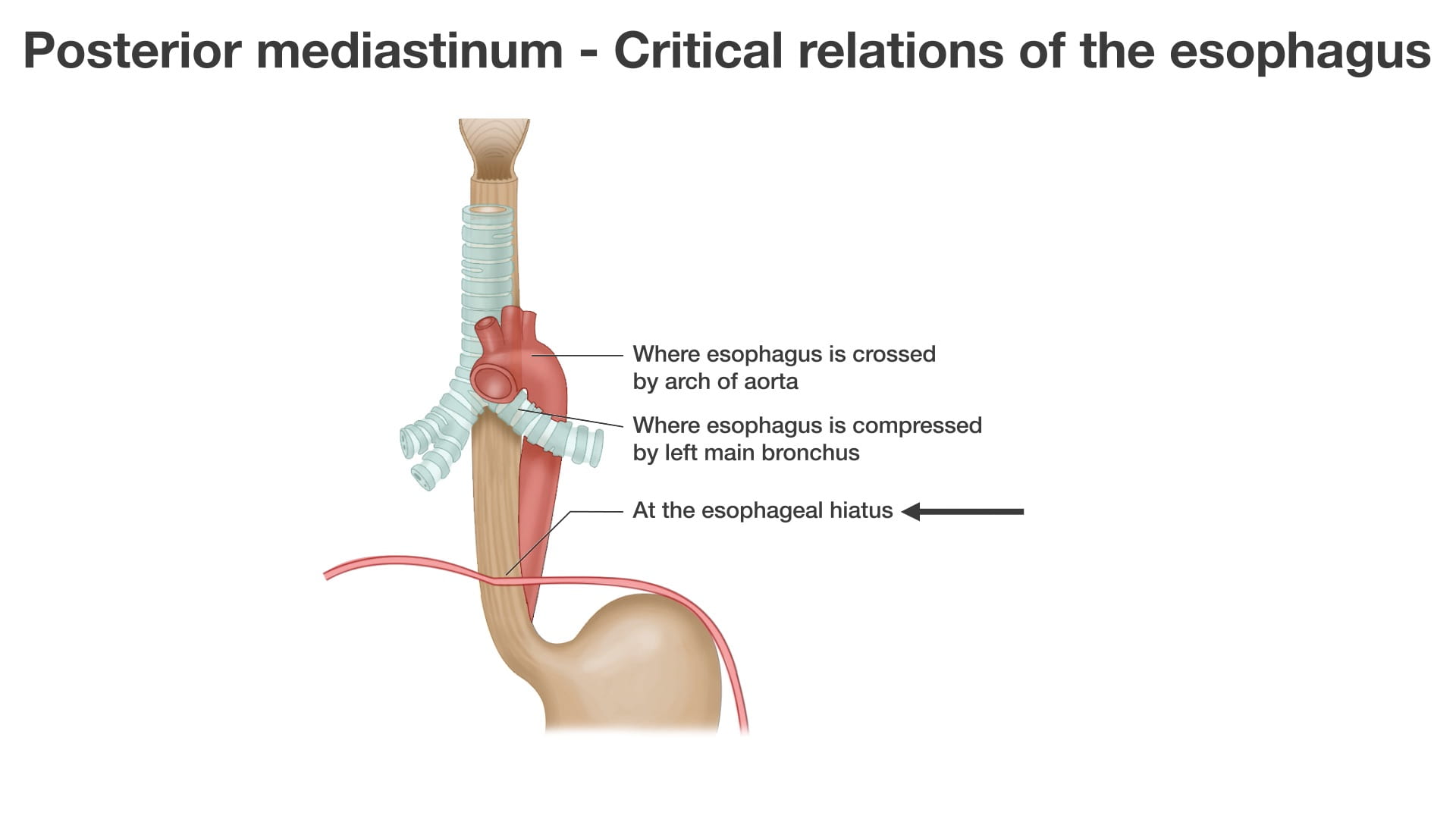
  4. Describe the spatial relationships of the thoracic aorta, esophagus and thoracic duct in the posterior mediastinum.
  5. Name the principal mediastinal structures at the level of the sternal angle. 
  6. Describe the targets of medial branches and lateral branches (rami communicantes) of the sympathetic chain.
  7. Describe the positions of the aorta, esophagus and inferior vena cava as they pass through the diaphragm, include the vertebral level.
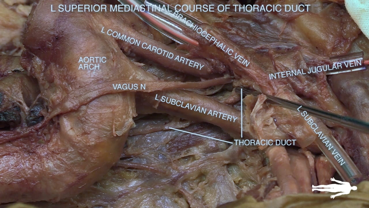
  8. Describe the position and drainage of the thoracic duct.

Lecture List

Mediastinum and Great Vessels, Superior Mediastinum and Thoracic Outlet, Apex of Pleural Cavity, Great Vessels, Posterior Mediastinum and Diaphragm

Mediastinum and Great Vessels

Mediastinum Gallery

Cervicothoracic Junction

Exposure of Superior Mediastinum

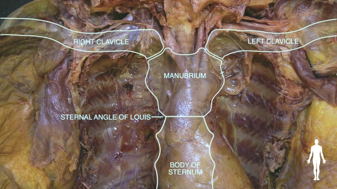
Using an oscillating saw and chisel, divide the first rib close to the manubrium and sternoclavicular joint to remove the manubrium.

Mobilize the clavicles by sharply dissecting their muscular attachments. Loosening the acromioclavicular (AC) joints with a chisel may facilitate this mobilization.

Reflect the clavicles laterally.

Labels (left to right): Inferior, manubrium, superior
2.1a) Manubrium and clavicles

Great Veins

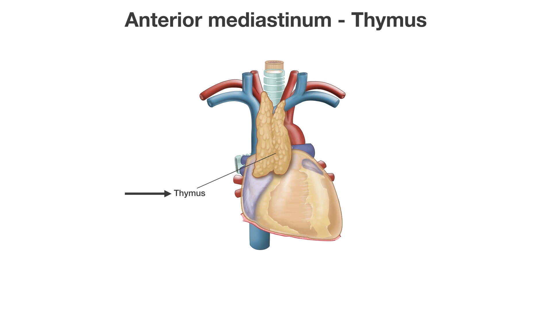
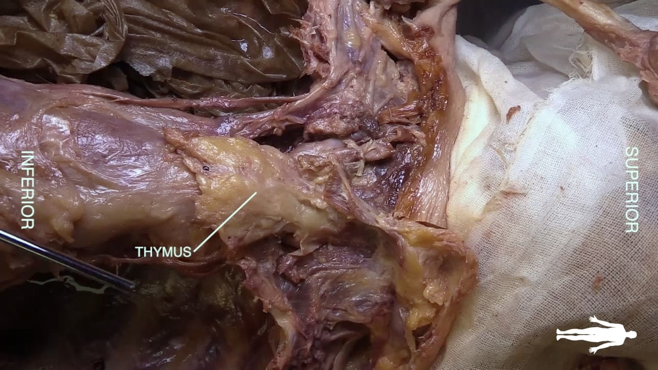
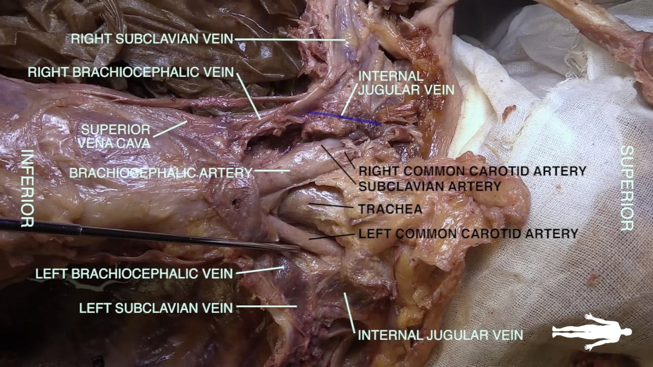
Identify the thymic remnant as the retrosternal fat pad and elevate it.

Identify the left subclavian, left internal jugular and left brachiocephalic veins.

Locate the right internal jugular, right subclavian and right brachiocephalic veins and the superior vena cava (SVC).

Question 1: What is the function of the thymus?

Subclavian Artery and Vein

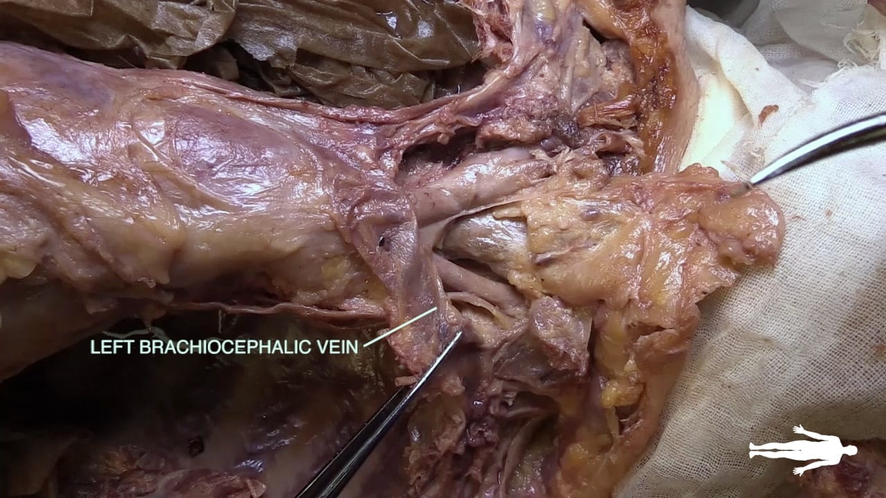
Divide the left brachiocephalic vein medially and reflect laterally to expose the subclavian artery.

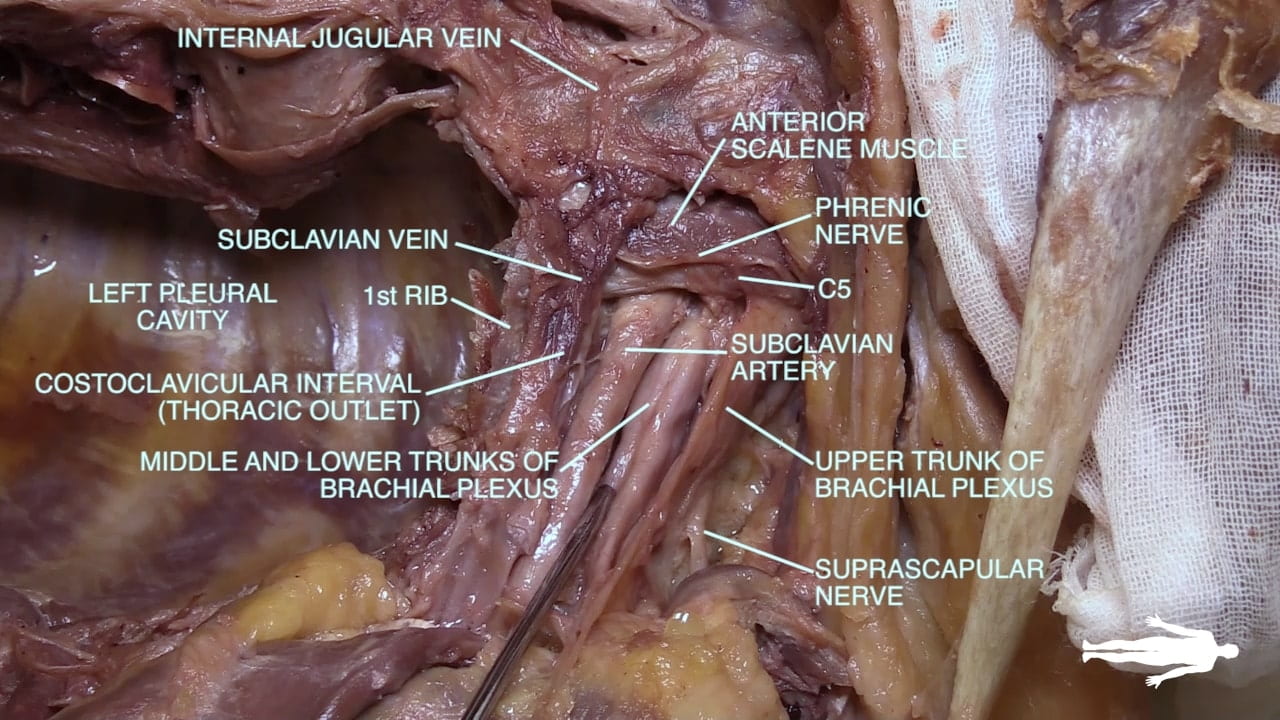
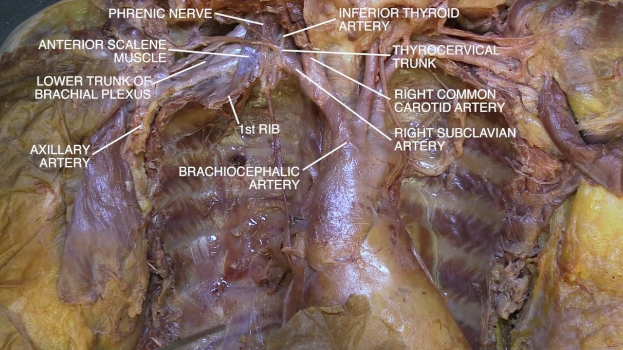
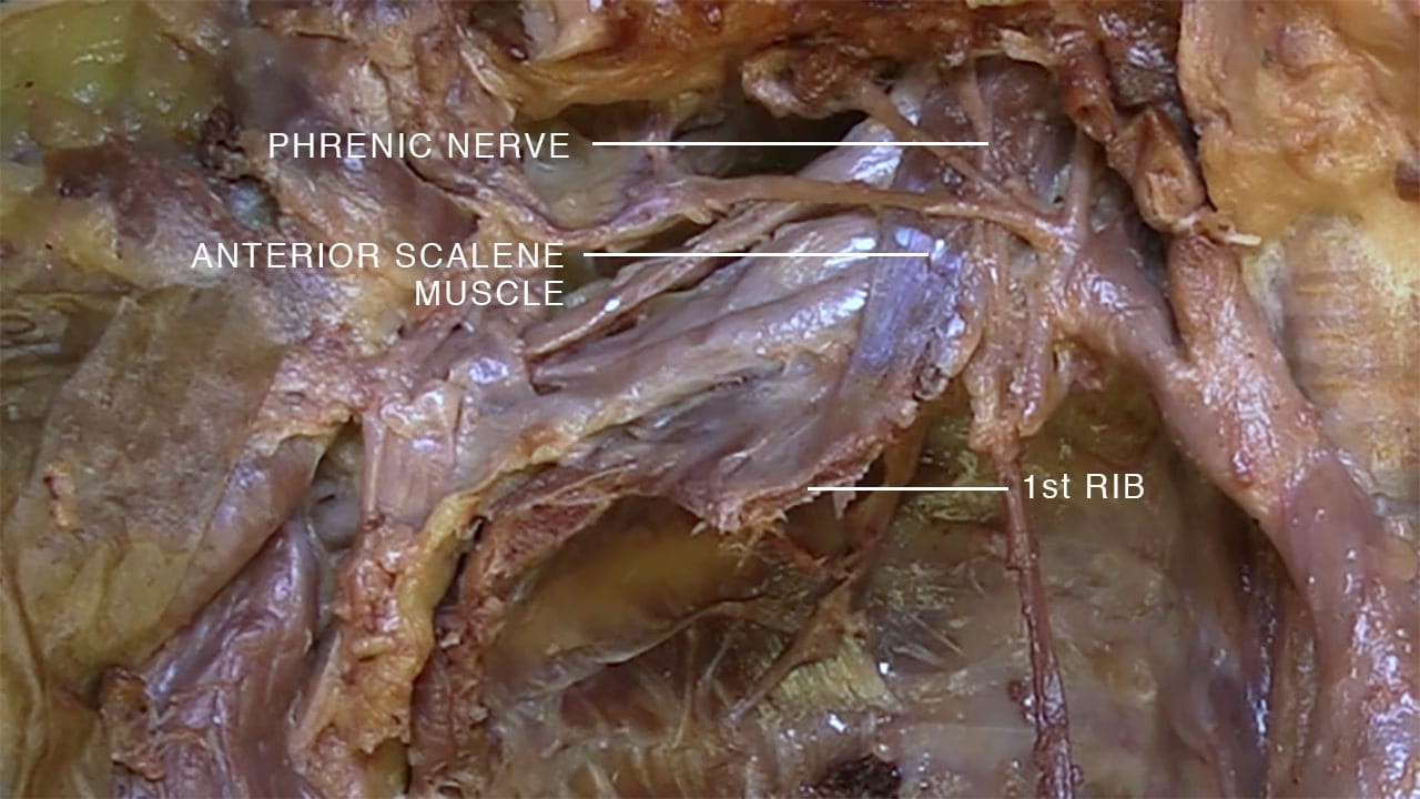
Note the relationship of the subclavian artery and vein to the first rib and to the anterior scalene muscle (vein anterior, artery posterior, interposed by anterior scalene).

Thoracic Outlet

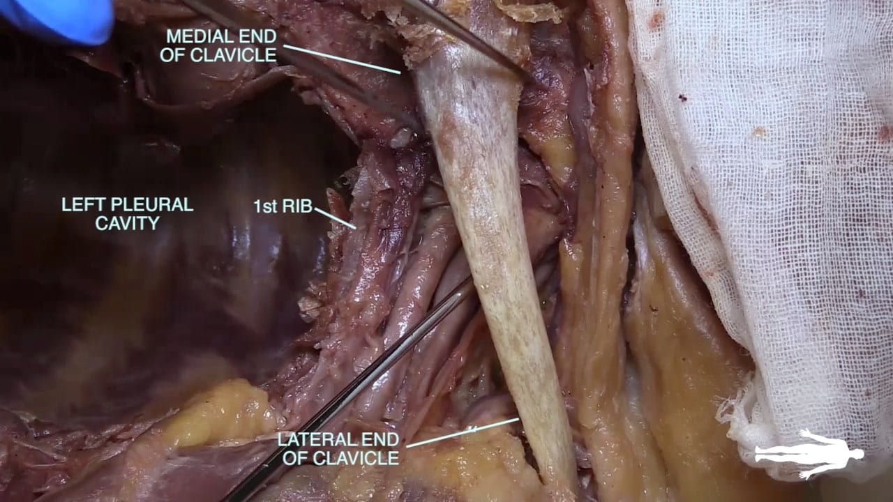
Place one clavicle back in position to gain an appreciation of the thoracic outlet (costoclavicular interval). The right side may have been disturbed from the preservation process.

With the clavicles laterally rotated, find the subclavian vessels and first rib again. Locate the trunks of the brachial plexus.

Note the phrenic nerve on the anterior surface of the anterior scalene muscle.

Apex of Pleural Cavity

Sympathetic Chain

Remove the costal and vertebral pleura from the superior and medial chest wall to expose the intercostal vessels and nerves. You may need to retract the subclavian artery superiorly.

Identify T1 spinal nerve passing over the first rib to join the C8 spinal nerve.

Identify the intercostal neurovascular bundle along the inferior side of a rib along with its components: vein, artery and nerve.

Find the sympathetic chain and look for lateral branches to the intercostal nerve (rami communicantes) and medial branches to the heart and lung.

Labels (top to bottom): Aorta, subclavian artery, stellate ganglion, sympathetic chain, C8, lower trunk, second ganglion, T1, rami communicans, vein, artery and nerve of 2nd intercostal space, visceral (cardiopulmonary branches), costal parietal pleura, left pleural cavity
3.1a) Spinal nerves and sympathetic chain

Great Vessels

Arch of Aorta

Reflect the right and left brachiocephalic veins.

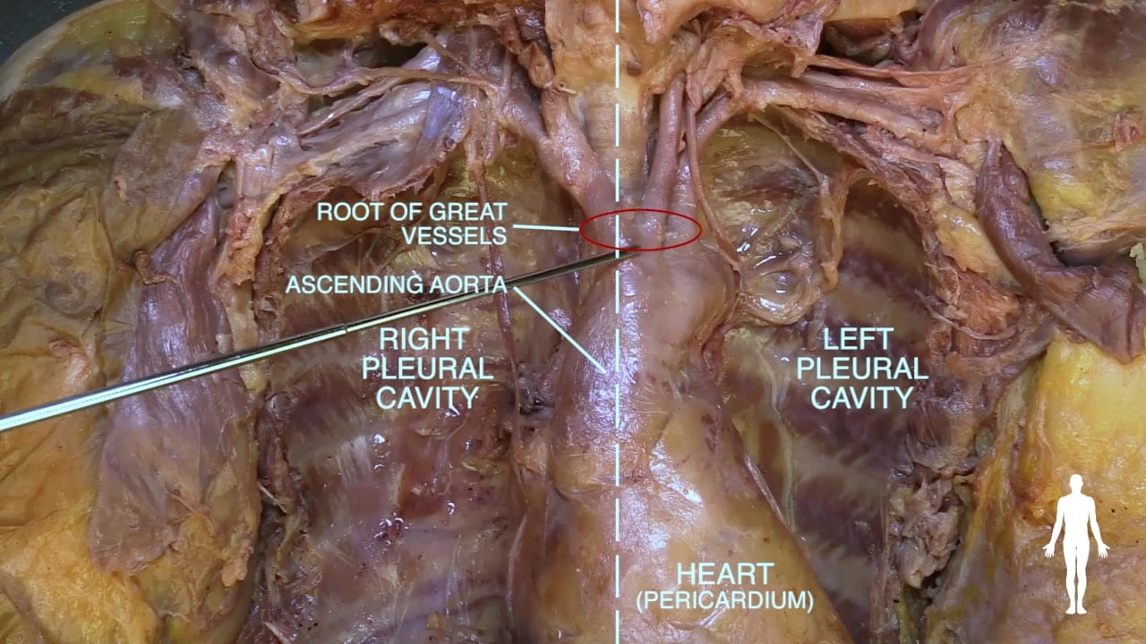
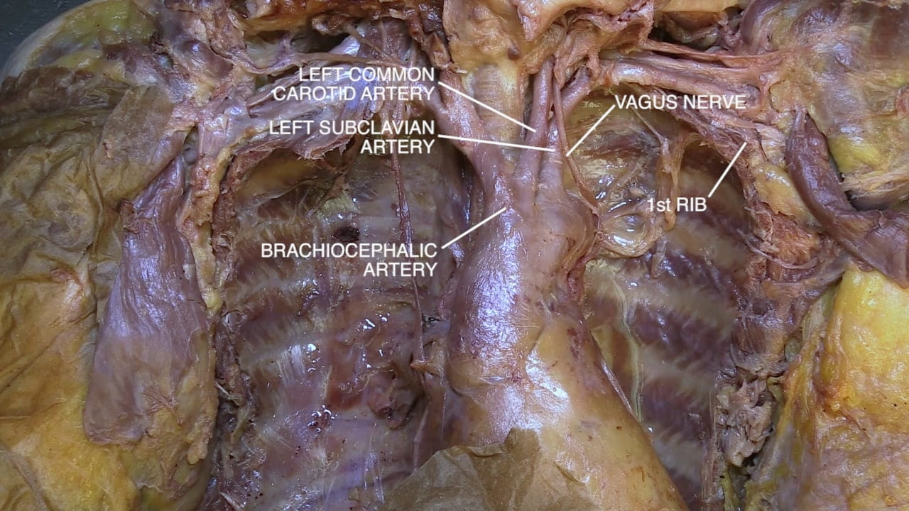
Re-identify the arch of the aorta and locate the origins (root) of the great vessels: brachiocephalic, left common carotid and left subclavian arteries.

Follow the brachiocephalic superiorly and identify its right common carotid and right subclavian branches. Note the subclavian artery is posterior to the anterior scalene. The subclavian artery becomes axillary artery after it passes over the first rib.

Identify the phrenic nerve on the anterior surface of the anterior scalene.

Posterior Superior Mediastinum

Return to the root of the great vessels. Identify the right common carotid artery and subclavian artery. Note the vagus nerve passes anterior to the subclavian artery.

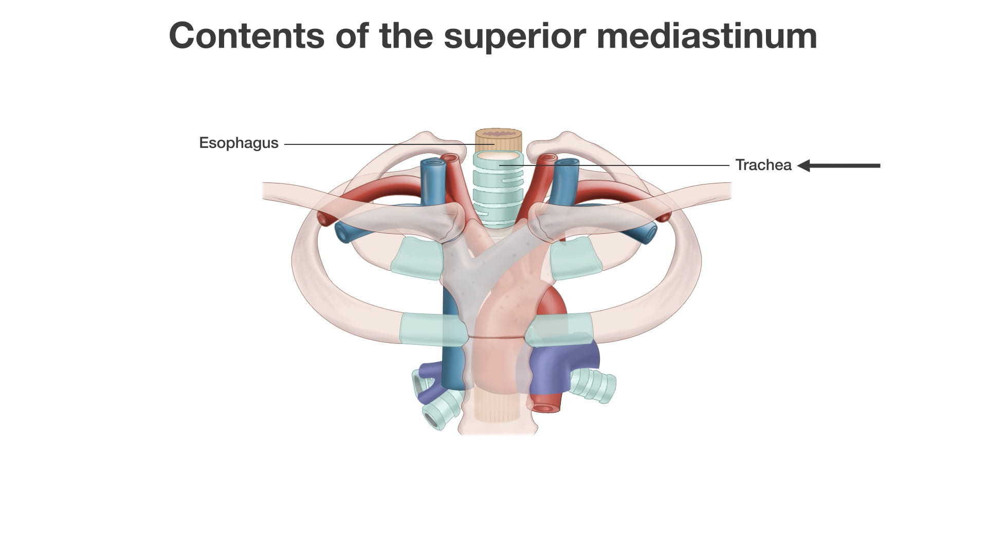
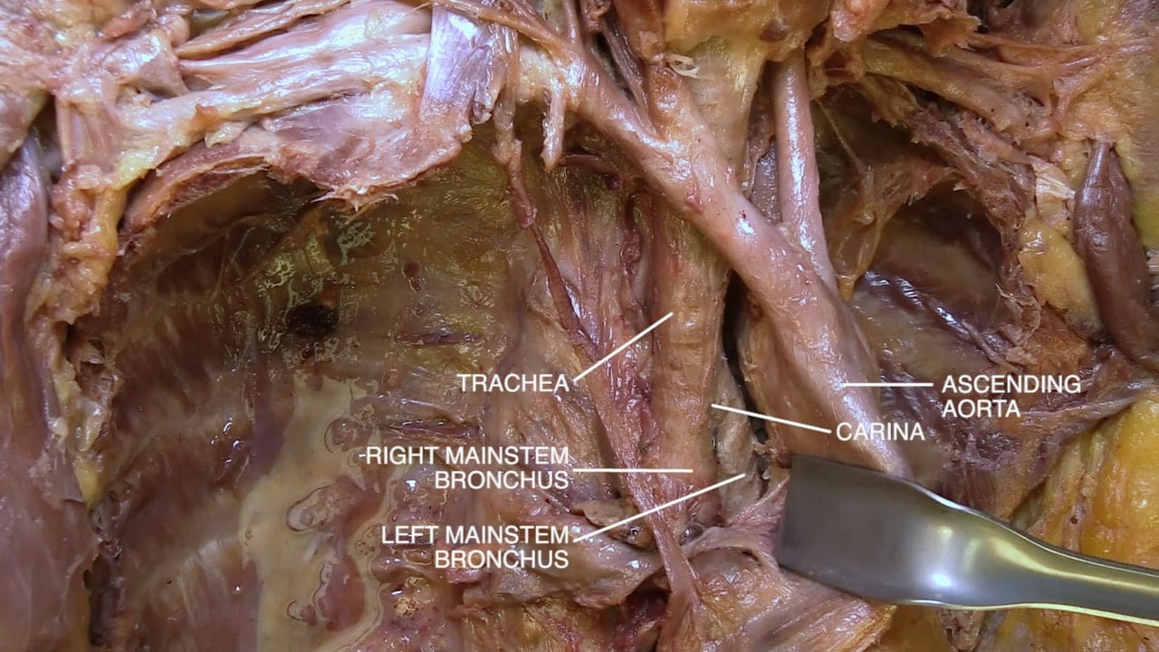
Retract the ascending aorta to the left to identify trachea and its bifurcation, the carina.

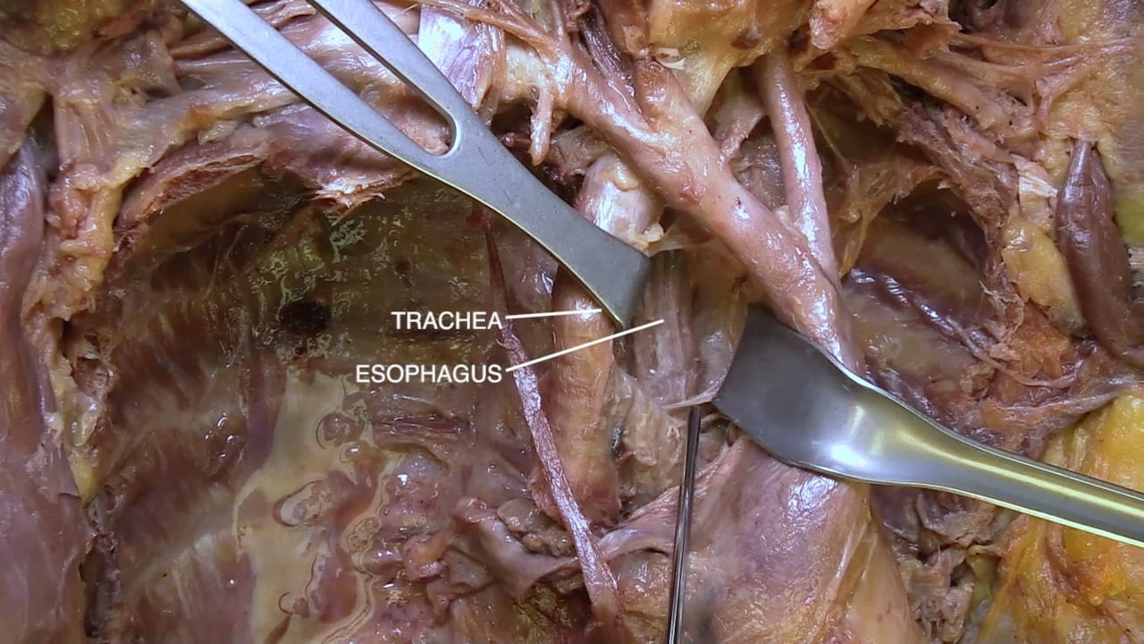
Retract the trachea to the right and identify the esophagus deep to it.

Vagus Nerve and Ligamentum Arteriosum

Examine the aortic arch and surrounding structures from the left.

Identify the pulmonary trunk, left pulmonary artery and mainstem bronchus. Identify ligamentum arteriosum, left pulmonary artery and aorta.

Identify the vagus nerve and follow its branching. Note the close relationship between the ligamentum arteriosum and left recurrent laryngeal branch of the vagus.

Also re-identify the phrenic nerve.

Labels (Top to bottom): Vagus nerve, recurrent laryngeal branch, arch of the aorta, cardiac sympathetic nerve, phrenic nerve, hilus of the lung, ligamentum arteriosum, left pulmonary artery, descending aorta, ascending aorta, left mainstem bronchus, pulmonary trunk, heart (pericardium)
4.3a) Aortic arch

Posterior Mediastinum and Diaphragm

Posterior Mediastinum

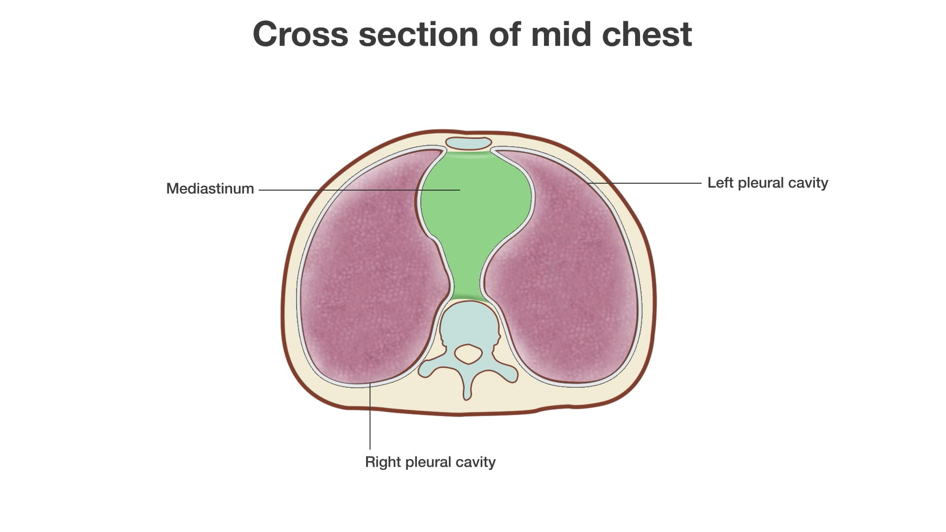
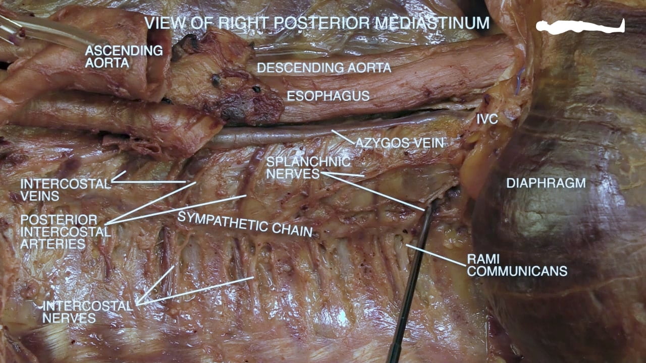
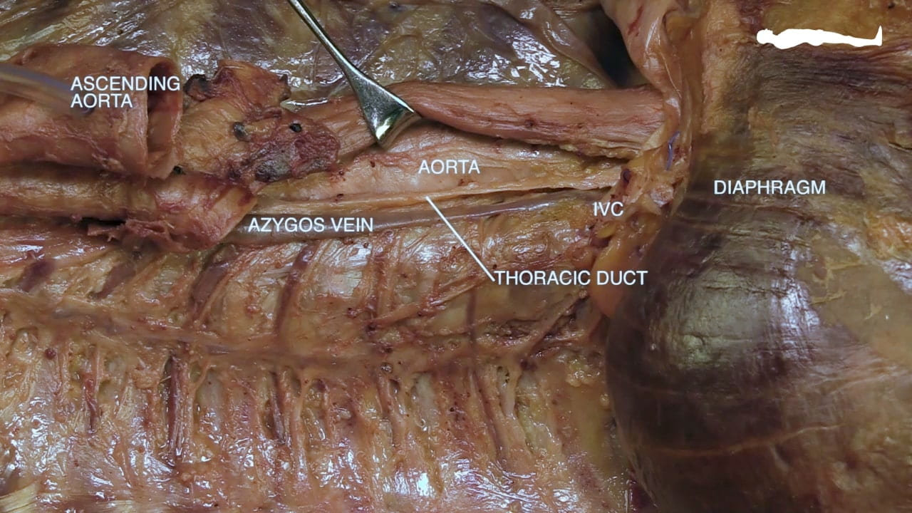
View the posterior mediastinum from the right. Identify the thoracic aorta, esophagus and azygos vein.  Locate the thoracic duct posterior to the esophagus and to the right of the aorta.

Remove as much pleura as possible from the vertebral column and chest wall to expose the mediastinum.

Identify an intercostal vein, artery and nerve.

Expose the sympathetic chain. Identify the thoracic splanchnic nerves and rami communicantes.

Diaphragm

Examine the superior surface of the diaphragm.

Identify right and left hemidiaphragms. Note the central tendon of diaphragm and the attachment of the pericardium.

Locate the inferior vena caval, esophageal and aortic hiatuses.

Question 2: What are the vertebral levels of these hiatuses?

Title: Superior view of diaphragm Labels (left to right): L hemidiaphragm, (stomach underlying), (spleen underlying), aorta (passes between crura t12, central tendon, esophageal hiatus (t10), IVC (t8), esophagus, r hemidiaphragm, (liver underlying)
5.2a) Superior view of diaphragm

Review Quiz