Chest Cavity and Lungs
Lab Summary
Learn chest anatomy from chest wall to lung including breast structure and its important relationships to muscle, lymphatics and nerves. The structure of the rib cage along with its external and external architecture are presented. Pleural, lungs and their connections are taught.
Lab Objectives
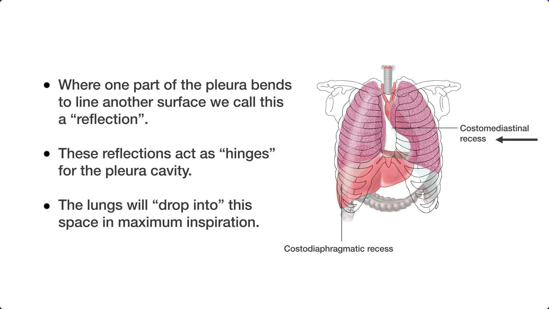
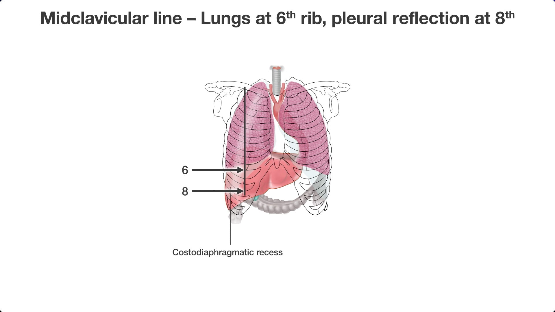
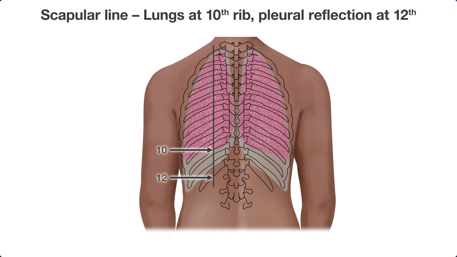
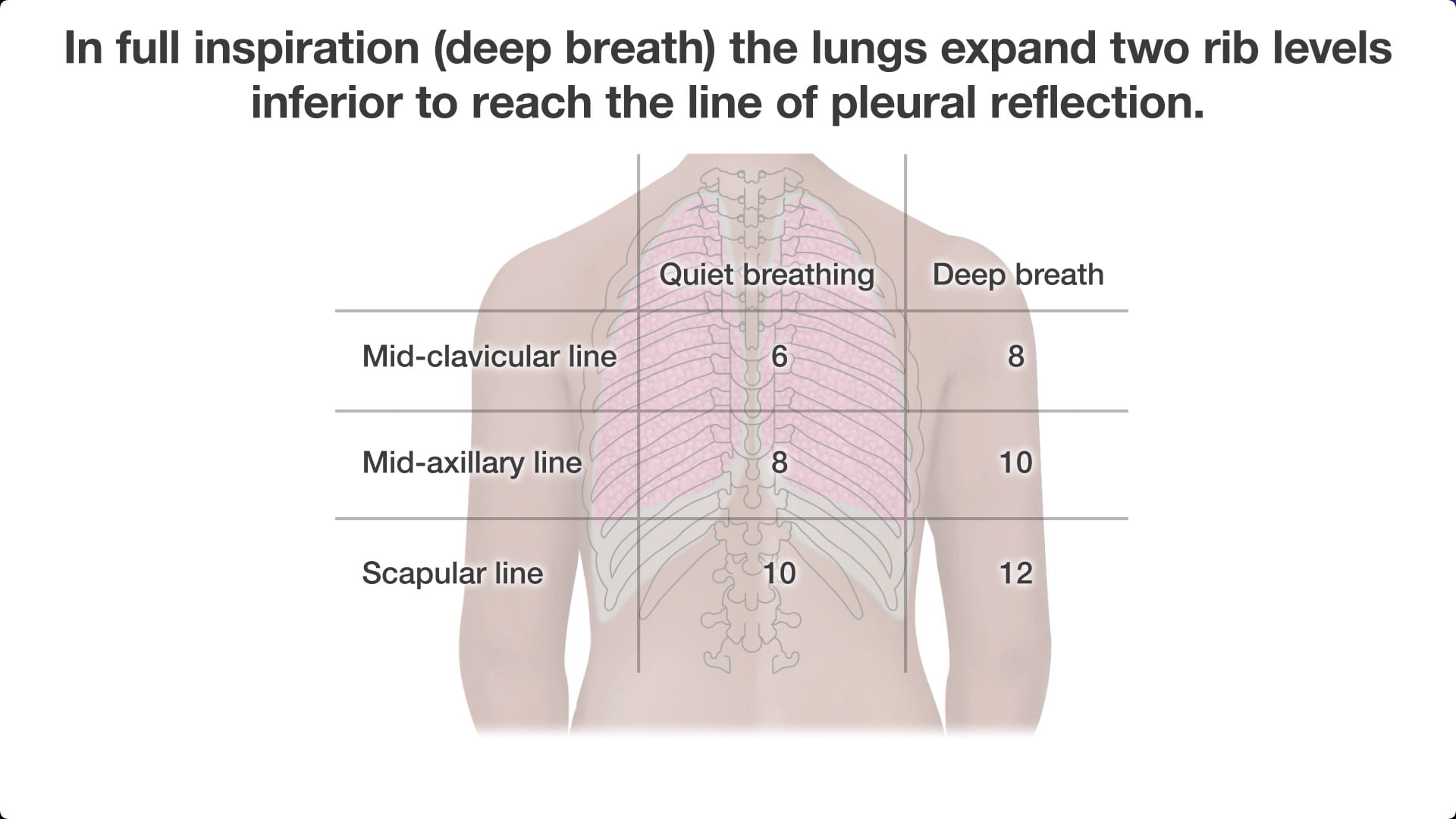
- Describe and demonstrate on your donor the pleural recesses (costodiaphragmatic, costomediastinal). Also be able to describe their surface landmarks.
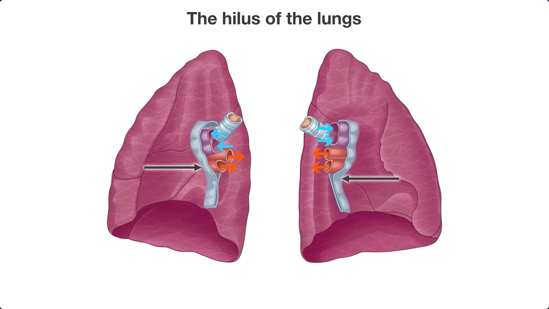
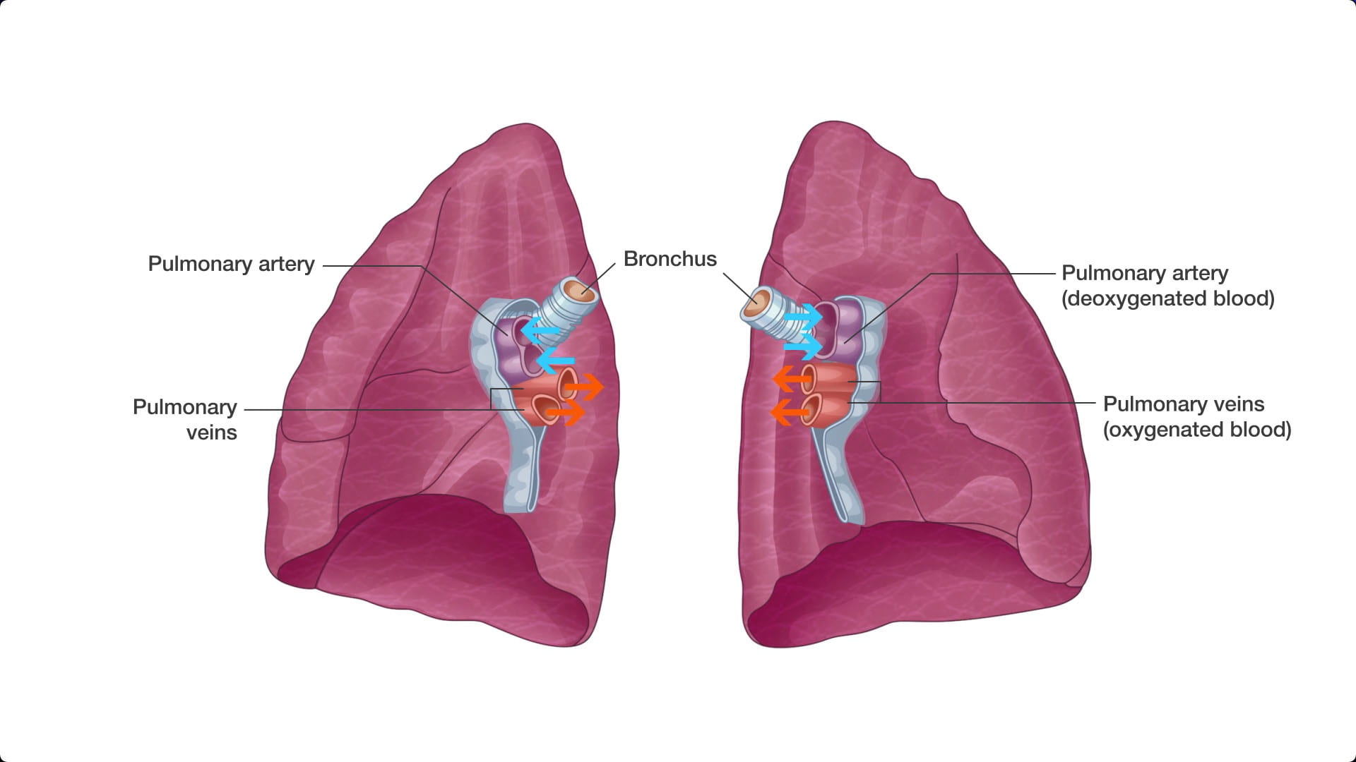
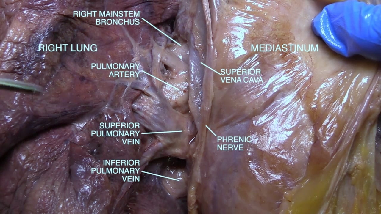
- Describe / diagram / demonstrate on your donor the relationship of the main structures at the hilus of the lungs.
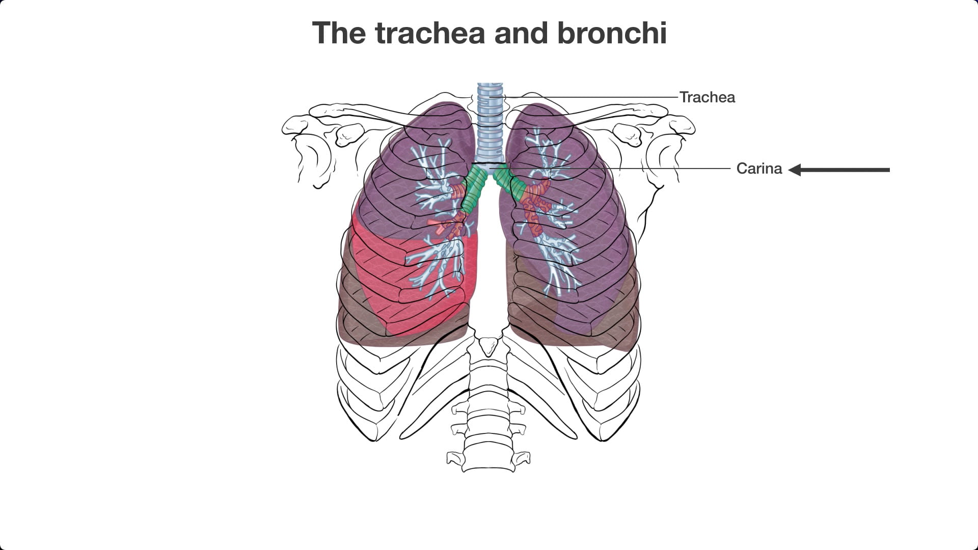
- Describe the positions of the lobes of the lung and the bifurcation of the trachea and their relationships to the surface landmarks.
- Describe the course and position of the internal thoracic (mammary) artery.
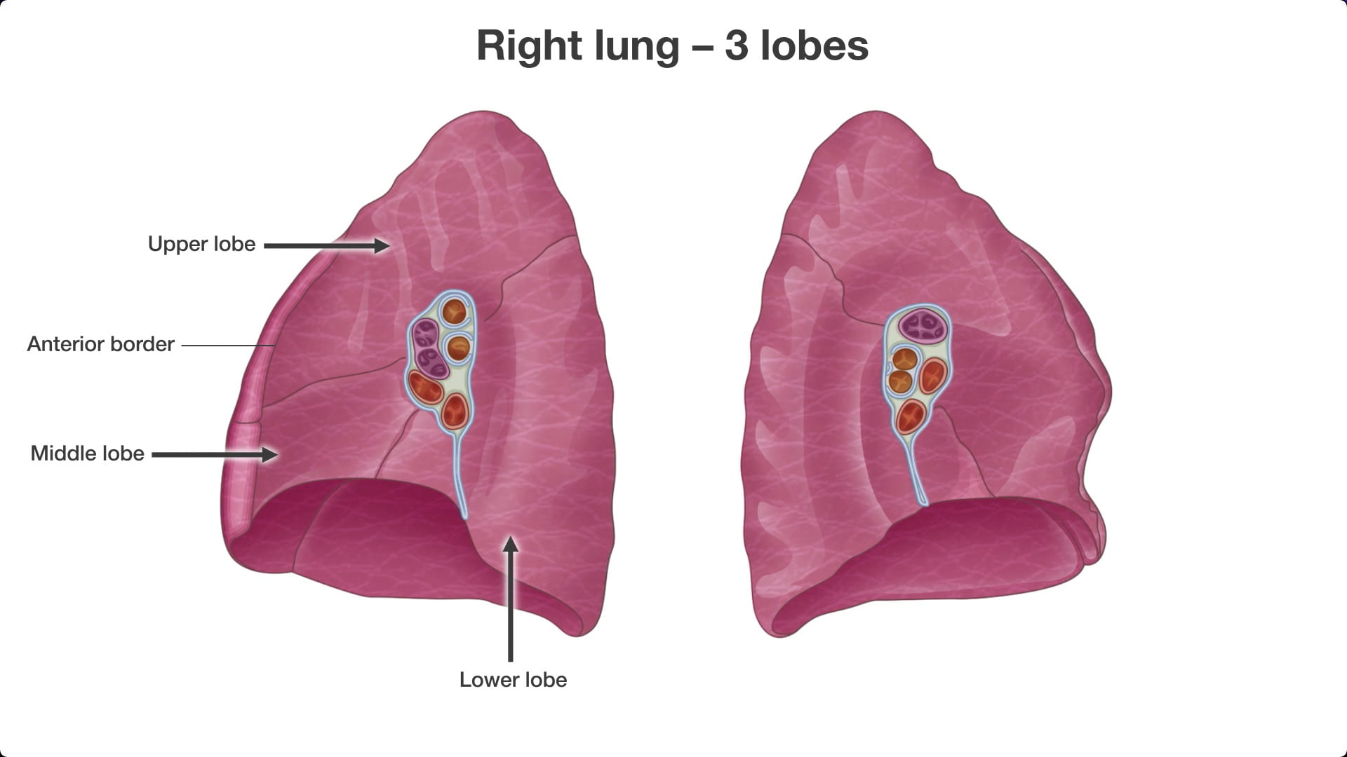
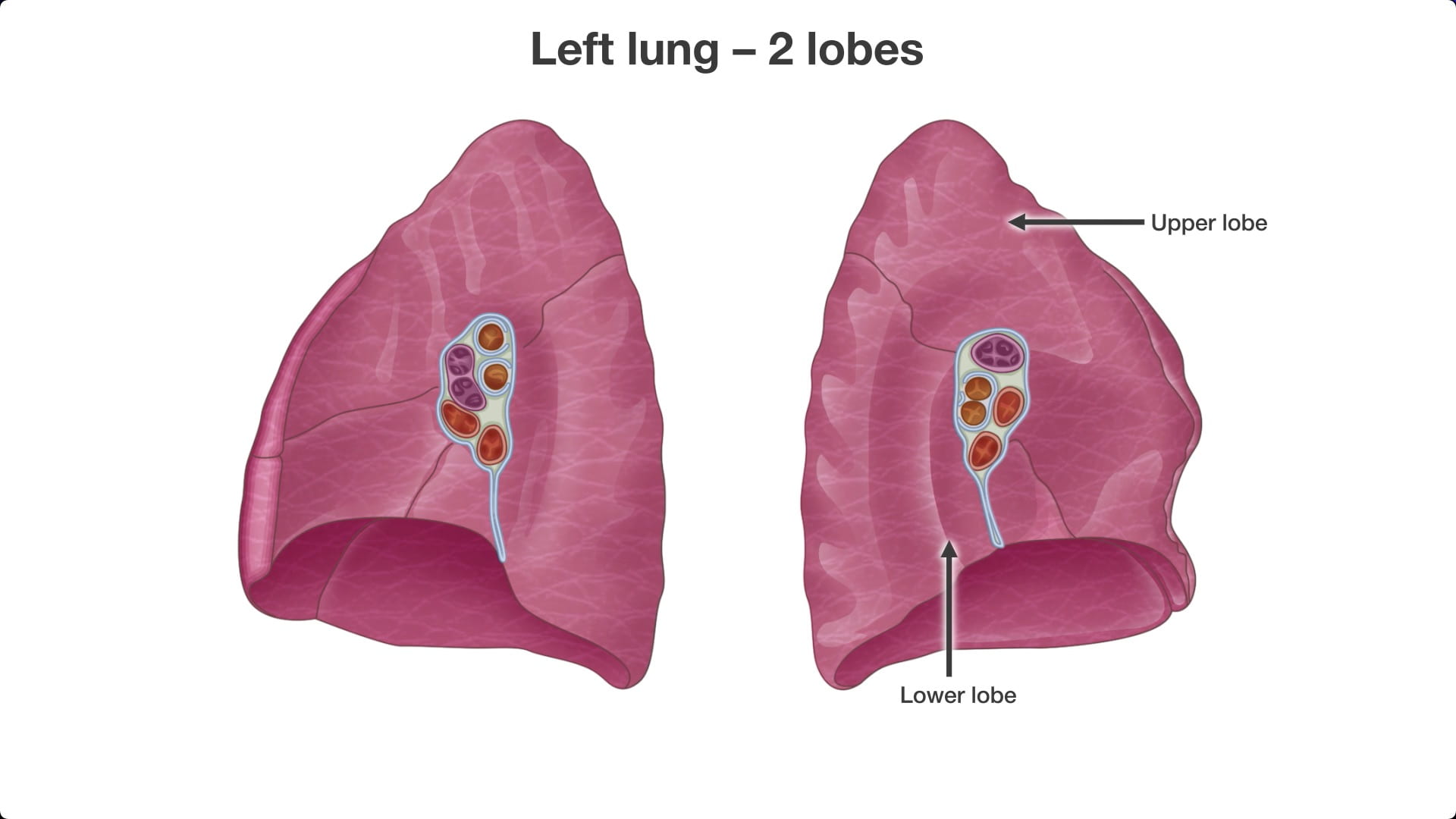
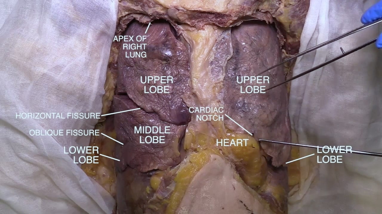
- Describe the lobes and fissures of the lungs.
- Describe the course of and branches of the vagus and phrenic nerves near the hilus of the lung.
- Describe the relationship between the branching of pulmonary arteries, veins and bronchi.
Lecture List
The Thorax Chest Wall, Lungs, Chest Plate Removal, Lung Removal, Lung Dissection
1. Thoracic Structure
1.1. Thoracic Structure Gallery
2. Introduction to Lungs
2.1. Introduction to Lungs Gallery
3. Chest Plate Removal
3.1. Exposing the Chest
Remove the chest plate by incising skin and superficial fascia in the midline along the length of the sternum.
Incise around the nipple to leave it in place attached to the chest wall.
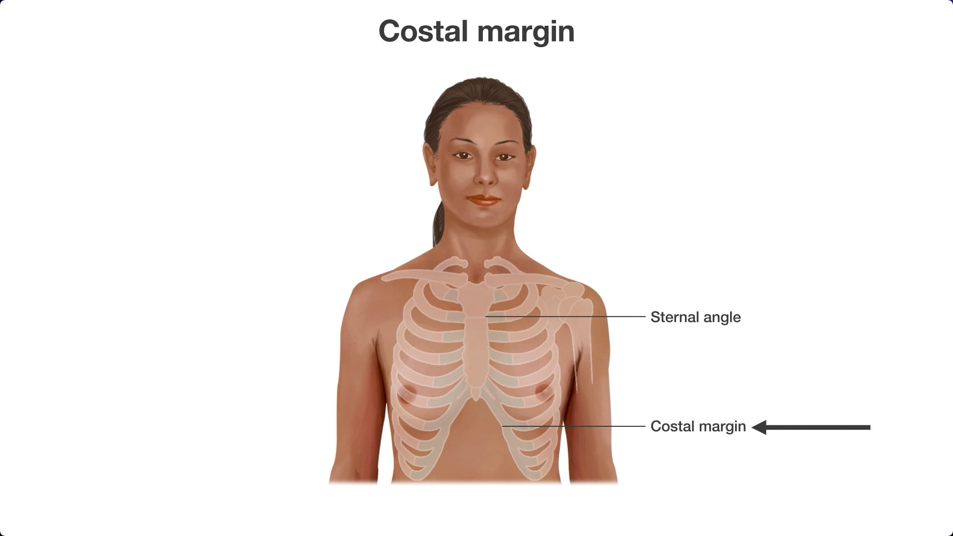
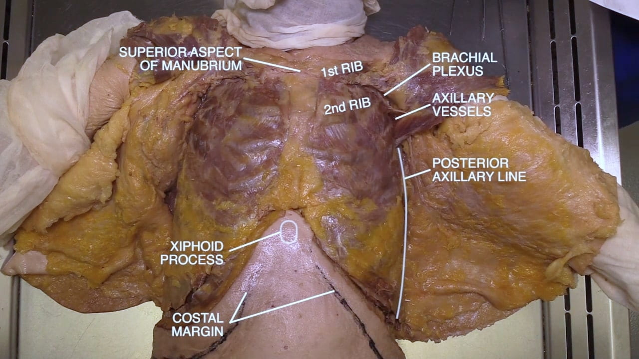
Make lateral incisions just below clavicles extending inferior to axilla to posterior axillary line. Inferiorly, make lateral incisions along the costal margin to the posterior axillary line.

3.2. Superficial Fascia, Pectoralis Major
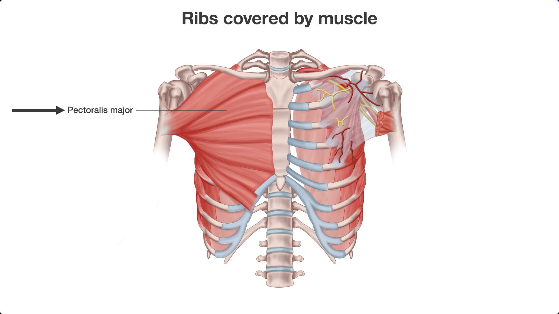
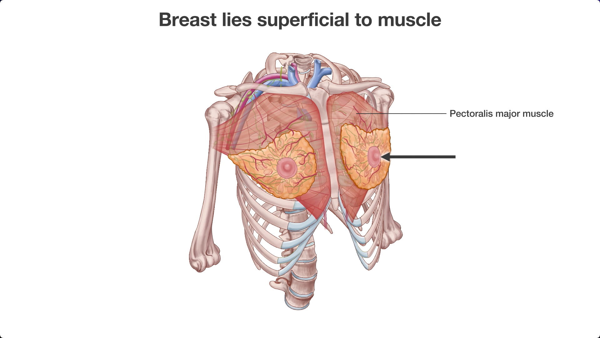
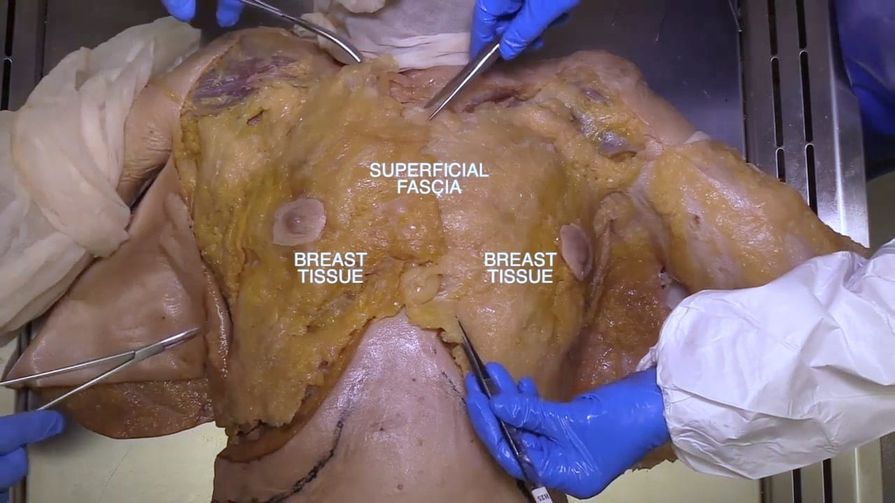
If you have a female donor, examine the extent of breast tissue. Note the breast is superficial to the pectoralis major and extends to axilla.
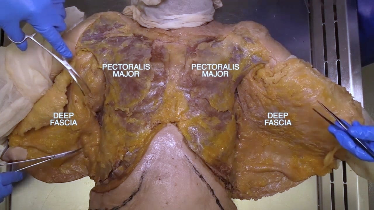
Divide the clavicular and sternal attachments of the pectoralis major muscles. Reflect these muscles laterally.
Question 1: Where is the lymphatic drainage of the breast?
Axillary lymph nodes.
3.3. Removal of Chest Plate
Using the oscillating saw, cut the manubrium and the first and second ribs just below the clavicle.
Extend this cut laterally and inferiorly along the posterior axillary line.
The inferior cut should be 5cm above the costal margin.
Gradually elevate the chest plate from inferior to superior while separating pleura and pericardium from the chest wall. Note, the cut ribs are sharp.
Where necessary, divide soft tissue with sharp dissection.

3.4. Internal Surface Chest Plate
Examine the interior surface of the chest plate.
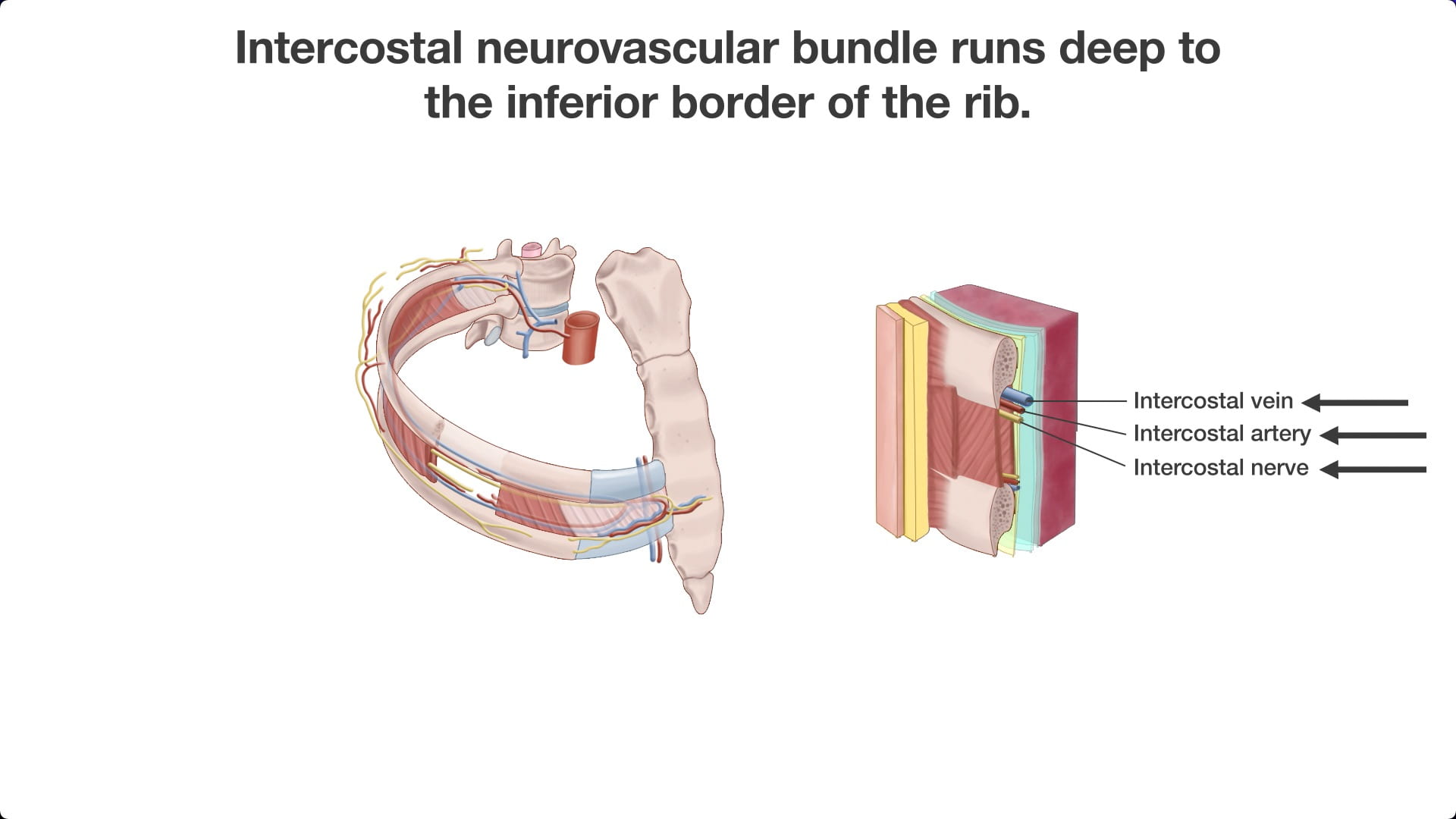
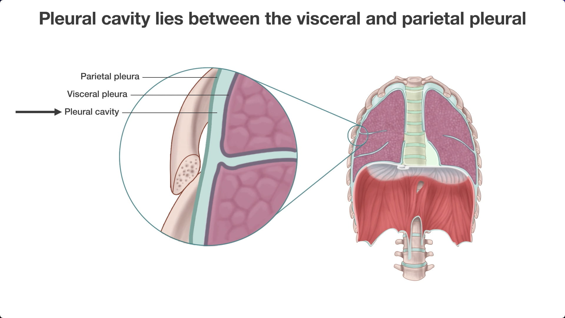
Identify the costal parietal pleura lining the ribs and intercostal spaces.
Locate the internal thoracic (internal mammary) vessels lateral to the sternum.
Question 2: How could the internal thoracic artery be useful in coronary artery bypass graft surgery?
The left internal thoracic artery runs close to the heart and can be anastomosed to a coronary artery.

3.5. Chest Cavity
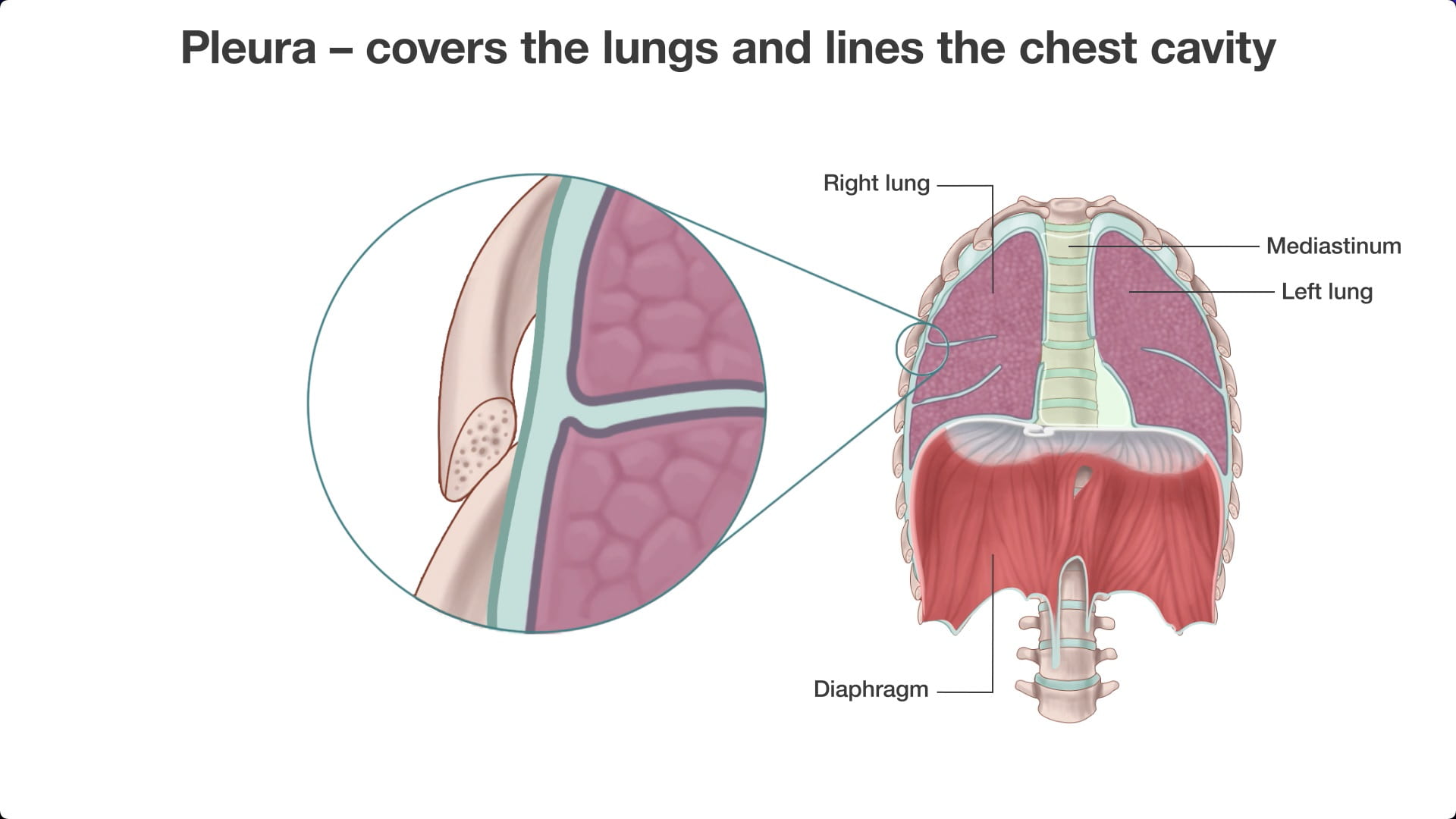
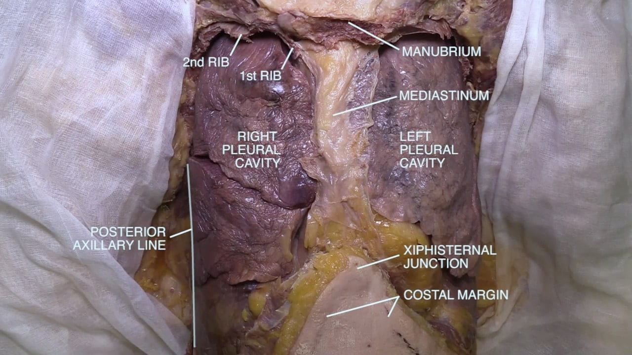
Note the right and left pleural cavities and the mediastinum.
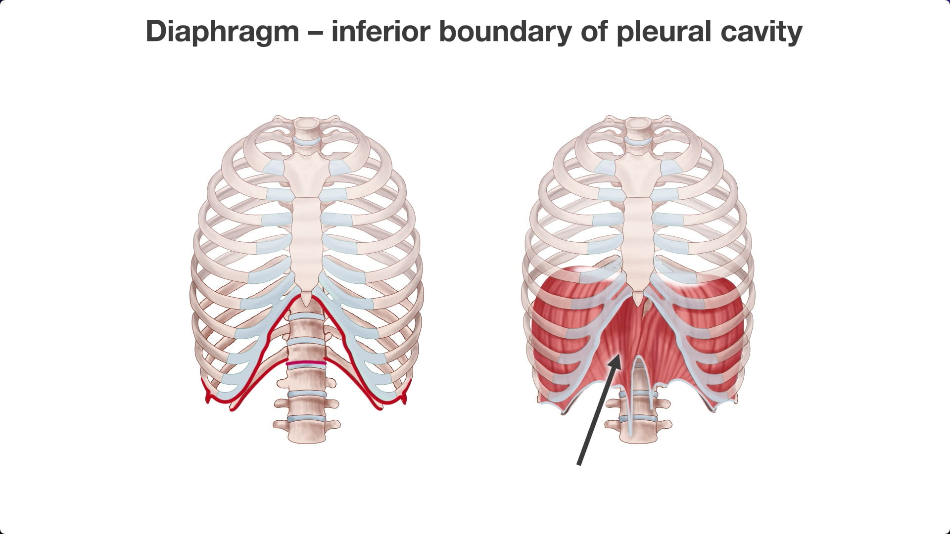
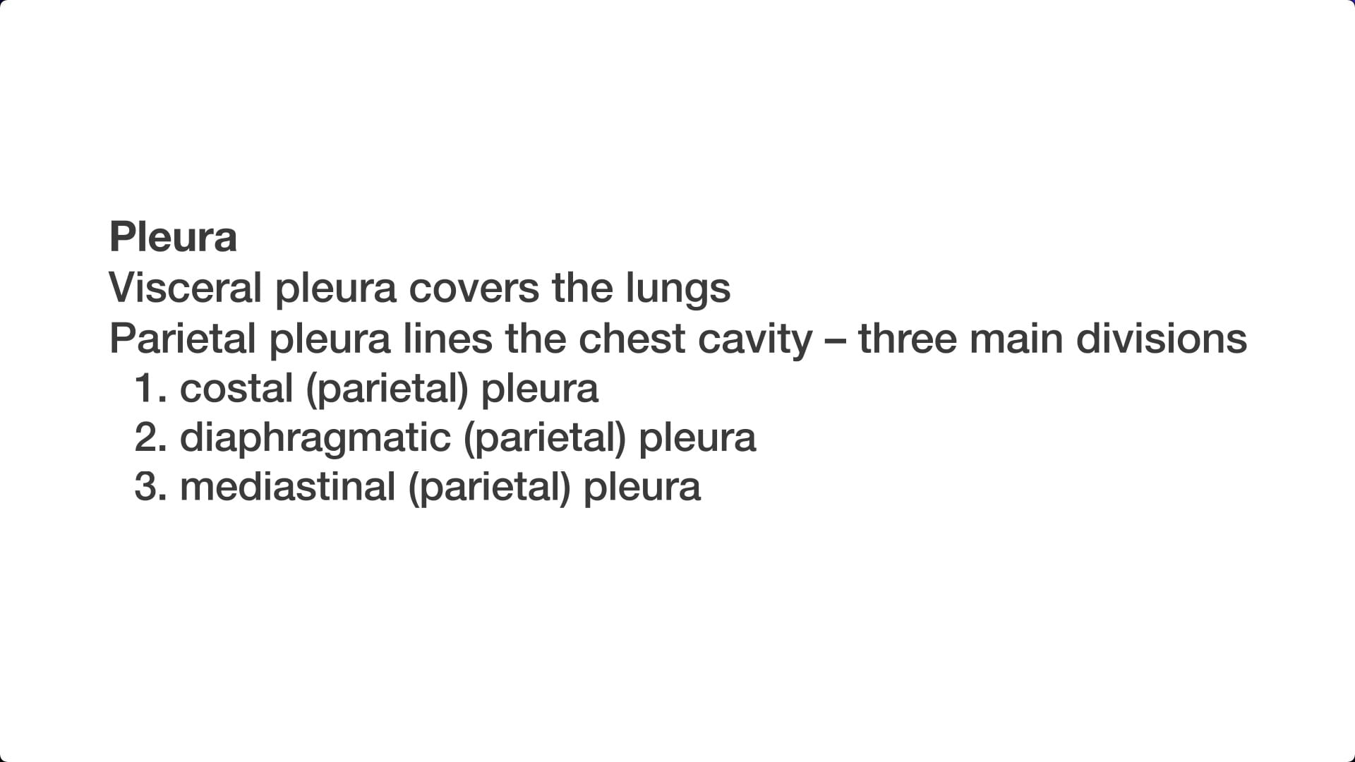
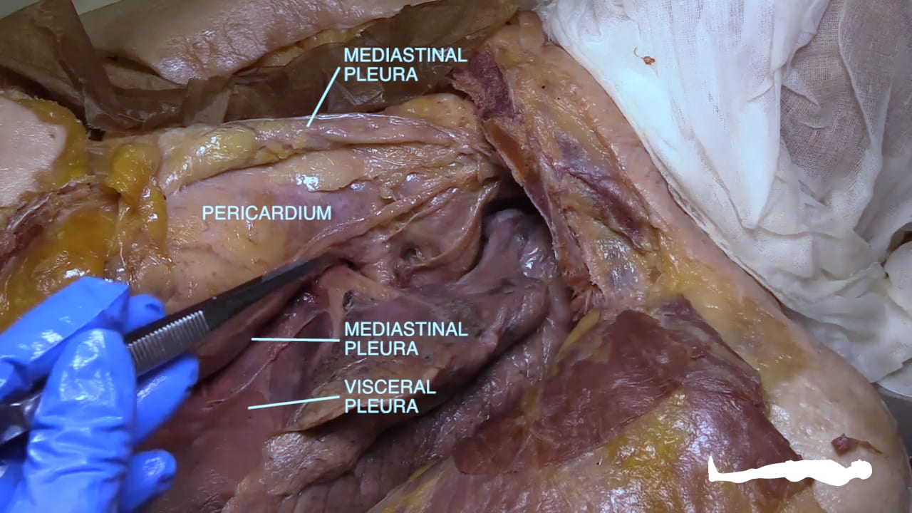
Identify the parietal pleura (costal, diaphragmatic and mediastinal divisions) and visceral pleura.
Identify the right and left lungs along with their lobes.
Identify the pericardium. Note the relationships of the heart to the diaphragm and lungs.
4. Lung Removal
4.1. Right Pneumonectomy
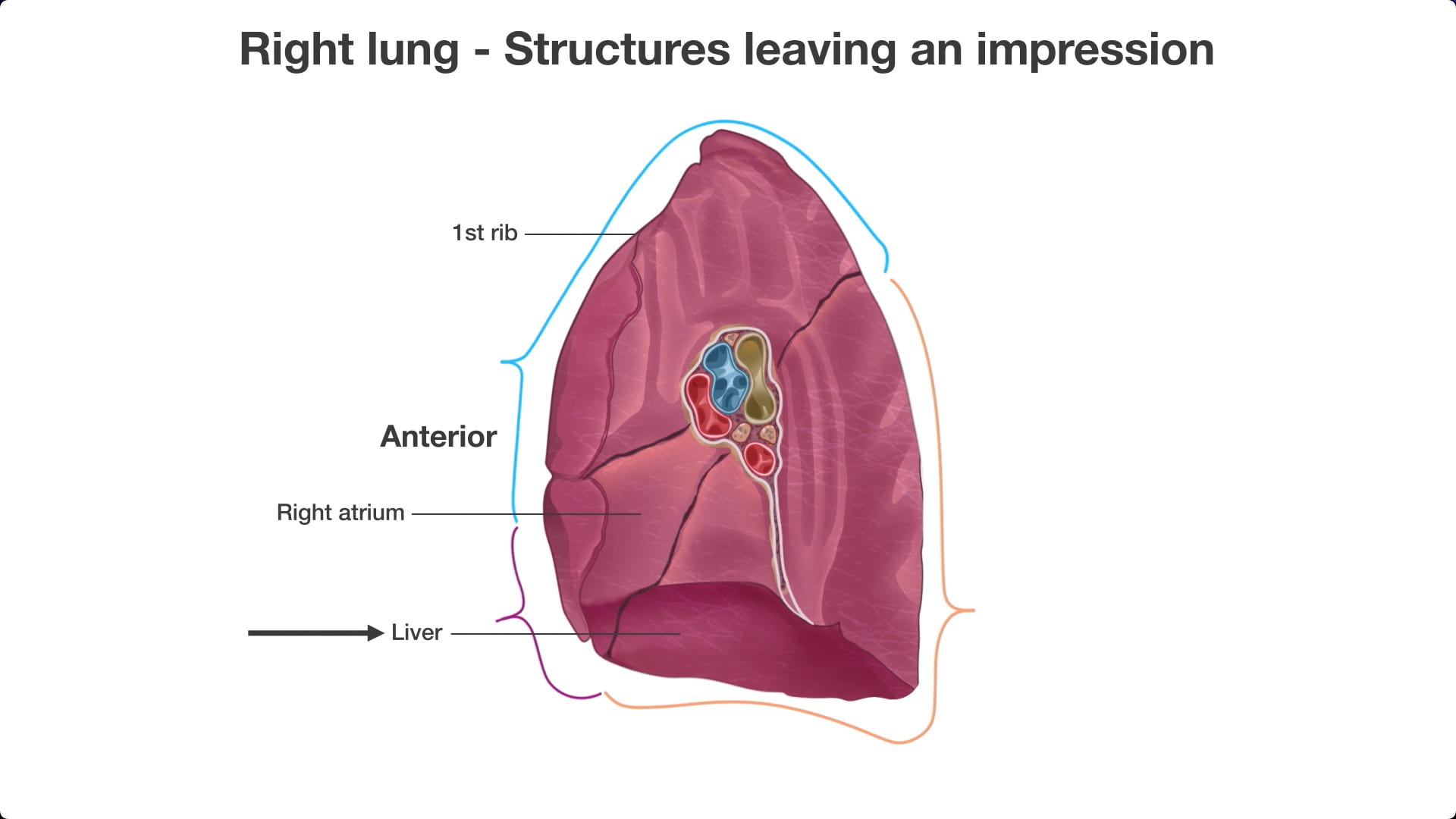
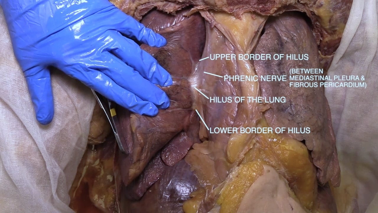
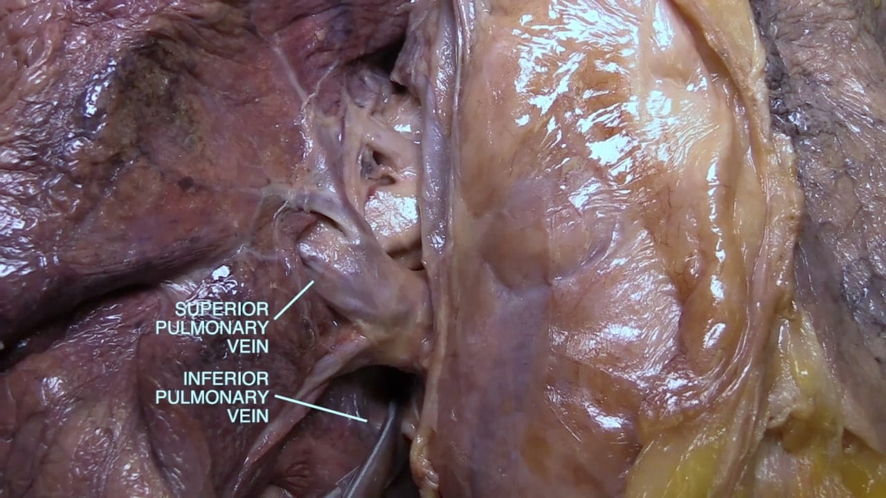
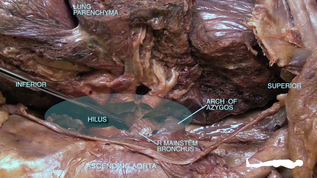
Manually retract the medial surface of the right lung laterally to expose the hilus.
At the hilus of the lung, the mediastinal pleura is reflected onto the lungs as visceral pleura.
Anterior to the hilus, identify the phrenic nerve, which runs between pleura and pericardium to reach the diaphragm.
4.2. Right Pneumonectomy
Divide the pleura at the hilus.
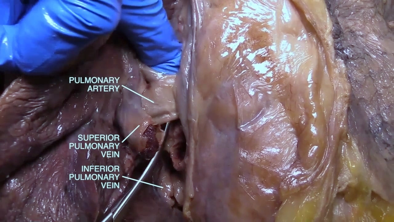
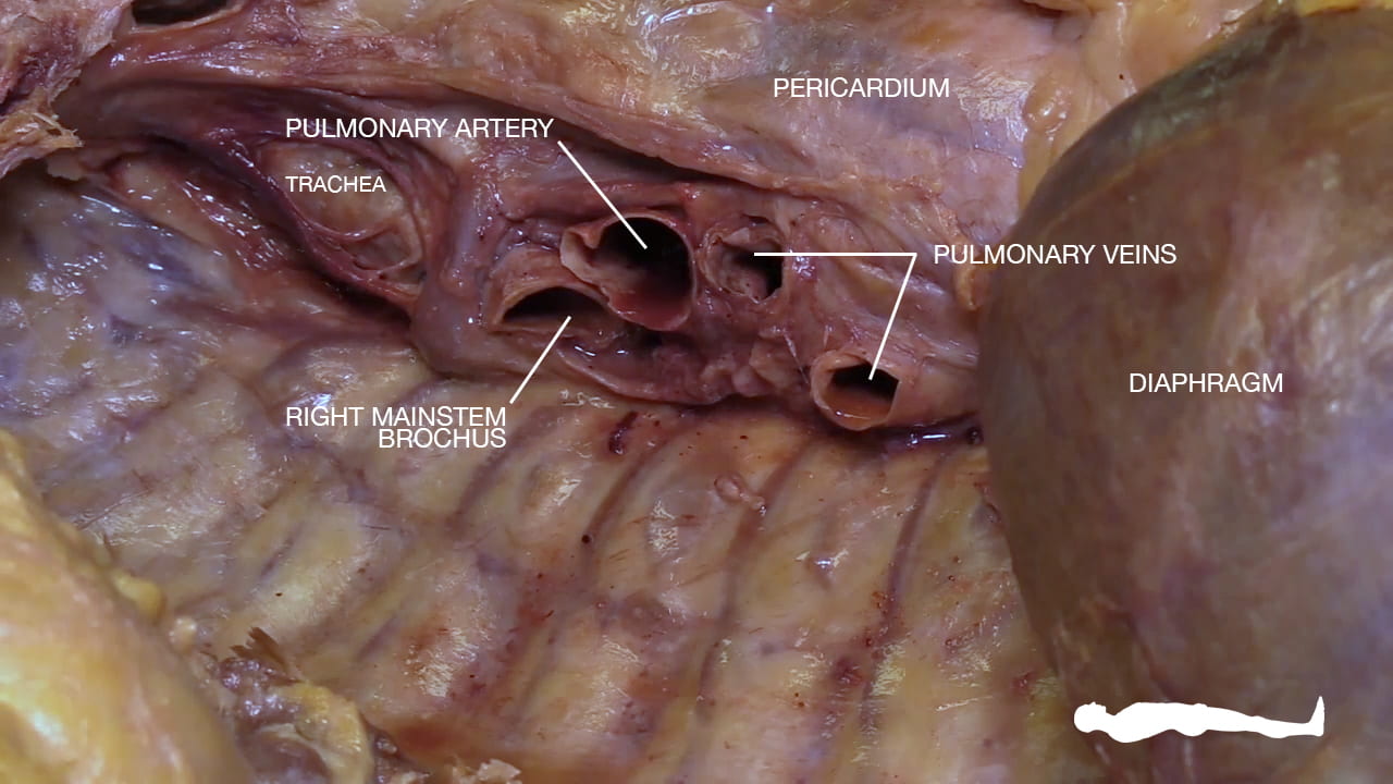
Locate and divide superior and inferior pulmonary veins.
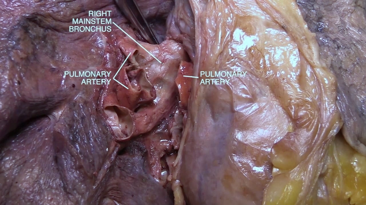
Next divide the right pulmonary artery.
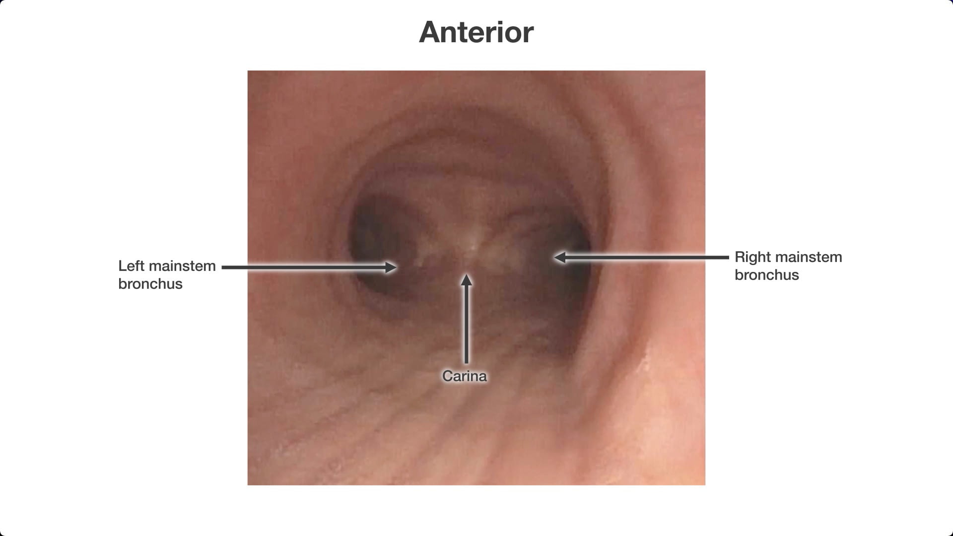
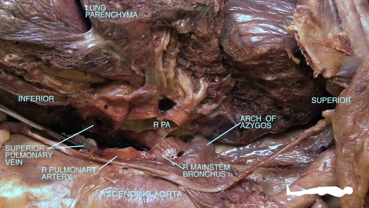
Posterior to the right pulmonary artery, locate and divide the mainstem bronchus.
Divide the posterior pleura and remove the lung.
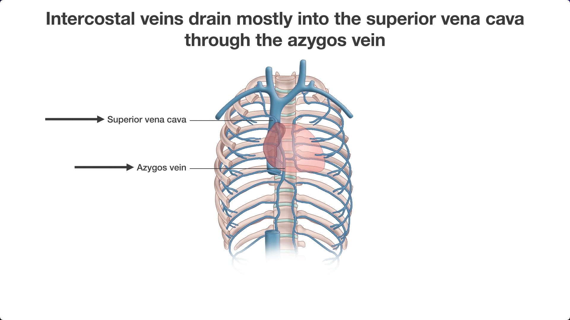
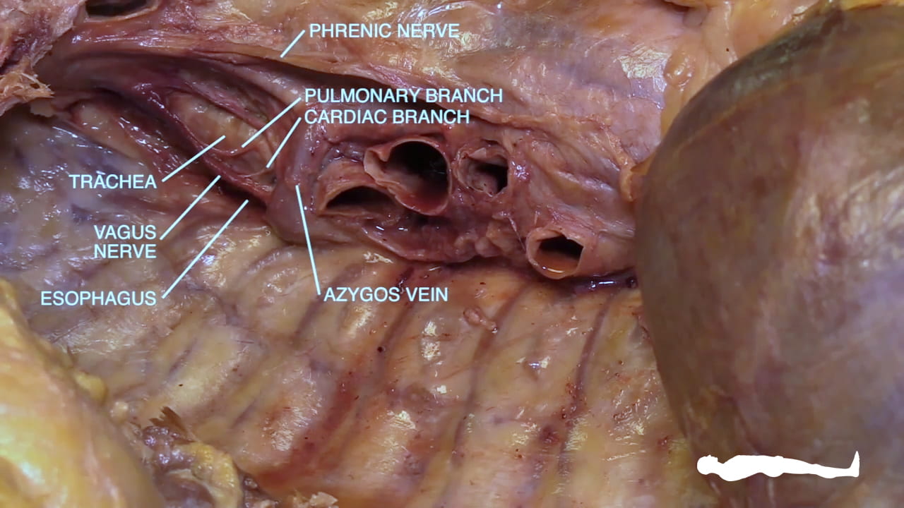
Re-identify the phrenic nerve. On the mediastinum, locate the azygos vein and the right vagus nerve superior and posterior to the hilus.
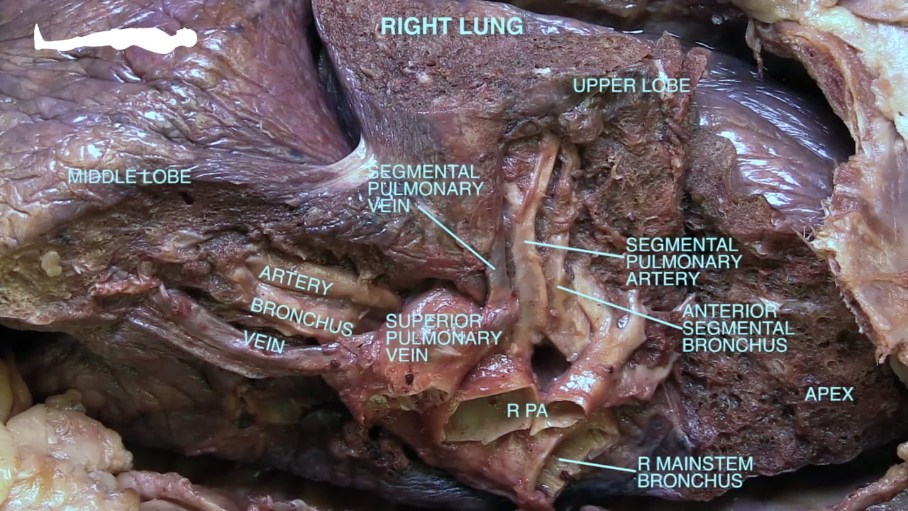
Identify the mainstem bronchus, pulmonary artery and pulmonary veins on the medial surface of the lung and mediastinum.
4.3. Left Pneumonectomy
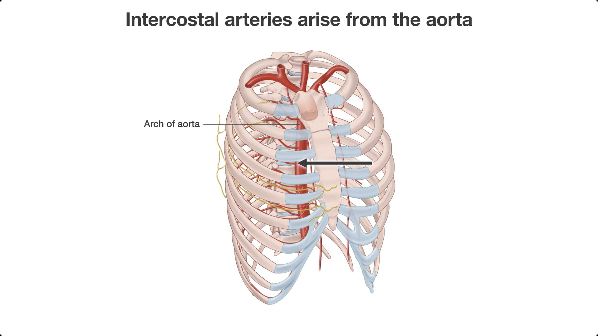
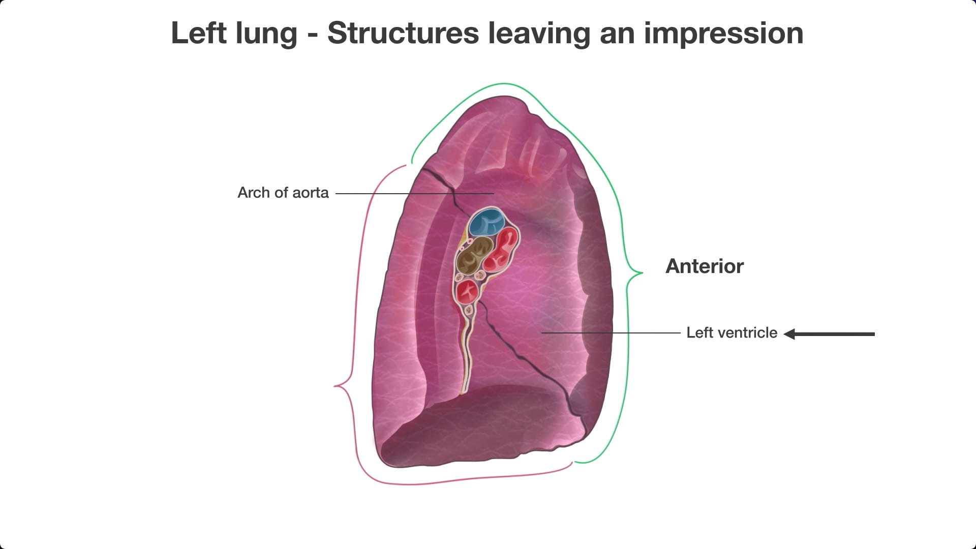
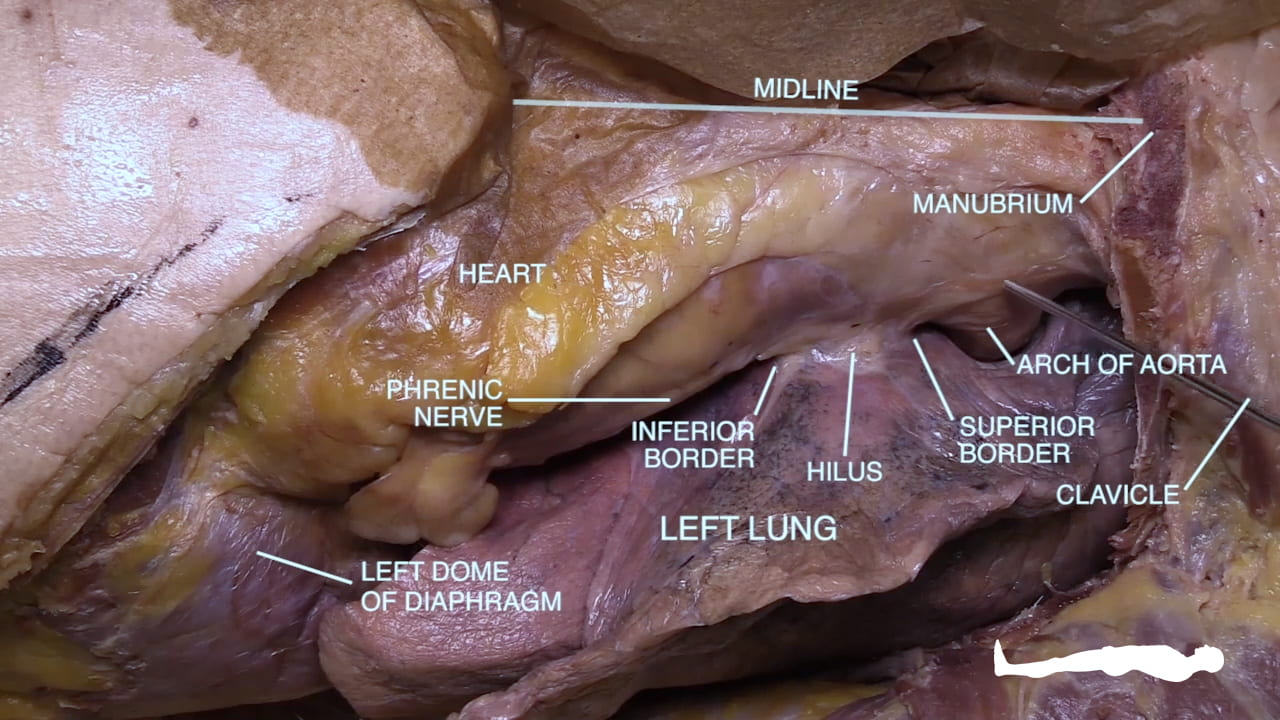
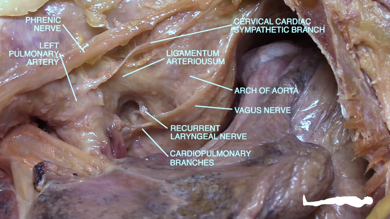
Manually retract the medial surface of the left lung laterally to expose the hilus. Note the relationships of the left lung to the diaphragm, pericardium and arch of aorta.
Identify the phrenic nerve.
Locate the left vagus nerve lateral to the arch of the aorta.
Locate the ligamentum arteriosum between the left pulmonary artery and the arch of the aorta.
Locate the left recurrent laryngeal branch of the vagus nerve as it passes posterior to the ligamentum arteriosum. Locate cardiac and pulmonary branches of the vagus nerve.
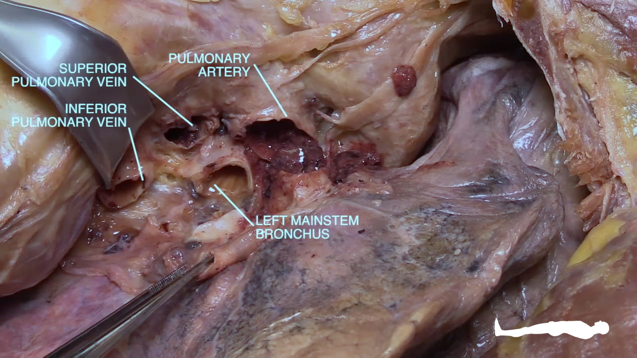
Divide the pleura at the hilus.
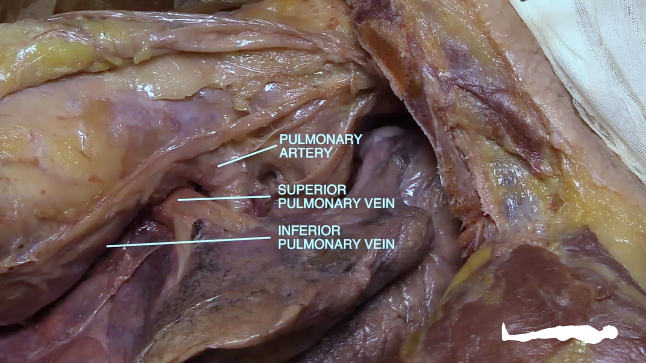
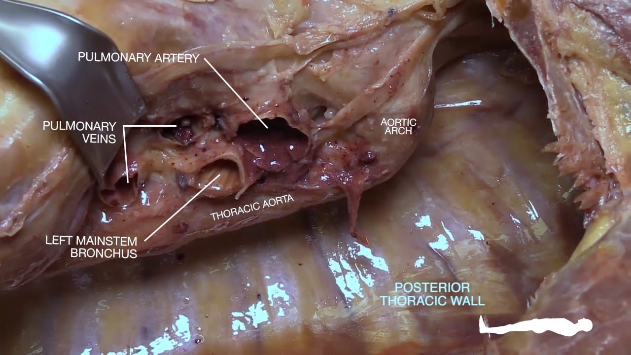
Divide the superior and inferior pulmonary veins then divide pulmonary artery. Next divide mainstem bronchus.
Incise the posterior pleura and remove the lung.
Identify the mainstem bronchus, pulmonary artery and pulmonary veins on the medial surface of the lung and mediastinum.
Question 3: Why are the phrenic nerves at risk for surgery in this area?
The phrenic nerve passes anterior to the hilus of the lung.
Question 4: What embryologic structure does the ligamentum arteriosum represent?
The ductus arteriosus which allows blood from the right ventricle to bypass the lungs.
5. Right Lung Dissection
5.1. Lobar Bronchi
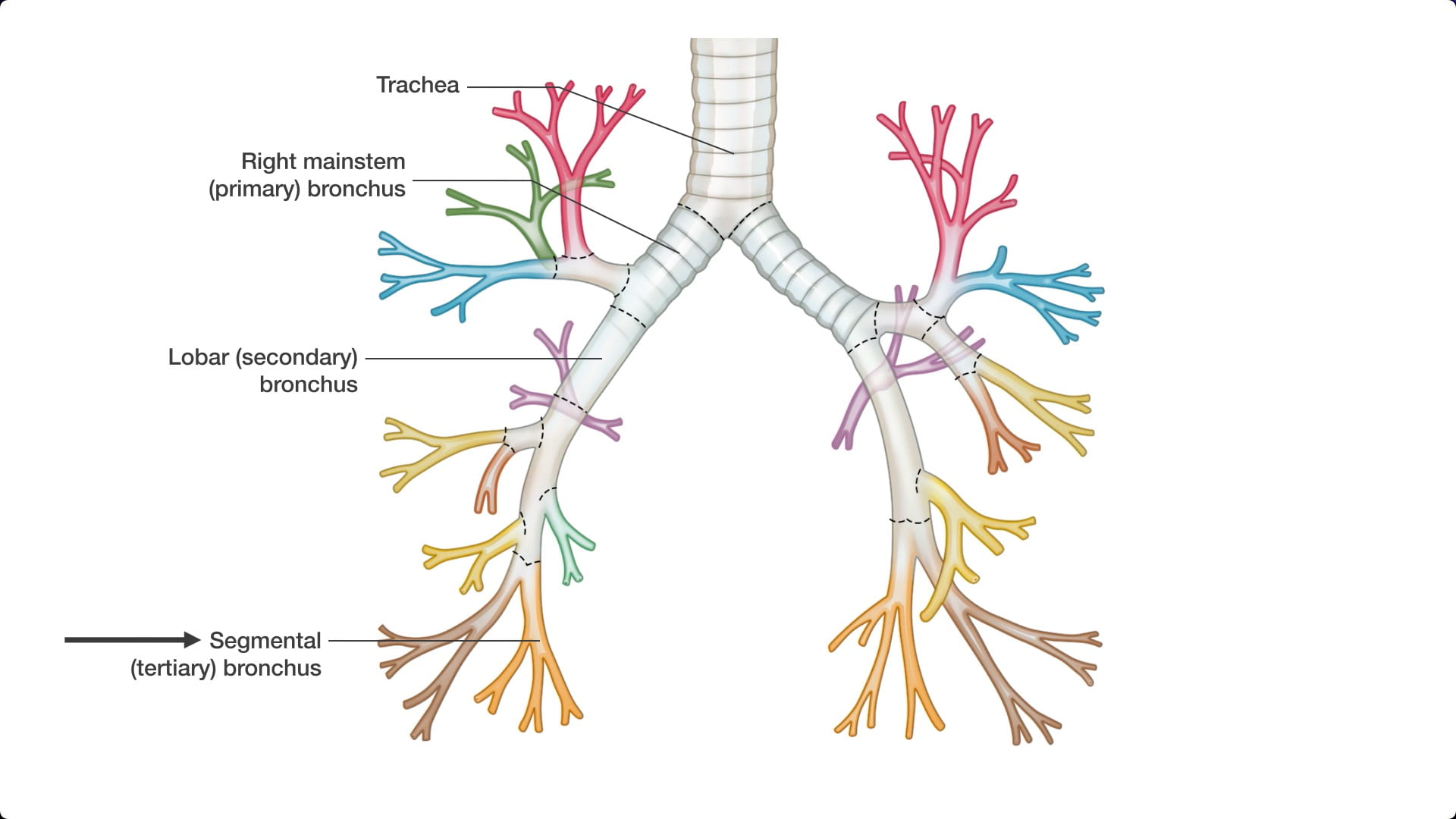
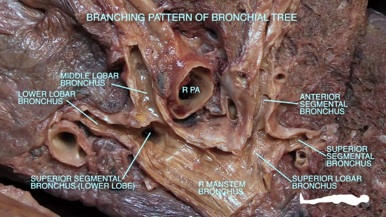
Bluntly dissect from the mainstem bronchus to lobar bronchi. Also dissect the accompanying pulmonary arteries.
Place the lung back in the chest cavity and note the relationship of bronchus, artery and veins from hilus to lung.
Open the bronchi to examine their internal structure.
Question 5: What is a segmental bronchus?
A tertiary bronchus which, along with a branch of the pulmonary artery, supplies a portion of the lobe called the bronchopulmonary segment.
5.2. Examining Recesses
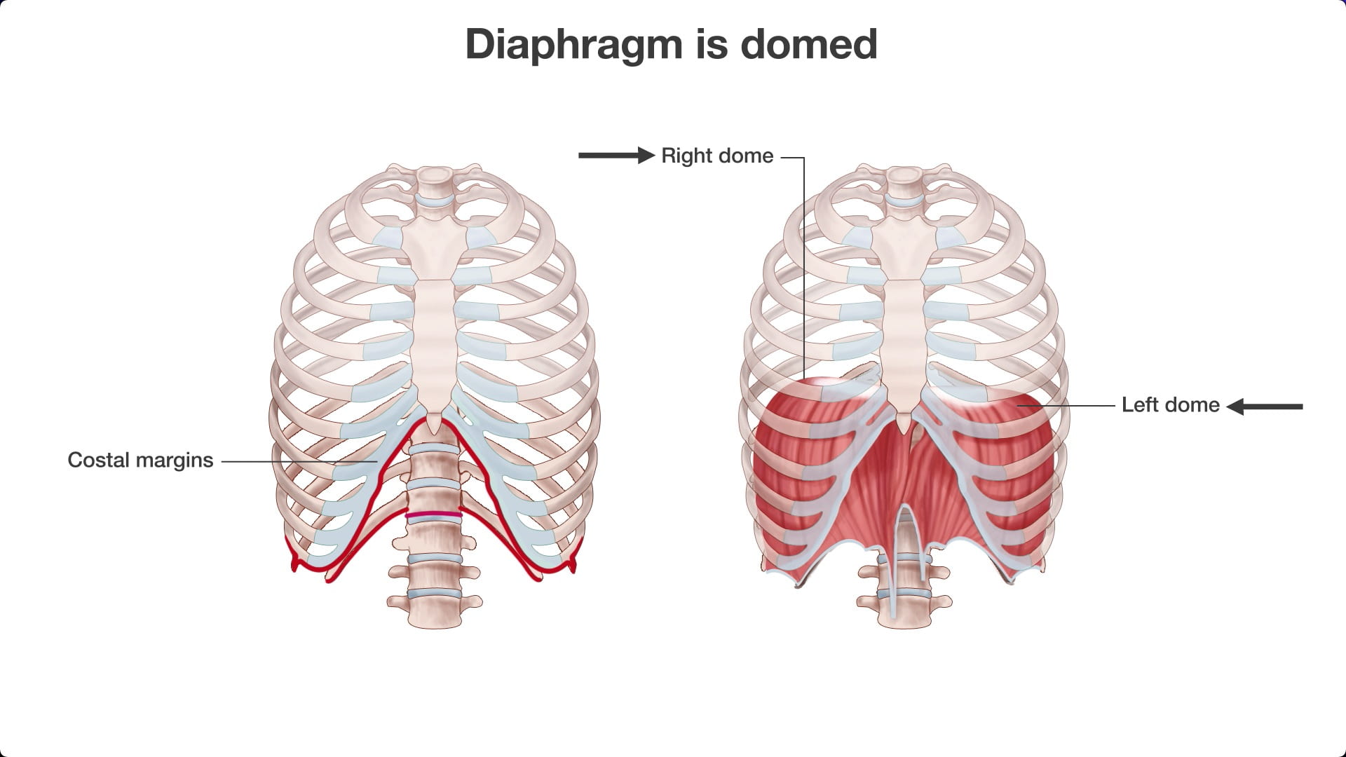
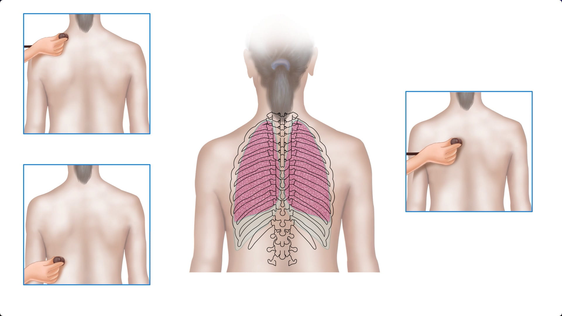
Place your hand in the costodiaphragmatic recess in the midclavicular, midaxillary and scapular lines.
Look at a skeleton in order to name the corresponding rib levels and compare to the chest film in figure 5.2a.
Place your hand in the cupola (chest cavity apex) on the donor or a skeleton. Note the relationship to the medial clavicle.

