Brachial Plexus

Lab Summary

The brachial plexus is taught in this lab from nerve roots to trunks, divisions, cords and branches. The relationships of these structures to scalene muscles, axillary artery and upper extremity musculature is presented. Nerves along chest wall, long thoracic and thoracodorsal nerves, vulnerable to axillary node dissection are emphasized for their surgical vulnerability. Peripheral nerves and vasculature are discussed as they course along the arm and elbow joint.

Lab Objectives

  1. Identify roots, trunks, divisions, cords and branches of the brachial plexus.
  2. Describe the relationship of the roots of brachial plexus to anterior and middle scalene muscles.
  3. Locate the phrenic nerve and describe its relationship to anterior scalene muscle.
  4. Relate the cords of brachial plexus to axillary artery.
  5. Identify the suprascapular, musculocutaneous, median, ulnar, radial and axillary nerves.
  6. Locate the subscapular, circumflex humeral and deep brachial branches of axillary artery.
  7. At the elbow, describe the relationship of tendon of biceps and brachial artery.
  8. At the elbow joint, describe the course of median, ulnar and radial nerves as they pass to forearm.

Lecture List

Brachial Plexus, Proximal Brachial Plexus, Distal Brachial Plexus

Brachial Plexus

Anterior Chest Musculature

If not previously accomplished, carry out the following:

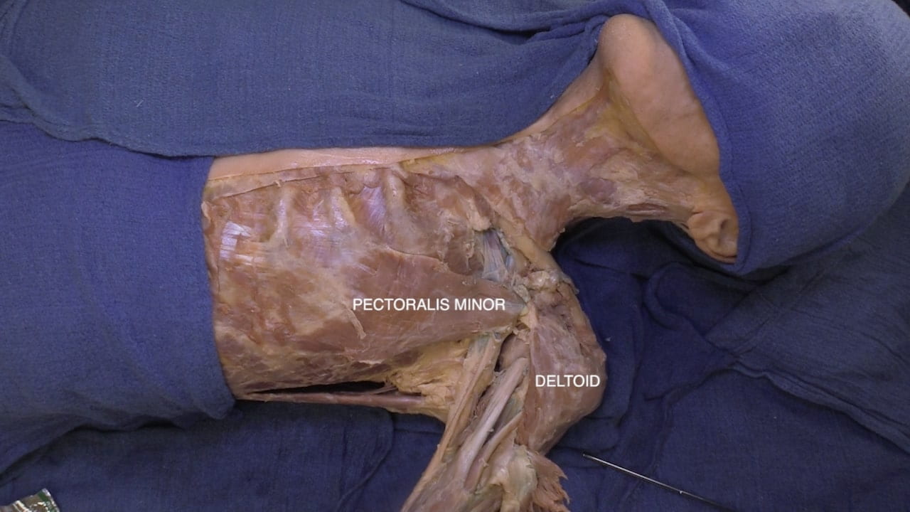
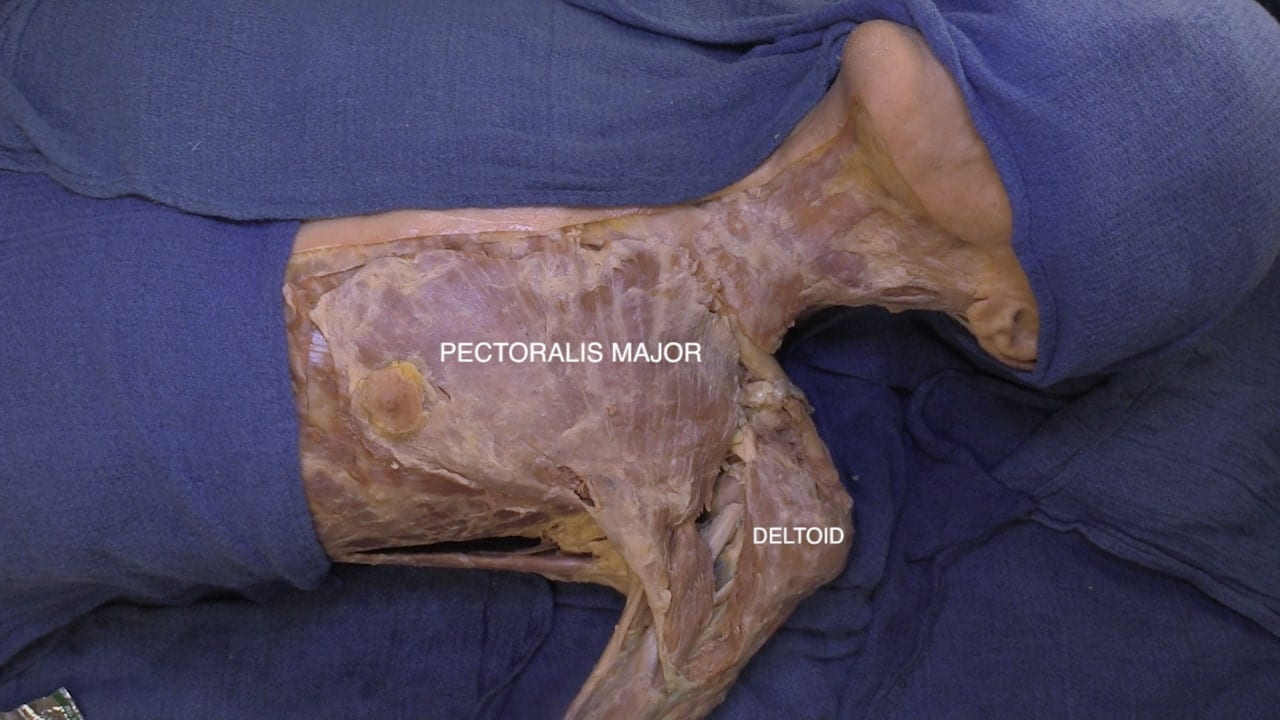
Reflect the pectoralis major to its attachment on the humerus and separate the deltoid from the clavicle and reflect laterally.

Divide the pectoralis minor at the coracoid process reflect medially.

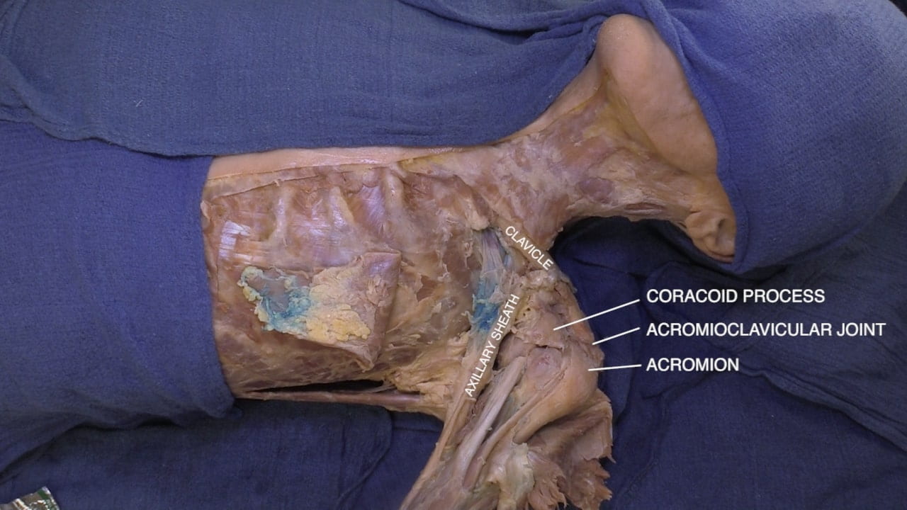
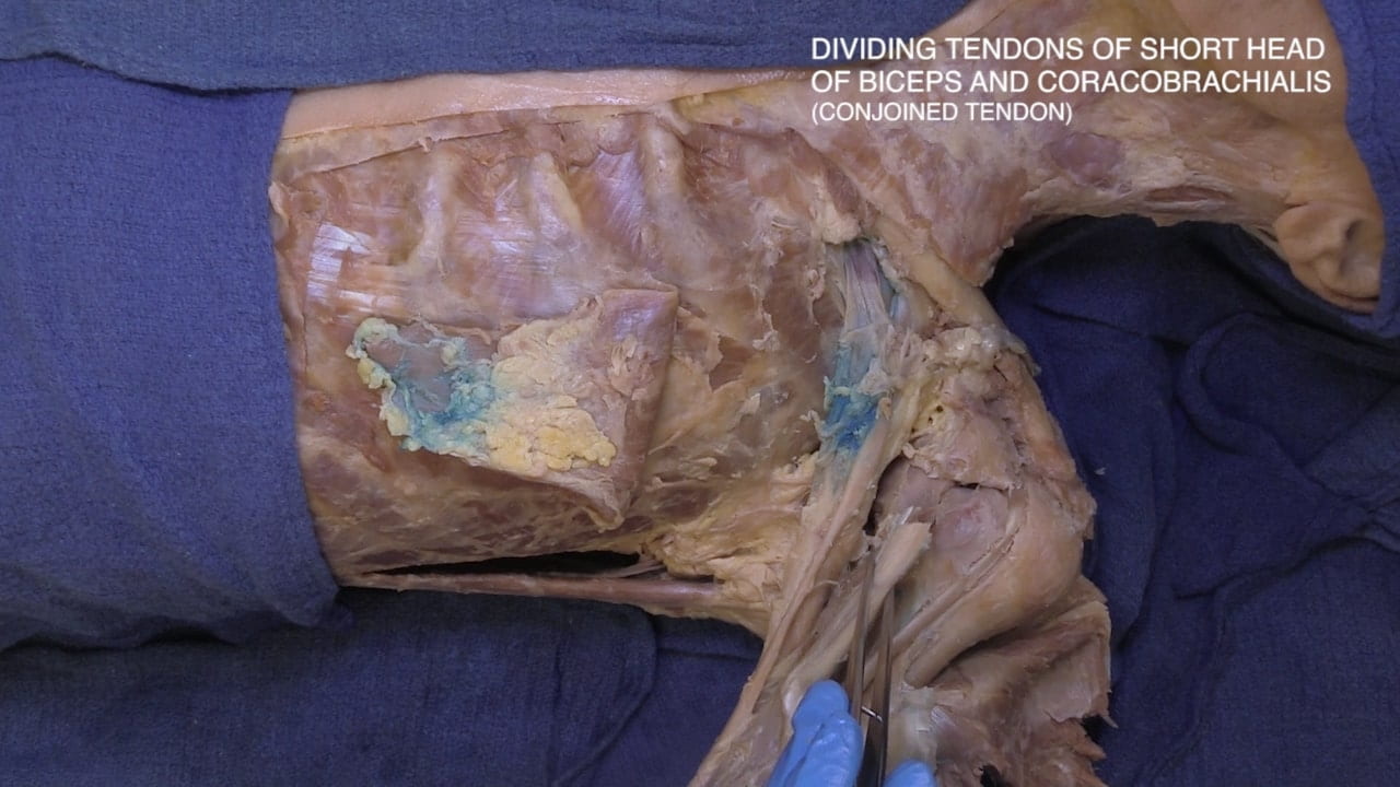
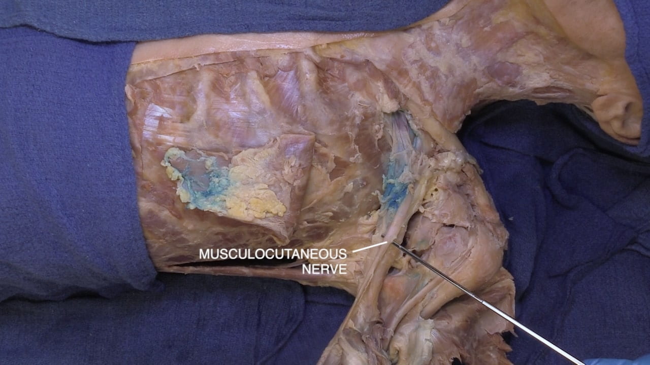
Divide and reflect the conjoined tendon (tendons of the short head of the biceps and the coracobrachialis). Locate the musculocutaneous nerve entering the coracobrachialis. Identify the axillary sheath in relation to the clavicle, acromion and coracoid process.

Axillary Vessels and Brachial Plexus

Open the axillary sheath proximal to the coracoid process. Identify the axillary vein and artery as well as a portion of the brachial plexus. Recall the axillary vessels become the subclavian artery and vein as they pass beneath the clavicle.

Labels (top to bottom): Subclavian artery, subclavian vein, axillary artery, axillary vein, brachial plexus, clavicle, cephalic vein
1.2a) Vasculature

Brachial Plexus and Axillary Vessels

Open the axillary sheath distal to the coracoid process.

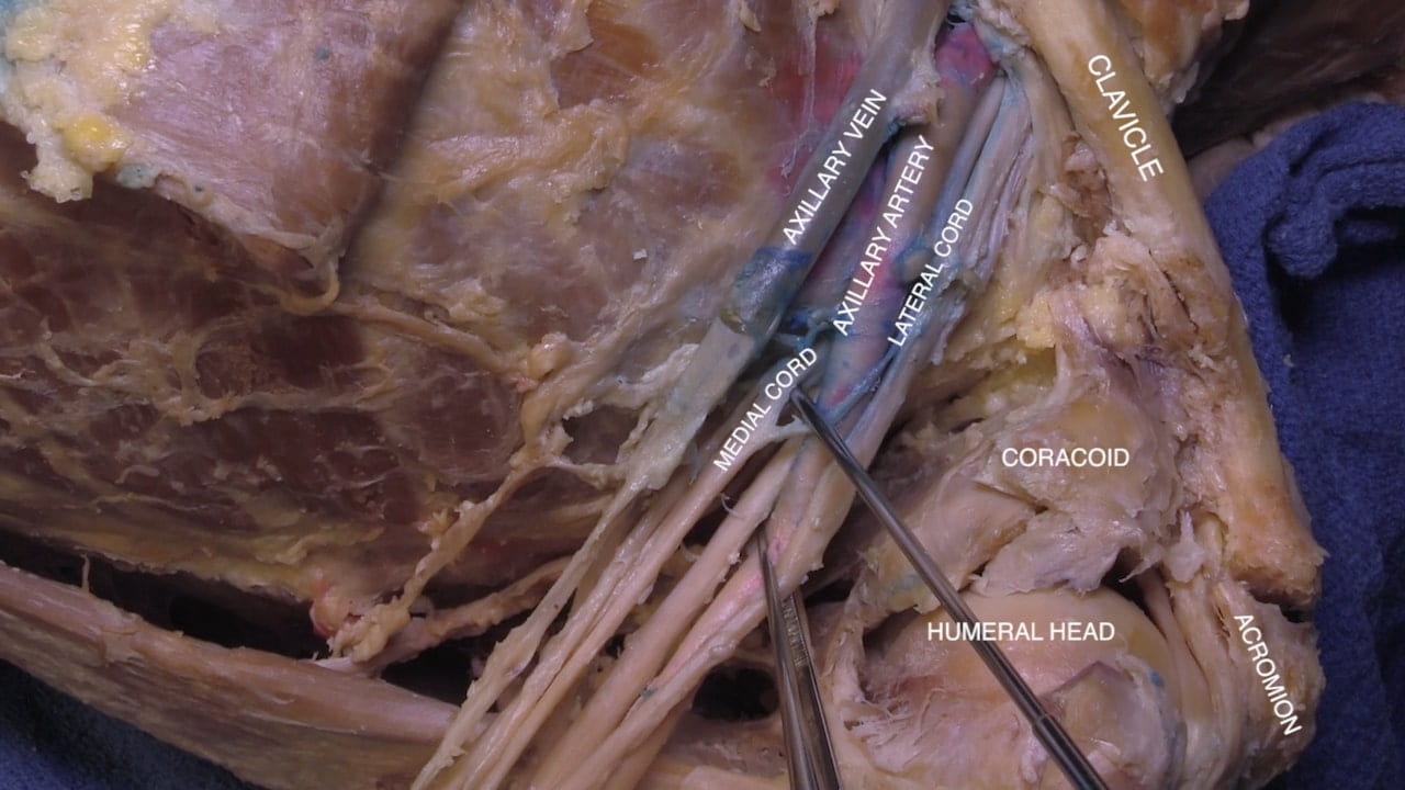
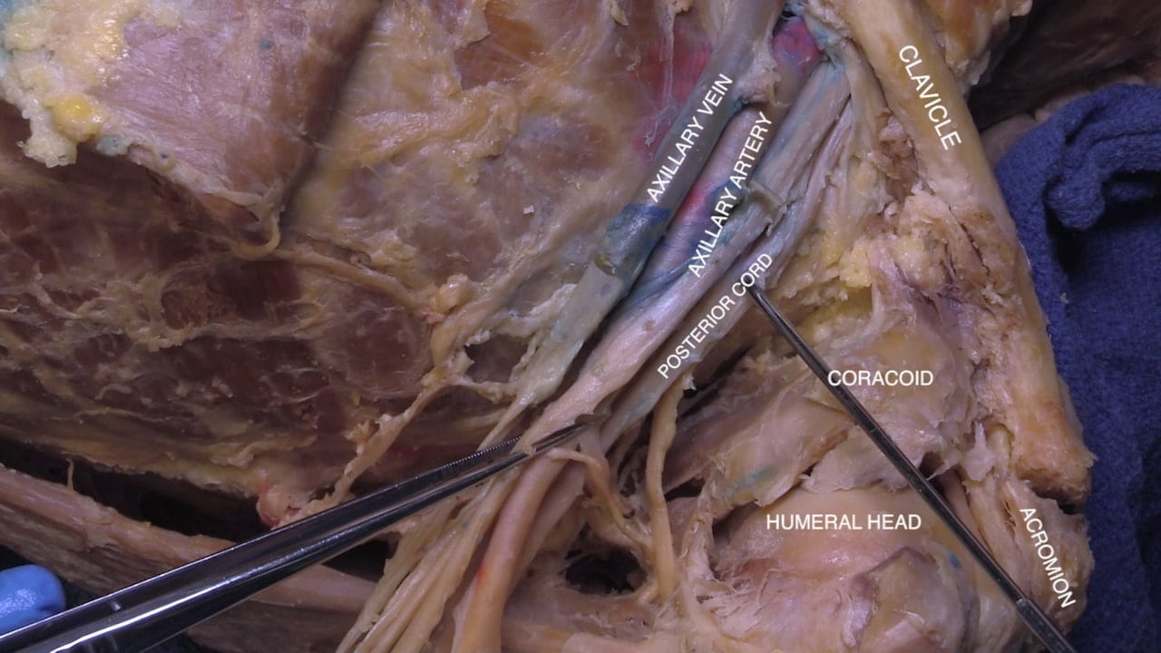
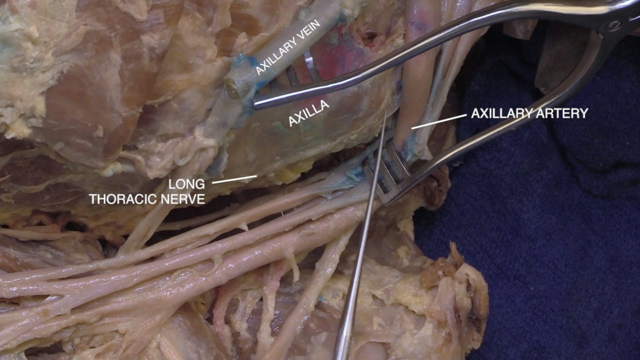
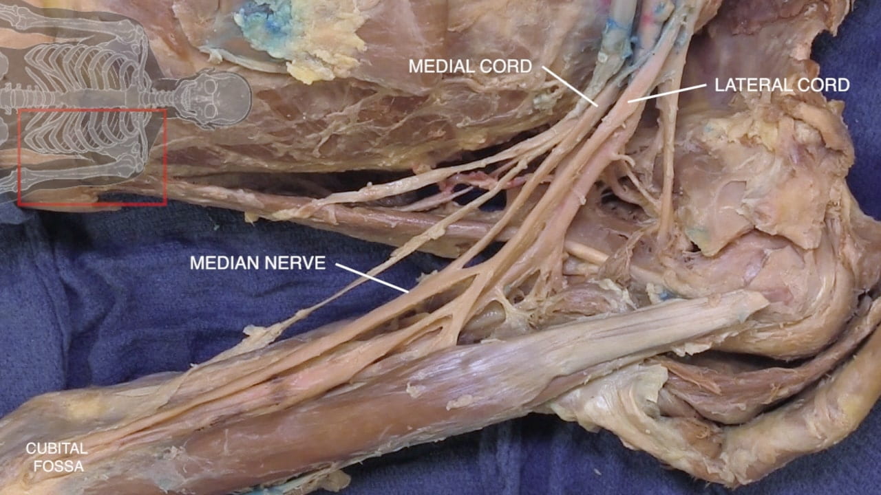
Identify the axillary artery. The cords of the brachial plexus are named for their relationship to the artery. Identify the medial and lateral cords. Reflect the axillary artery medially to locate the posterior cord.

Lateral Cord

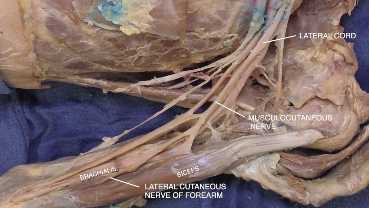
Follow the lateral cord distally.  Reidentify the musculocutaneous nerve to the biceps, coracobrachialis and brachialis muscles. Locate its distal branch, the lateral cutaneous nerve of the forearm.

Note the contribution of the lateral cord to the median nerve.

Labels (top to bottom): Lateral cord, median nerve, musculocutaneous nerve, lateral cutaneous nerve of forearm, biceps
1.4a) Lateral cord

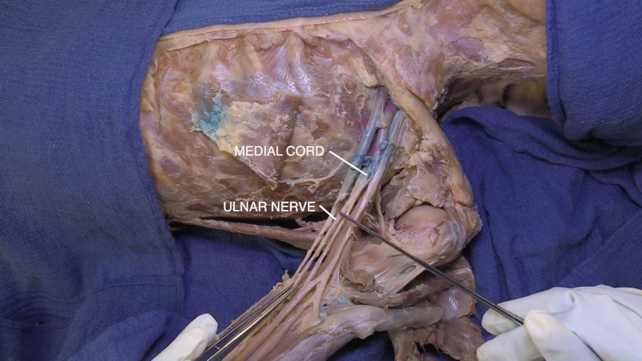
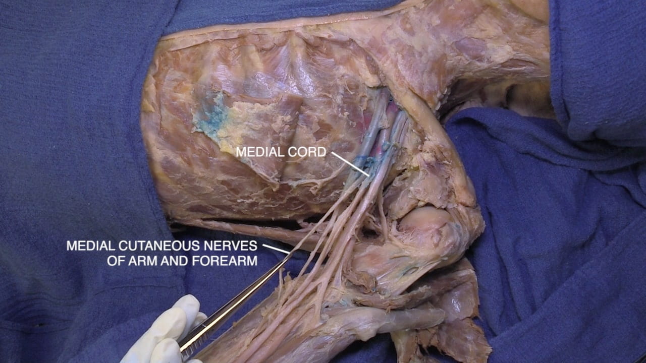
Medial Cord

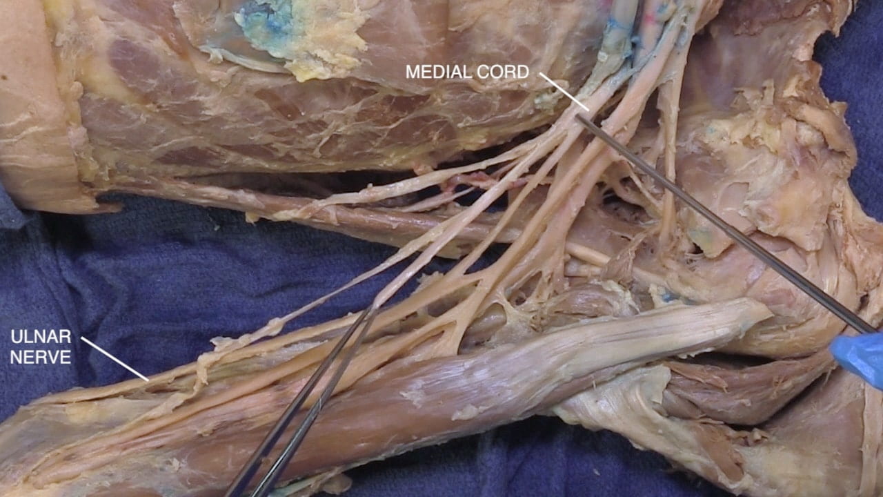
Locate the medial cord and identify the ulnar nerve by following its course distally. Also identify the medial cutaneous nerves of the arm and forearm as the remaining branches of the medial cord.

Note the contribution of medial cord to the median nerve.

Posterior Cord

Reflect the axillary vessels along with the medial and lateral cords medially.

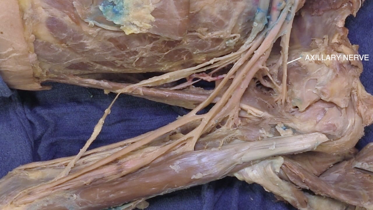
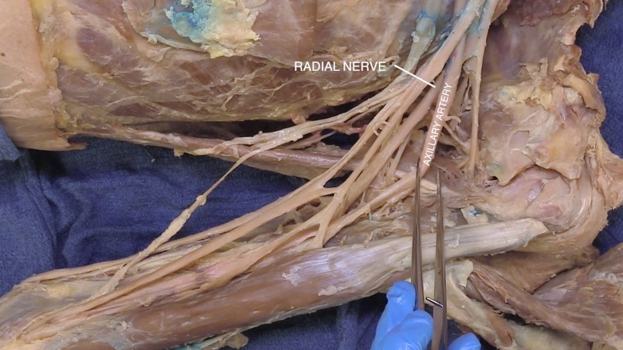
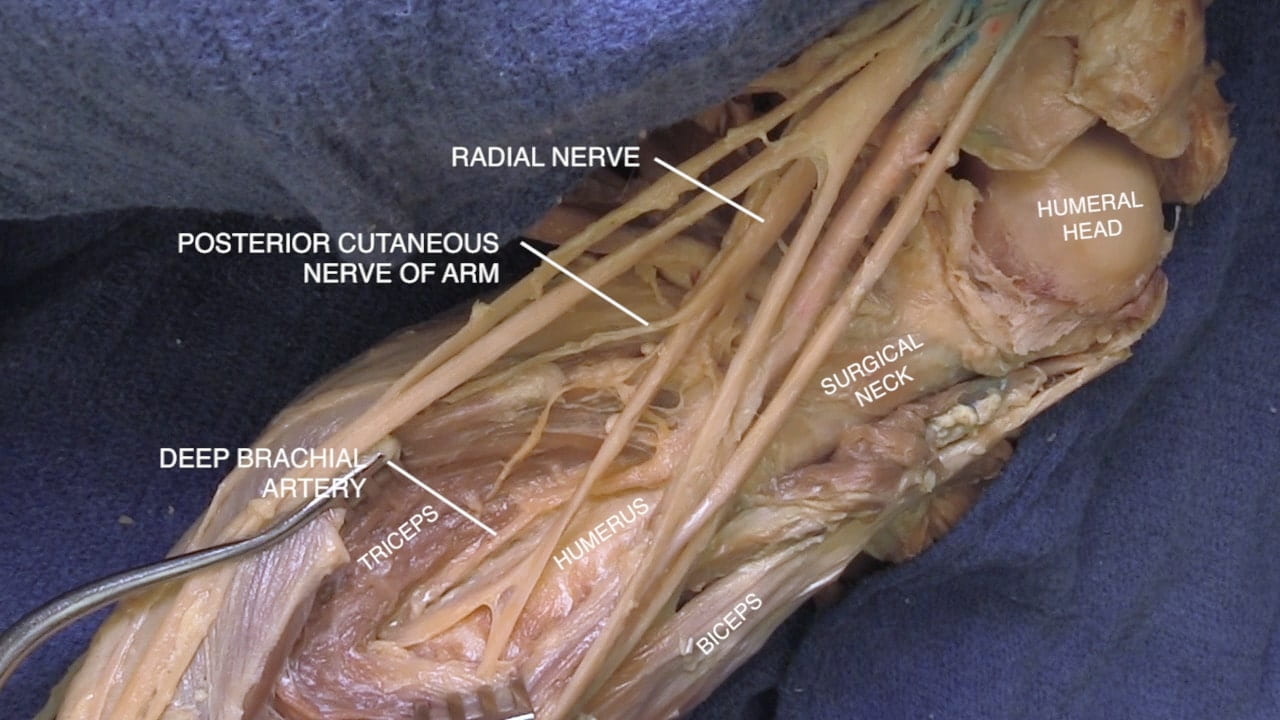
Identify the posterior cord and its major branches, the radial and axillary nerves.

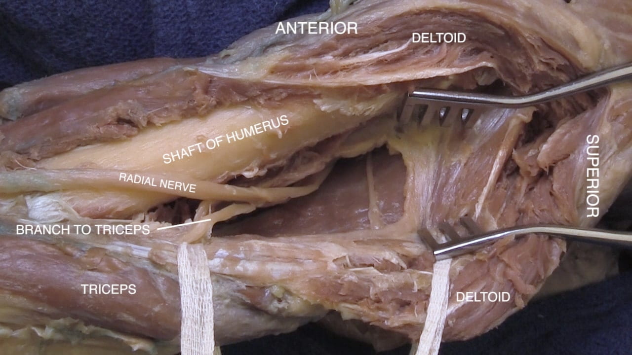
The axillary nerve will course around the neck of humerus and the radial nerve around the shaft of humerus.

Labels (top to bottom): Radial nerve, posterior cord, axillary nerve, lower subscapular nerve
1.6a) Posterior cord

Proximal Branches of Axillary Artery

Locate the circumflex humeral and subscapular branches of the axillary artery by following them to the deltoid and subscapularis.

Labels (top to bottom): Thoracoacromial artery, humeral head, axillary artery, posterior circumflex humeral artery, subscapular artery, anterior circumflex humeral artery, circumflex scapular artery, thoracodorsal artery
1.7a) Axillary artery branches

Long Thoracic and Thoracodorsal Nerves

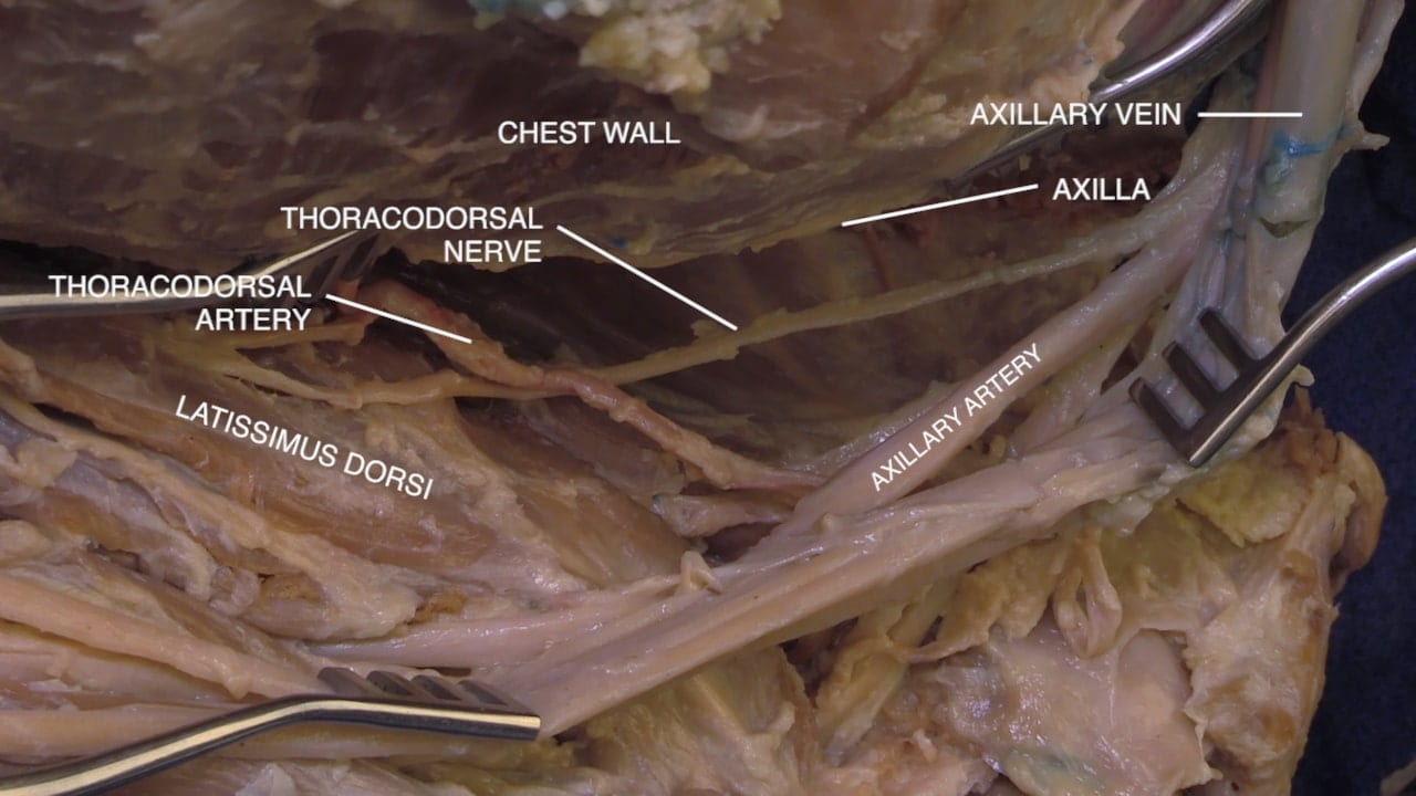
If the medial wall of the axilla is intact, identify the long thoracic nerve to the serratus anterior. This nerve arises from the roots of the brachial plexus and travels medially along the chest wall.

Similarly, identify the thoracodorsal nerve arising from the posterior cord and passing to the deep surface of the latissimus dorsi.

These two nerves are vulnerable with axillary node dissection.

Proximal Brachial Plexus

Scalene Muscles

Follow the brachial plexus medially. Identify the anterior and middle scalene muscles, which provide anatomic landmarks for the phrenic nerve, subclavian vessels and brachial plexus.

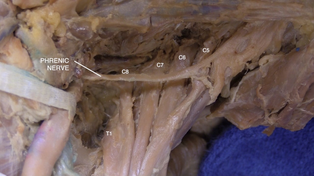
The phrenic nerve lies on the anterior surface of the anterior scalene.

The trunks of the brachial plexus pass between the anterior and middle scalene muscles. Identify the upper, middle and lower trunks.

Labels (left to right): Anterior scalene, trunks of brachial plexus, carotid sheath, phrenic nerve, middle scalene
2.1a) Anterior scalene

Trunks of the Brachial Plexus

Note the relationship of the subclavian vessels to the anterior scalene. The subclavian vein lies above the anterior scalene and the subclavian artery lies beneath it.

Look for the suprascapular nerve arising from the upper trunk. Follow this nerve posteriorly to the suprascapular notch. This nerve continues on the posterior surface of the scapula to the supraspinatus and infraspinatus.

Labels (left to right): Subclavian artery, subclavian vein, anterior scalene, lower trunk, middle trunk, upper trunk, middle scalene, suprascapular nerve
2.2a) Subclavian vessels

Phrenic Nerve

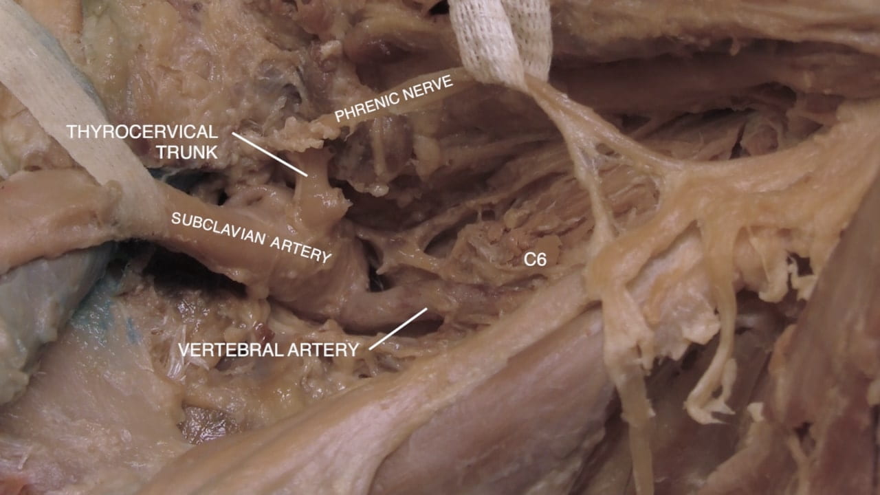
Detach the anterior scalene to reveal the C5, C6, C7, C8 and T1 spinal nerves (roots of brachial plexus).

With inferior retraction of the subclavian artery, the vertebral artery may come into view. The vertebral artery is a proximal branch of the subclavian that provides posterior circulation to the brain. At C6, the vertebral artery enters the transverse foramina of the cervical spine.

Distal Brachial Plexus

Brachial Plexus Branches

Re-identify the medial and lateral cord and their contributions to the median nerve. Follow the median nerve to the cubital fossa (antecubital fossa).

Follow the lateral cord and reidentify the musculocutaneous nerve and the lateral cutaneous nerve of the forearm.

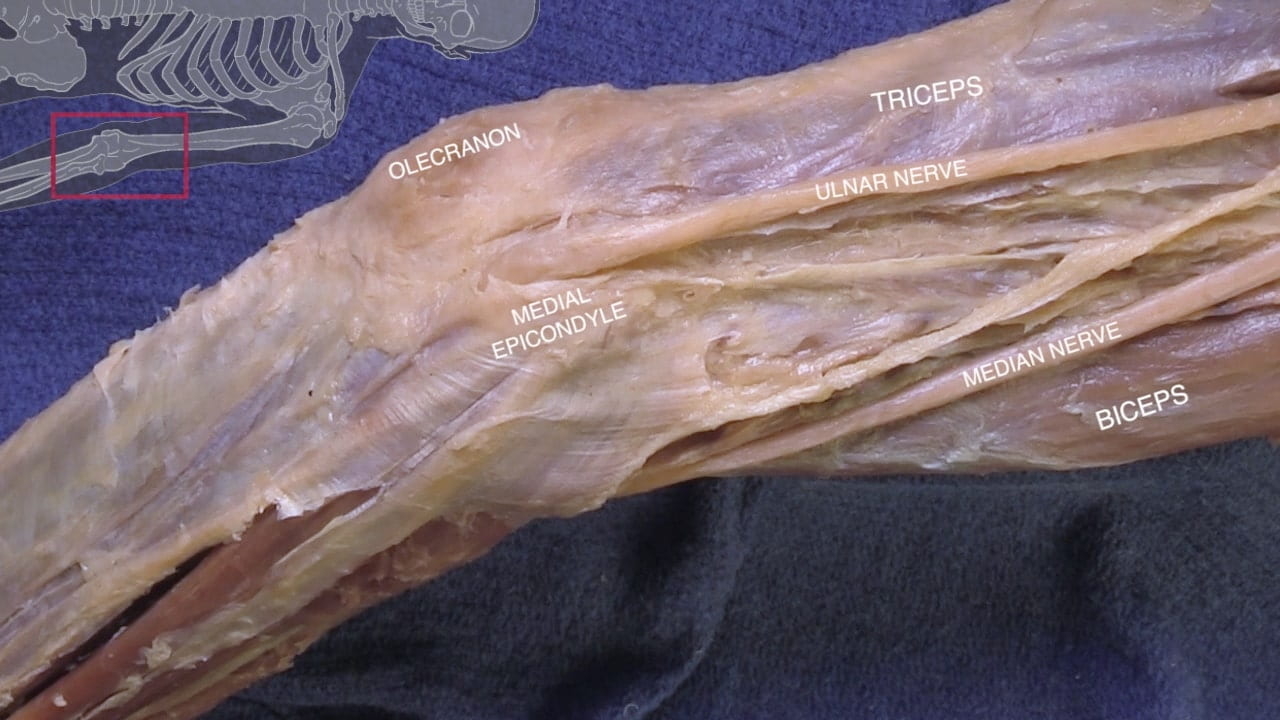
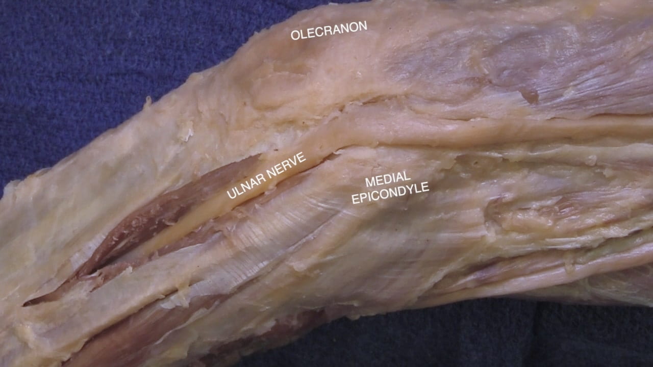
Follow the ulnar nerve from the medial cord to to the cubital tunnel, posterior to the medial epicondyle of the humerus.

Follow the axillary nerve around the neck of the humerus.

Cubital Fossa

Examine the cubital fossa and locate the:

  • Lateral cutaneous nerve of the forearm
  • Biceps tendon
  • Median nerve
  • Brachial artery
Labels (left to right): Median nerve, elbow, ulnar nerve, brachial artery, lateral cutaneous nerve, biceps
3.2a) Cubital fossa

Brachial Artery Branches

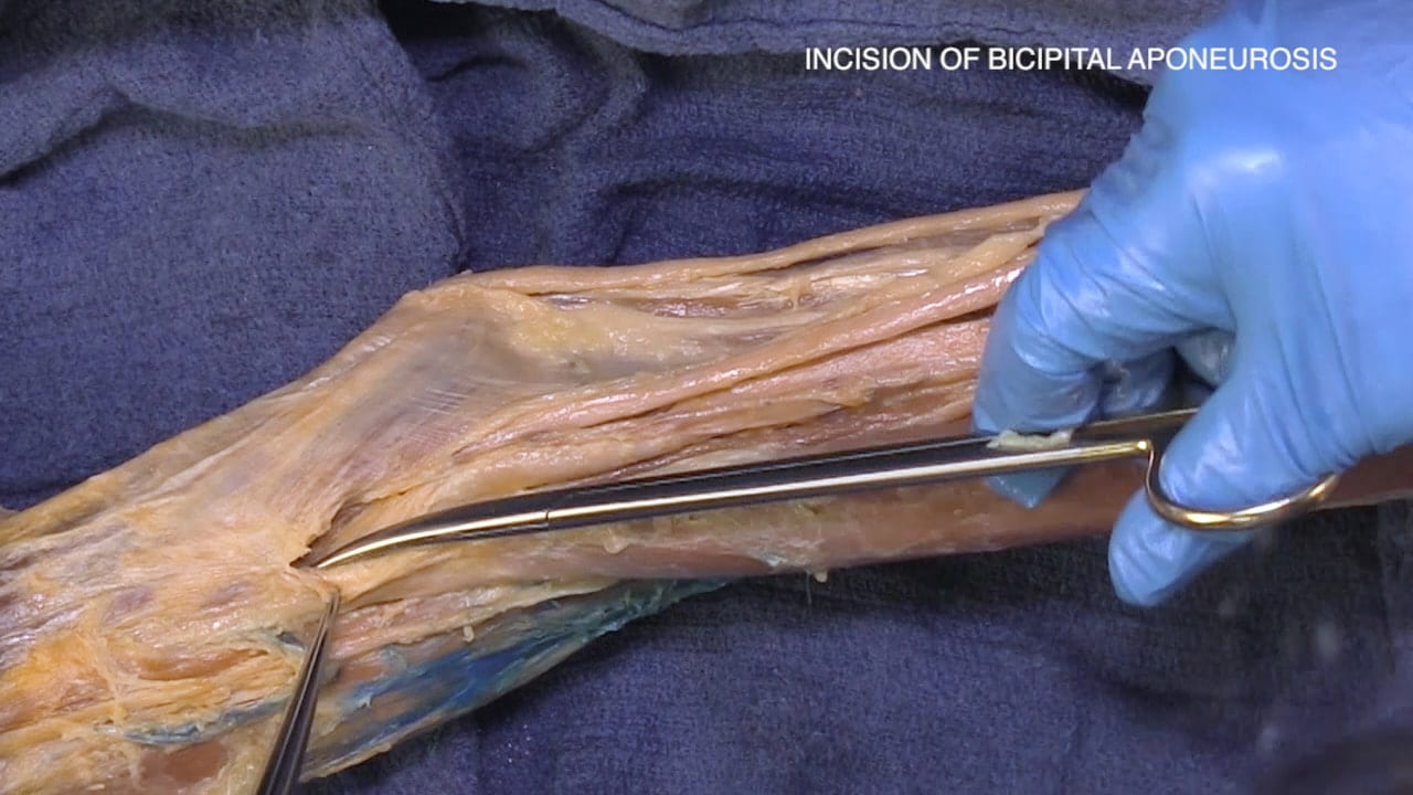
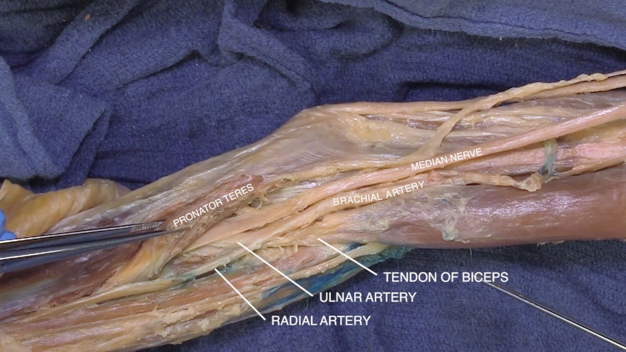
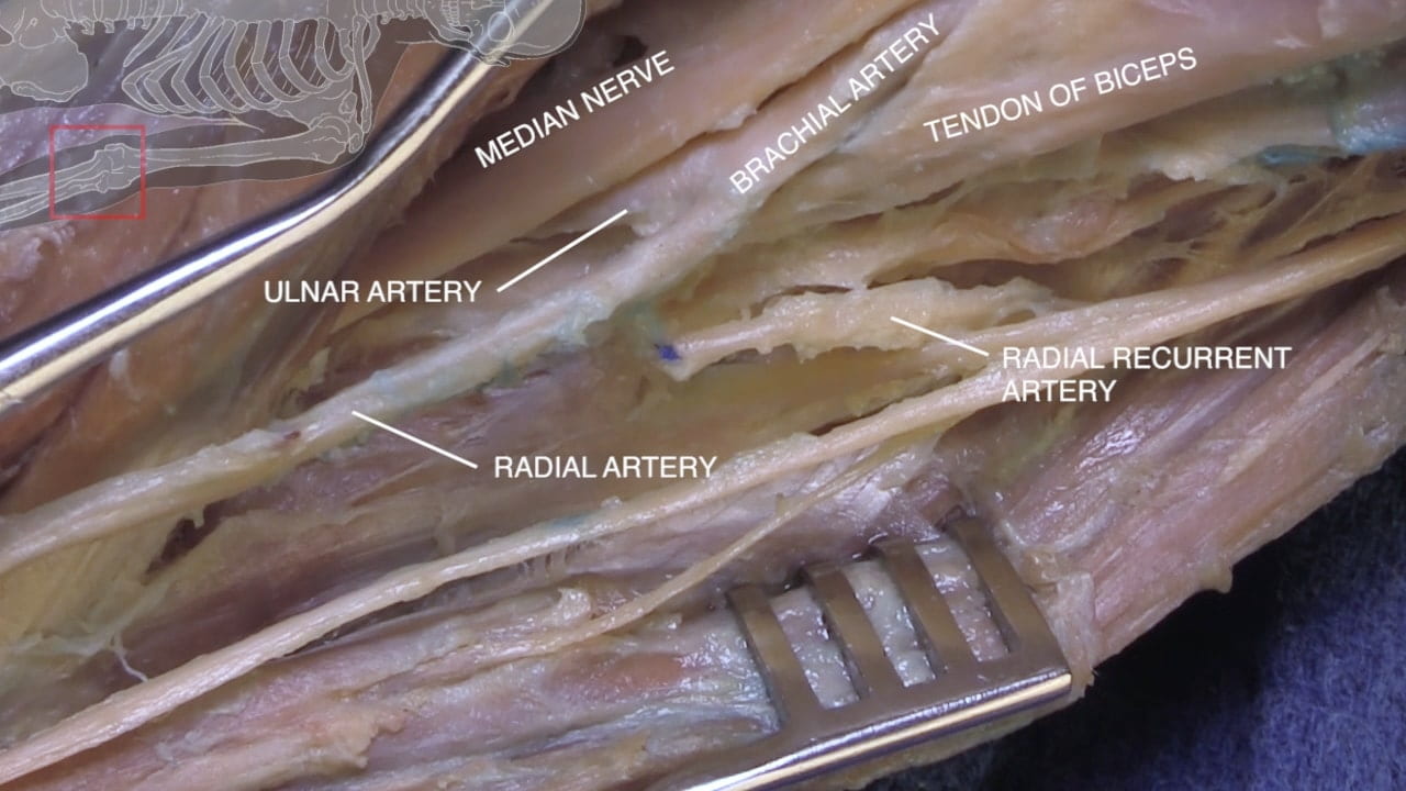
Incise the bicipital aponeurosis between the pronator teres and biceps tendon to follow the the brachial artery and median nerve into the forearm.

Identify the radial and ulnar branches of the brachial artery.

Follow the median nerve as it passes deep to the humeral head of the pronator teres.

Radial Nerve

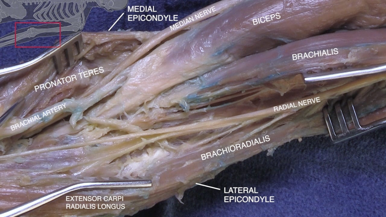
From a medial approach, separate the biceps and triceps to expose the humeral shaft. Follow the radial nerve from the posterior cord around the radial (spiral) groove in the humerus.

Branches of Radial Nerve

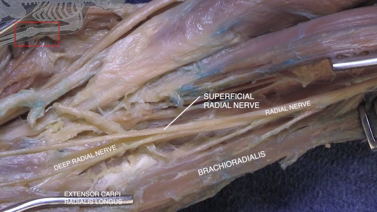
Lateral to the biceps tendon, identify the brachialis and brachioradialis muscles. Identify the superficial (sensory) and deep (motor) branches of the radial nerve emerging between brachialis and brachioradialis.

Ulnar Nerve

At the medial elbow, identify the ulnar nerve as passes in the ulnar groove between the medial epicondyle of the humerus and the olecranon process of the ulna. Incise the aponeurosis (cubital tunnel) to follow the nerve into the forearm.

3D Brachial Plexus

Review Quiz