Skull Base, Cavernous Sinus, Orbit

Lab Summary

In this lab structures of the skull base, cavernous sinus and orbit are taught. The trigeminal nerve branches in the middle fossa are included with the structures of cavernous sinus. The orbit and its contents are presented from superior and inferior perspectives. In situ base of the brain is taught from a complex dissection of skull base.

Lab Objectives

  1. Describe exit of cranial nerves from base of brain.
  2. Describe dural course of cranial nerves.
  3. Describe clinical significance of middle meningeal artery.
  4. Describe the course of the cranial nerves and internal carotid artery through the cavernous sinus.
  5. Describe motor innervation of extraocular muscles.
  6. Describe components of anterior chamber of eye.

Lecture List

Cranial Nerves at Dural Base, Ventral Brain, Trigeminal and Cavernous Sinus, Orbit, Eye Anatomy, Skull Base, Orbit 360°

Cranial Nerves at Dural Base

Interior Skull Base

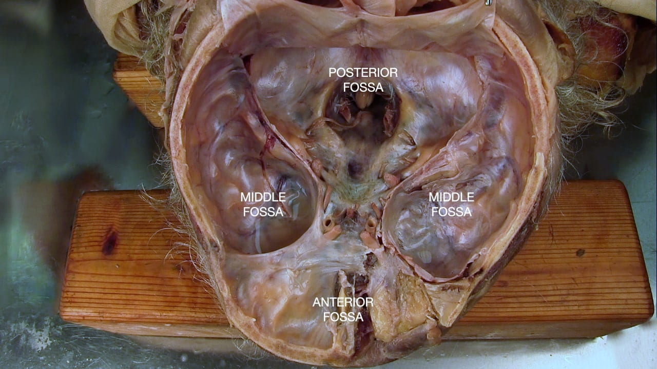
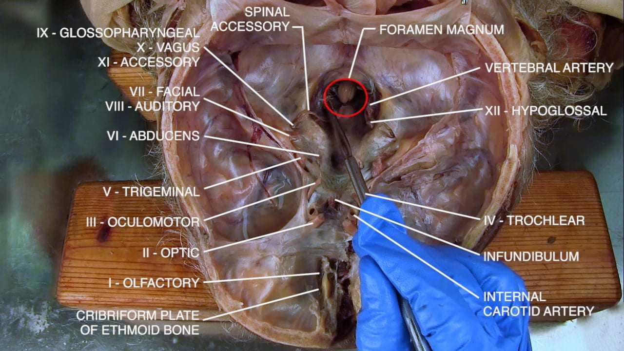
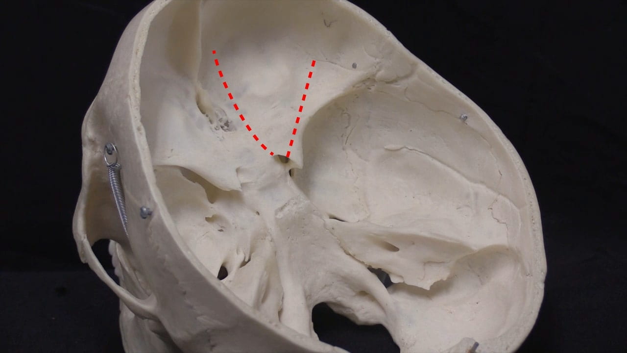
Place the donor in prone position. Identify the anterior, middle and posterior fossae (Fig. 1.1a).

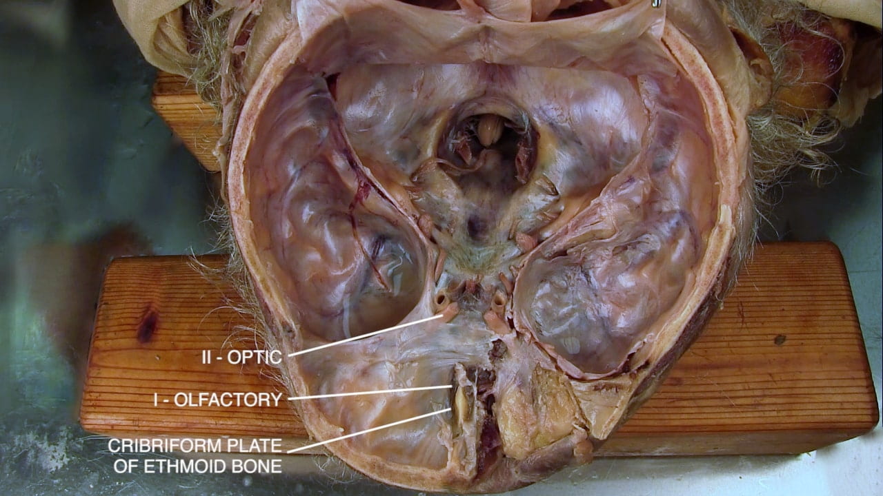
In the anterior fossa, note the cribriform plate, branches of olfactory nerve (I) and the bulge of orbital plate of the frontal bone (Fig. 1.1b).

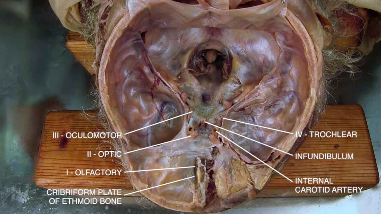
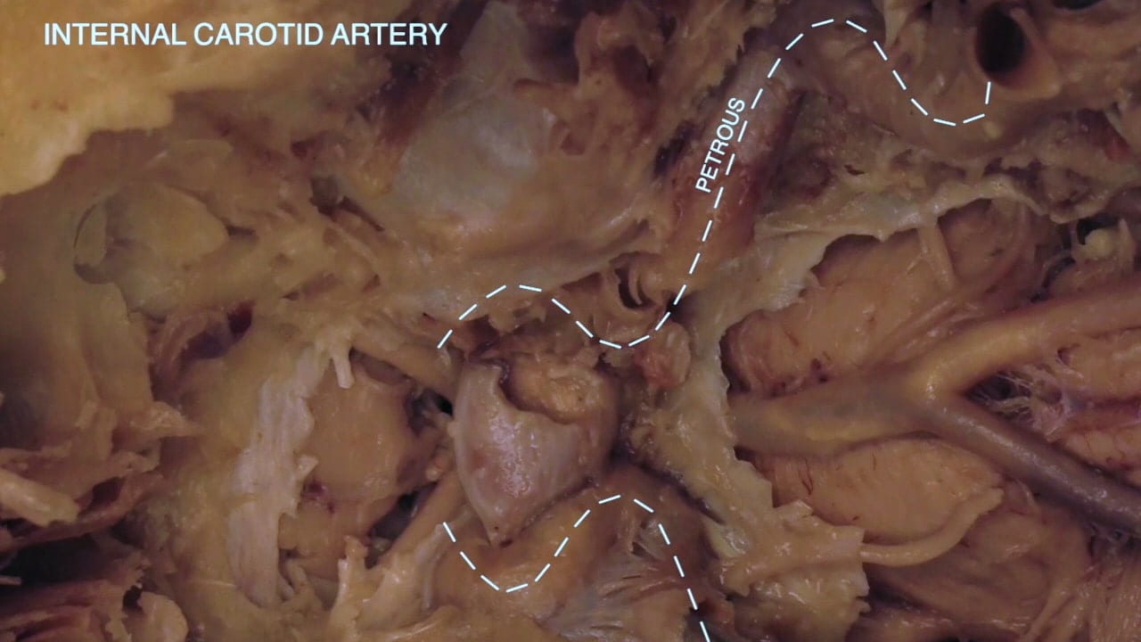
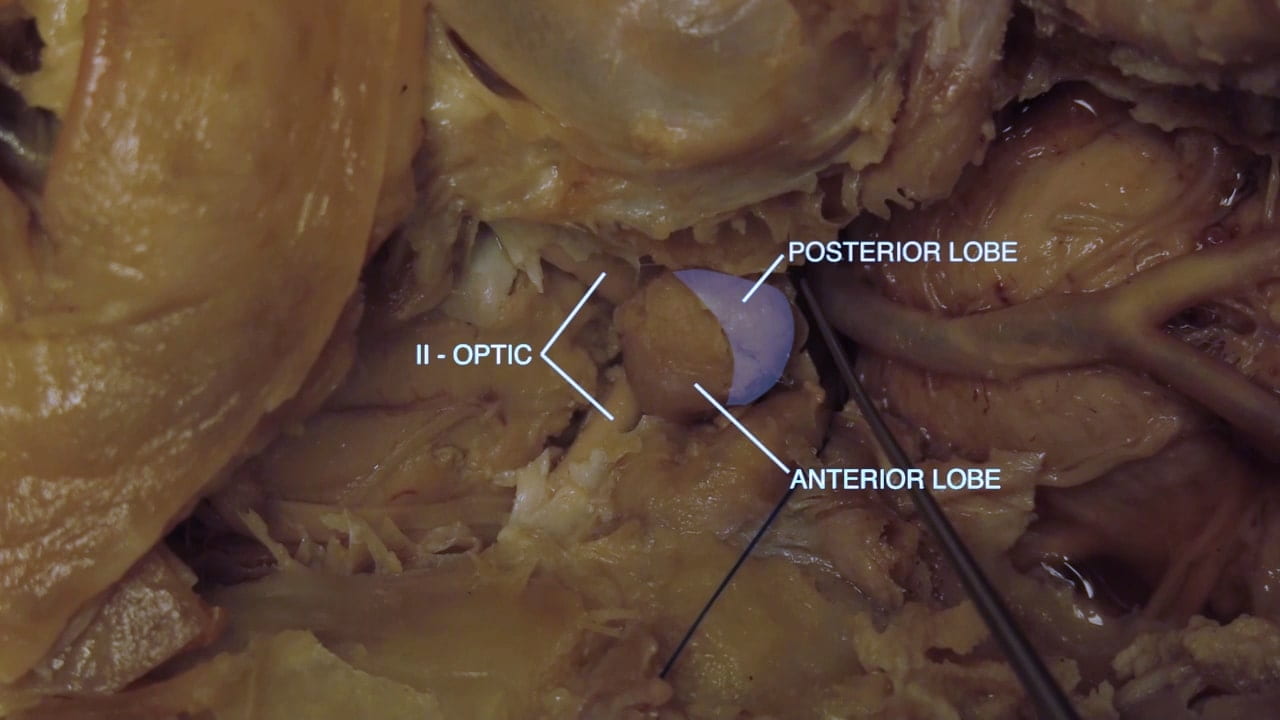
In the parasellar region, note the infundibulum in midline, optic nerves (II), internal carotid artery, oculomotor nerve (III) and trochlear nerve (IV) (Fig. 1.1c).

Cranial Nerves at Skull Base

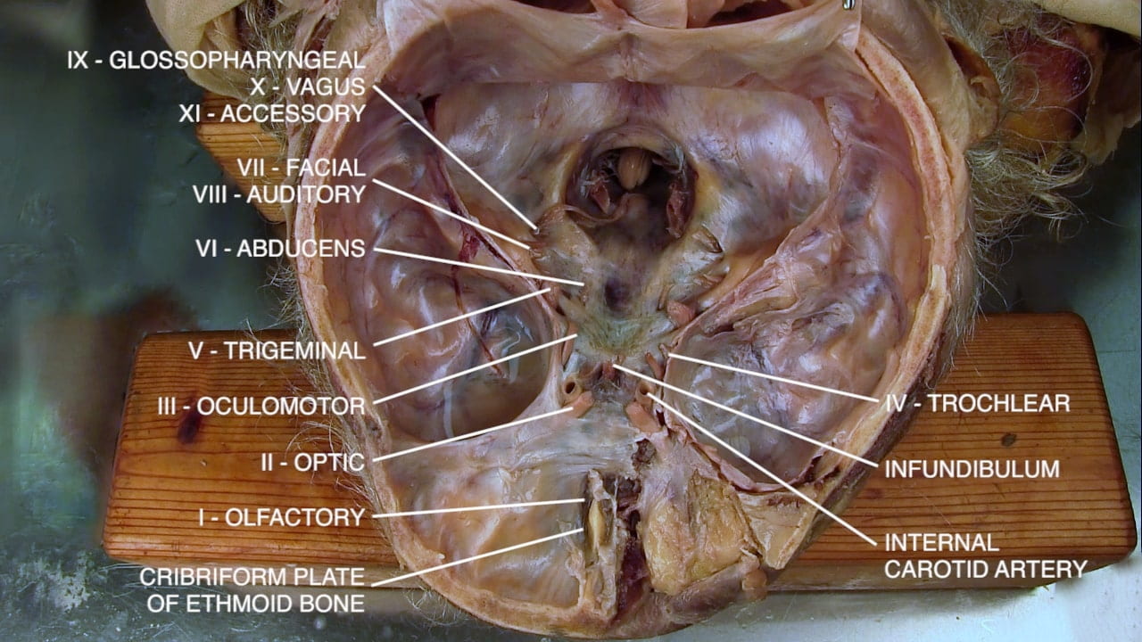
In the posterior fossa, note the trigeminal nerve (V) just behind the line of tentorium. Below it and slightly medial is the abducens nerve (VI). The facial (VII) and auditory nerves (VIII) are further caudal and lateral with an exit point at the internal acoustic meatus. The cranial nerves IX, X and XI, glossopharyngeal, vagus and accessory, are inferior to VII and VIII with an exit point at the jugular foramen (Fig. 2a).

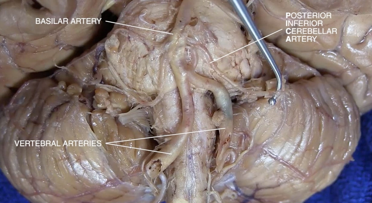
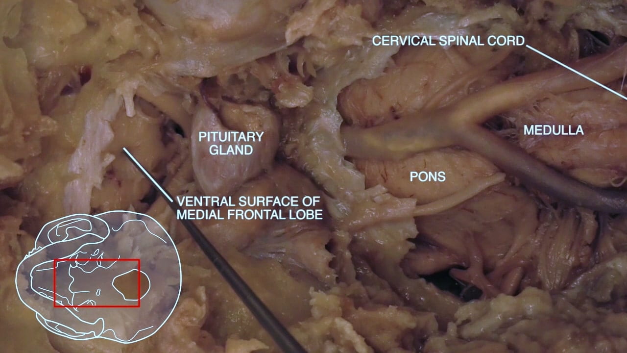
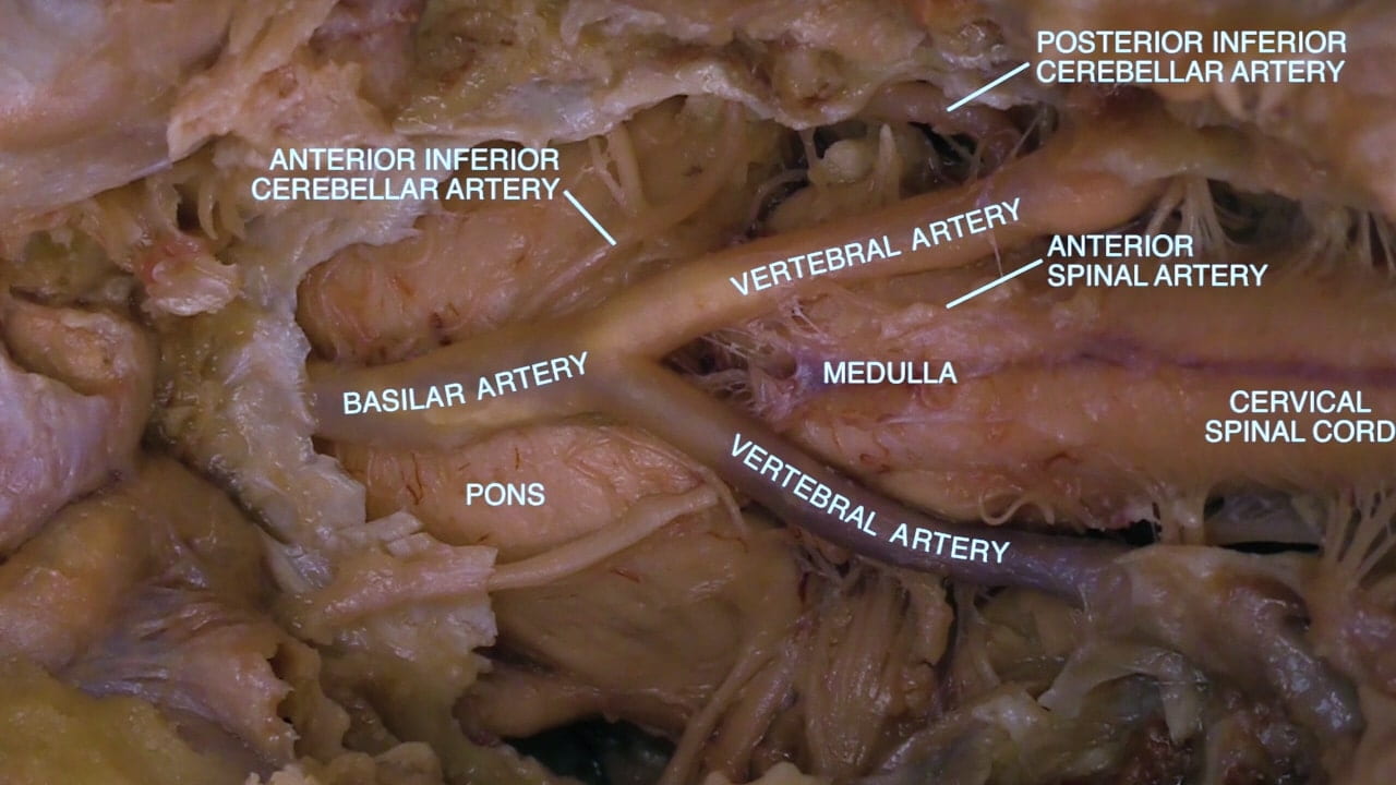
At foramen magnum, identify spinal cord and vertebral arteries. Laterally, locate spinal root of accessory nerve (XI) and hypoglossal nerve (XII) (Fig 2b).

Middle Meningeal Artery

In the middle cranial fossa, note the middle meningeal artery coursing through dura. Reflect the dura to show the impression of this artery in skull. Note the entry point of the middle meningeal artery into the dura at the foramen spinosum.

Question 1: What can be the consequence of traumatic disruption of middle meningeal artery?

Labels (top to bottom): Foramen spinosum, middle meningeal artery, middle fossa, lateral, anterior
1.3a) Middle fossa

Cranial Nerves at Base of Brain

Base of Brain

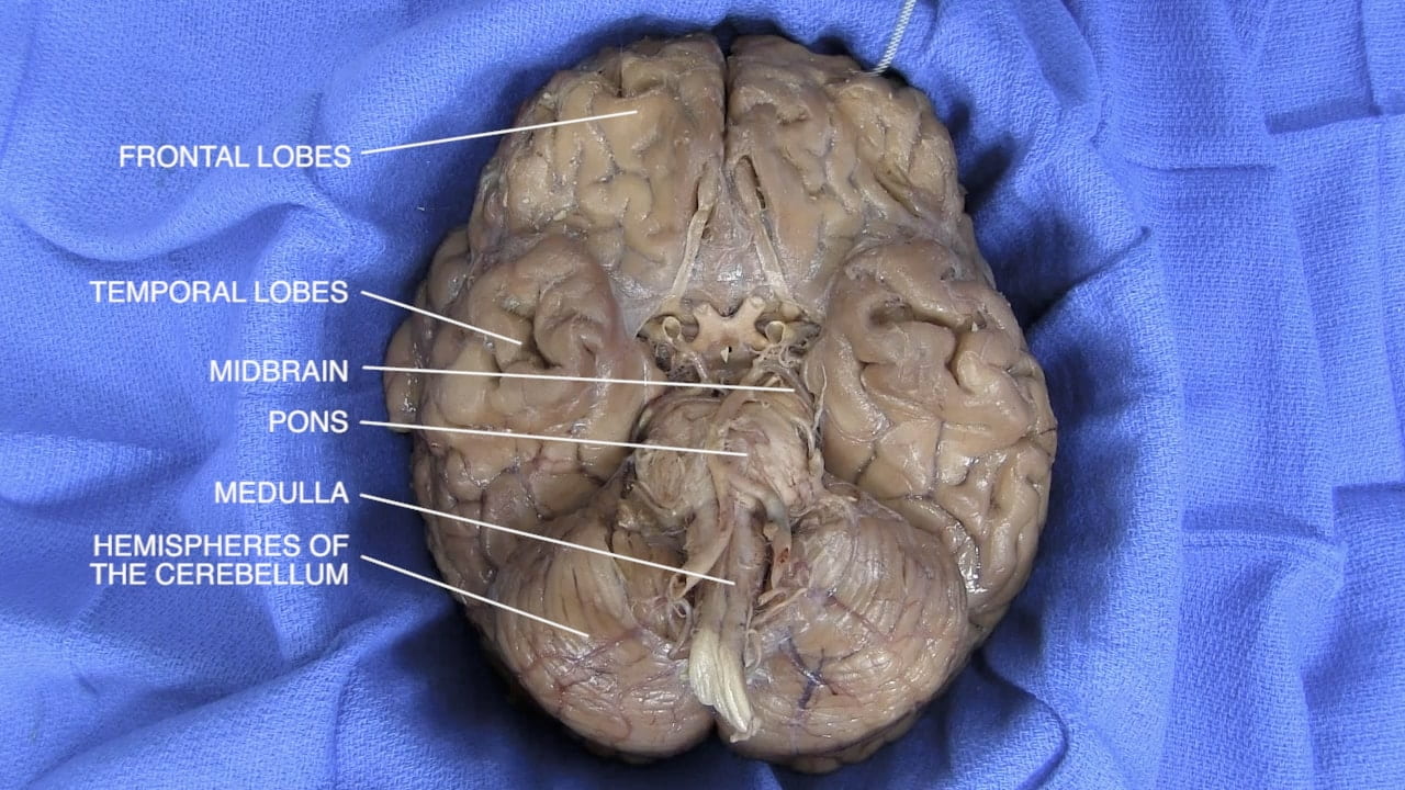
Examine the base of brain in relation to the skull base.

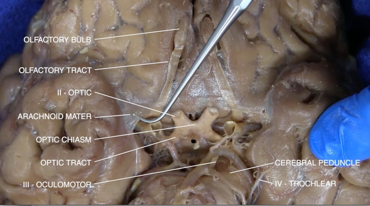
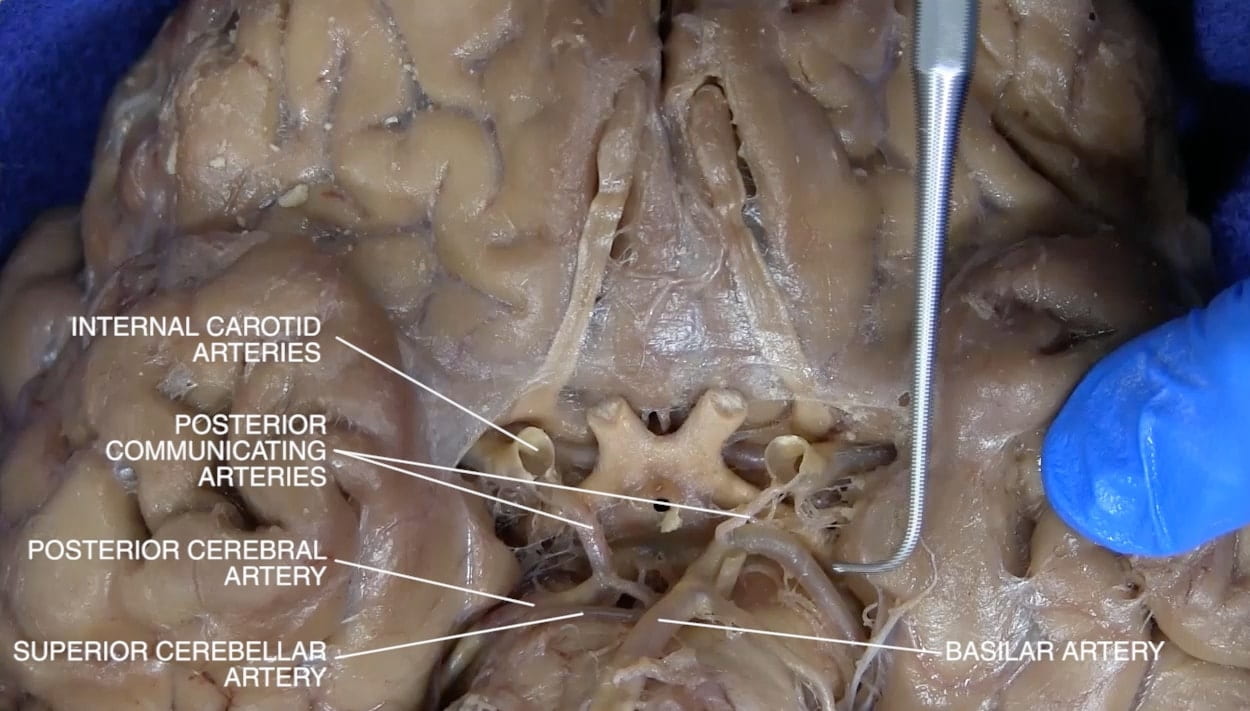
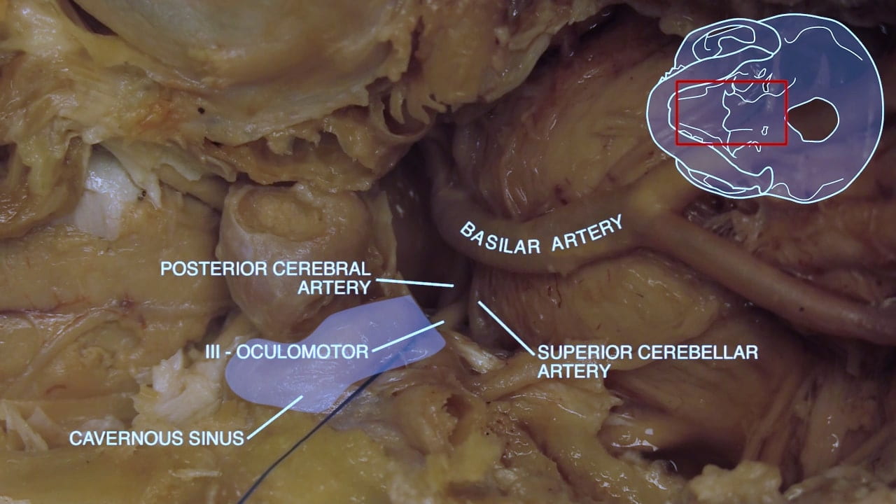
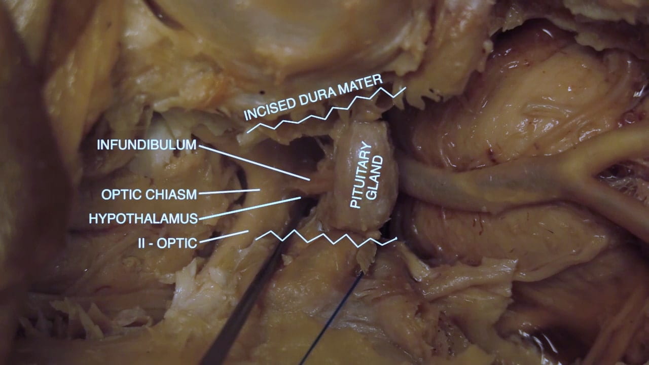
Note the frontal and temporal lobes, brainstem and cerebellum. Note the olfactory bulb and tract. Note the optic nerve, optic chiasm and optic tract and adjacent internal carotid artery. Note the oculomotor nerve (III) arising between the posterior cerebral and superior cerebellar arteries coursing medial to the temporal lobe. The trochlear nerve (IV) may be difficult to identify.

Circle of Willis

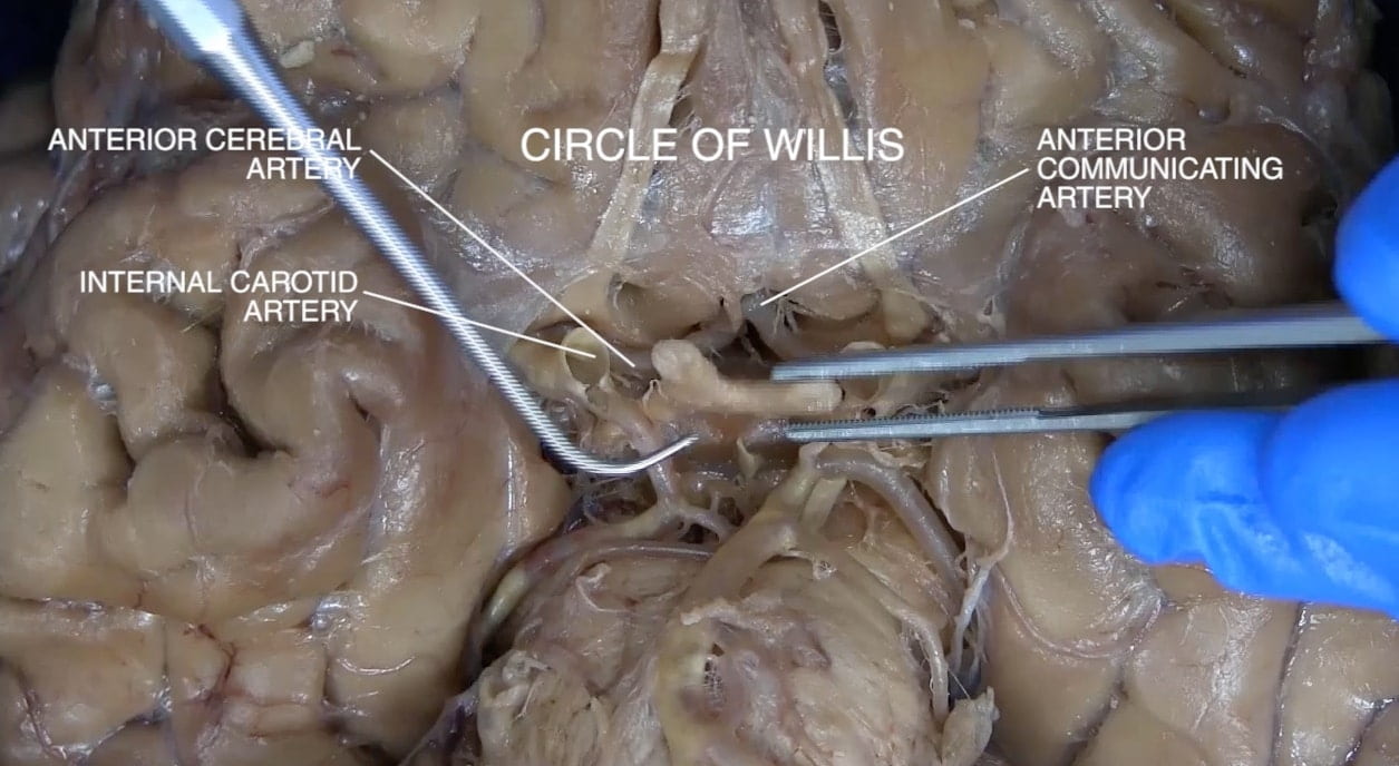
Examine the circle of Willis. Identify the internal carotid artery. Locate its middle and anterior cerebral branches. Identify the vertebral and basilar arteries. Locate the posterior cerebral and superior cerebellar branches of the basilar artery.

Identify the anterior communicating artery connecting the anterior cerebral arteries and the posterior communicating arteries connecting the posterior cerebral and internal carotid arteries.

Note individuals have considerable variation in the configuration of circle of Willis. Examine other donors.

Pontomedullary Junction

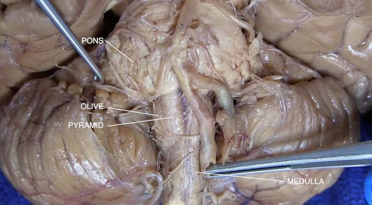
Examine the region of the pons and medulla.

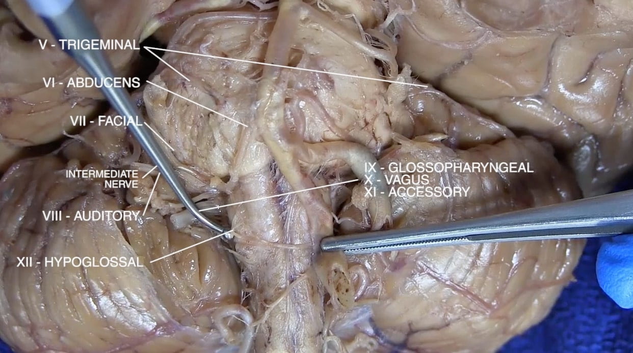
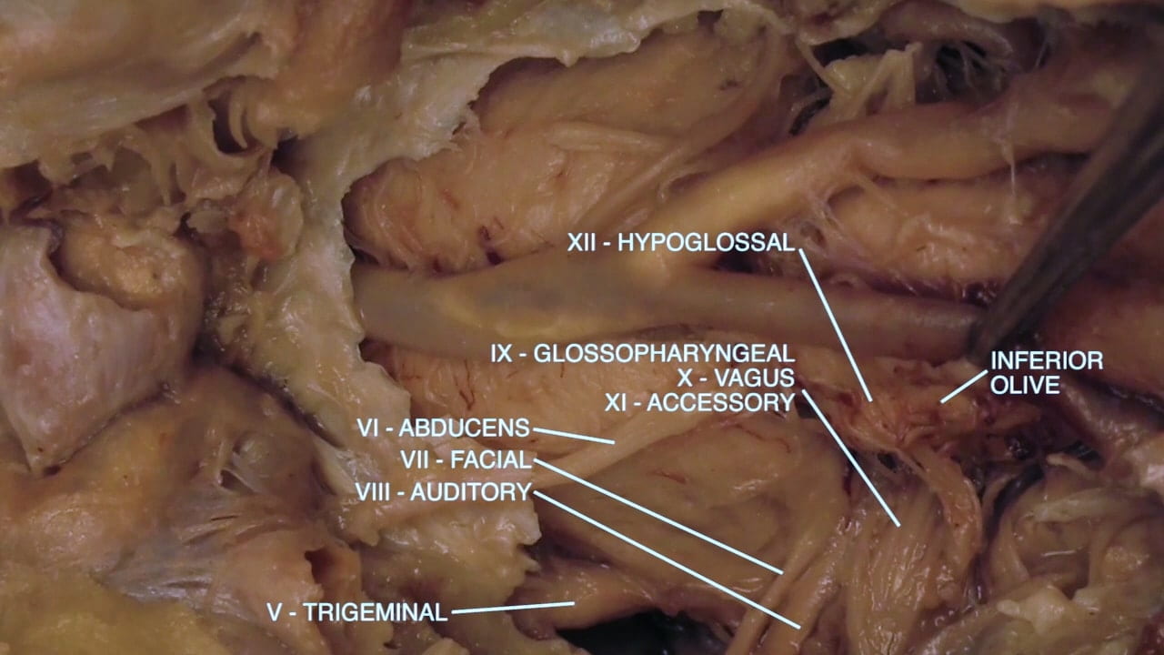
On the lateral pons, identify the trigeminal (V) nerve.

At the pontomedullary junction, locate the abducens (VI) nerve medially and the facial (VII) and auditory (VIII) nerves laterally.

On the lateral medulla, locate the bulge of the inferior olive. Posterior to the olive, locate strands of glossopharyngeal (IX), vagus (X) and accessory (XI) nerves. Anterior to the olive, between the olive and pyramid, locate strands of hypoglossal (XII) nerve.

Trigeminal and Cavernous Sinus

Trigeminal Nerve

Return to the skull base.

Orient by locating the trigeminal (V) nerve at the skull base. Remove the dura overlying the entry point of V nerve into dura. Sharply dissect this dura to expose the V nerve (semilunar ganglion) and branches of the V nerve: ophthalmic (V1), maxillary (V2) and mandibular (V3).

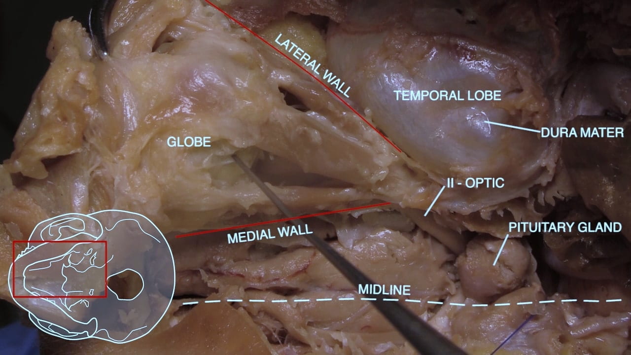
Extend sharp dural dissection medially to expose the cavernous sinus. Within the cavernous sinus, identify the oculomotor (III) and trochlear (IV) nerves.

Re-identify the optic nerve, internal carotid artery and infundibulum medial to the cavernous sinus.

Labels (top to bottom): trigeminal (semilunar) ganglion, IV - trochlear, III - oculomotor, lateral, infundibulum, V - trigeminal (branches): mandibular (V3), maxillary (V2), ophthalmic (V1), sellar diaphragm, internal carotid artery, II - optic, anterior
3.1a) Trigeminal ganglion

Cavernous Sinus

Reflect the oculomotor nerve (III) medially and ophthalmic branch (V1) laterally to locate the internal carotid artery and abducens nerve (VI) within the cavernous sinus.

Question 2: Estimate the distance from the cavernous carotid artery to the pituitary gland.

Labels (top to bottom): Lateral, III - oculomotor, V - trigeminal (ophthalmic branch), anterior
3.2a) Cavernous sinus

Orbitofrontal Dissection

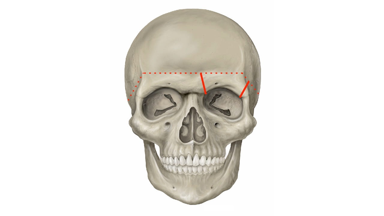
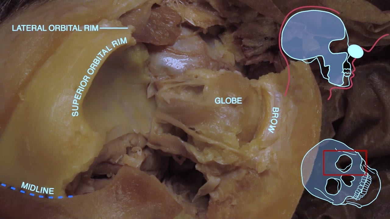
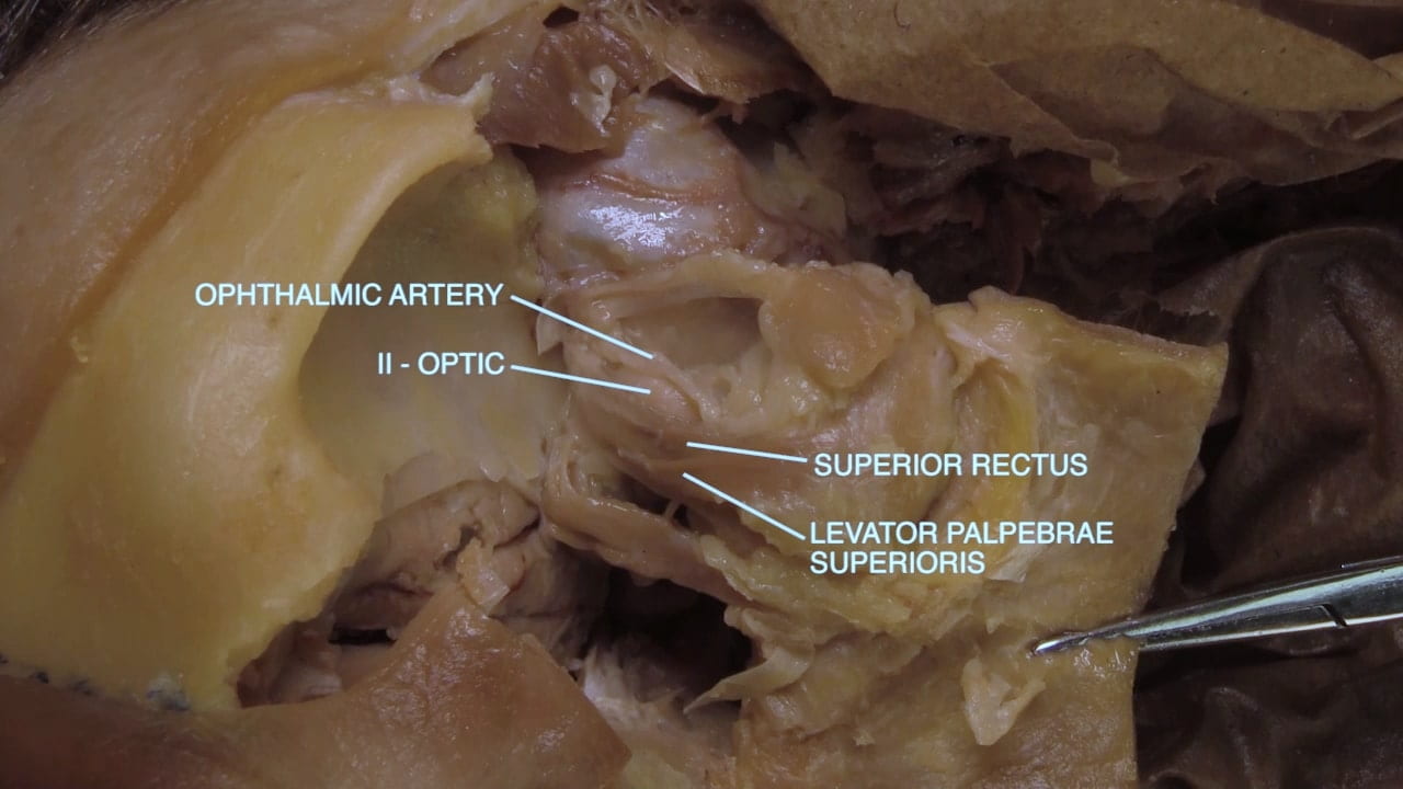
If not previously performed, make vertical cuts at the lateral and medial borders of the orbit to separate the superior orbital rim.  Elevate the orbital rim and orbital roof to expose contents of the orbit.

Remove the remaining bone of the orbital roof covering the superior orbit including exposure of the optic canal. Open periorbital fascia.

Orbit

Superior Orbit

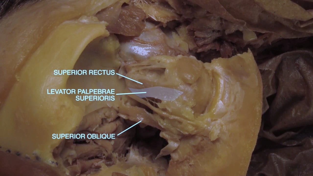
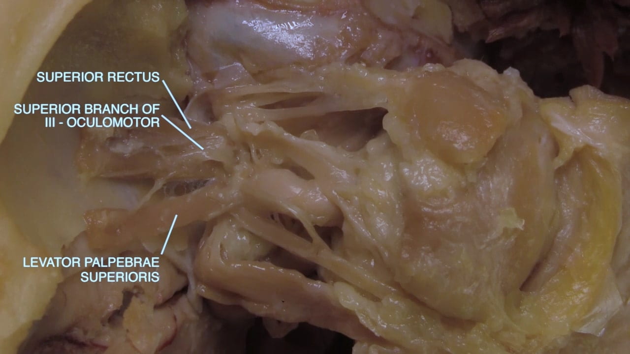
Identify in the superior orbit:

  • Levator palpebrae superioris
  • Superior rectus
  • Medial rectus
  • Lateral rectus
  • Superior oblique
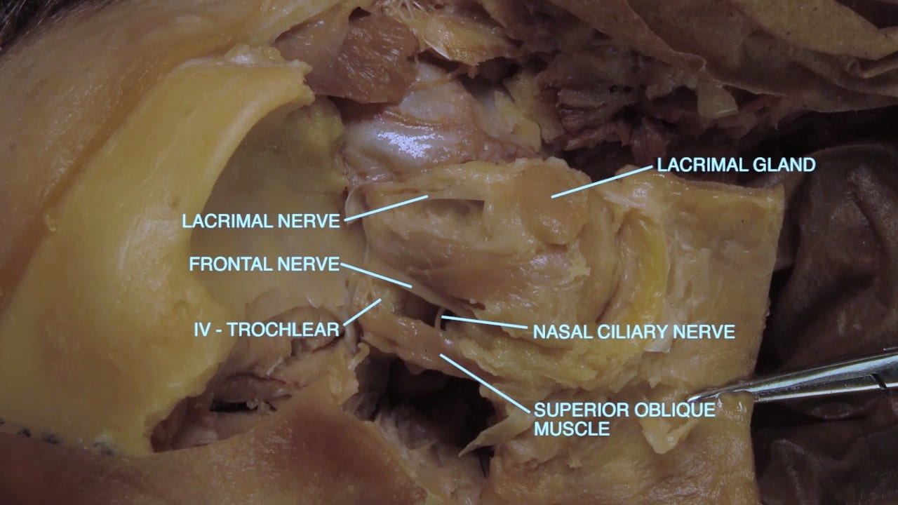
  • Lacrimal gland

Note the pulley arrangement of the superior oblique.

Also try to locate the lacrimal, frontal and nasociliary branches of the ophthalmic nerve (V1) and the trochlear (IV) and abducens (VI) nerves. There will be numerous small arteries and veins which make these nerves difficult to locate.

Labels (top to bottom): VI - abducens, lateral rectus, IV - trochlear, superior oblique muscle, lacrimal nerve, lacrimal gland, nasociliary nerve, frontal nerve, ethmoid air cells
4.1a) Superior orbit

Extraocular Muscle

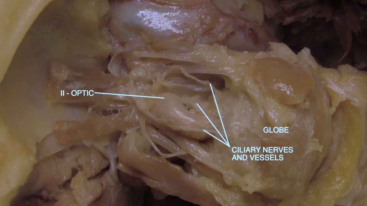
Separate the levator palpebrae superioris and superior rectus to identify the globe and optic nerve. Inject the globe with one to two cc of hyaluronic acid to restore turgor.

Divide the optic nerve and muscles close to the globe. Remove the eyeball for dissection.

Follow the steps in Dr. Forster’s video below, Anatomy of the Eye, to dissect the globe.

Labels (top to bottom): Lateral, optic canal, lateral rectus muscle, II -- optic, lacrimal gland, superior rectus muscle, superior oblique muscle, levator palpebrae superioris muscle, medial rectus muscle, anterior
4.2a) Superior orbit

Anatomy of the Eye

Advanced Dissection: Anterior Skull Base

View but do not perform this technically difficult exposure.

Anterior Skull Base Gallery

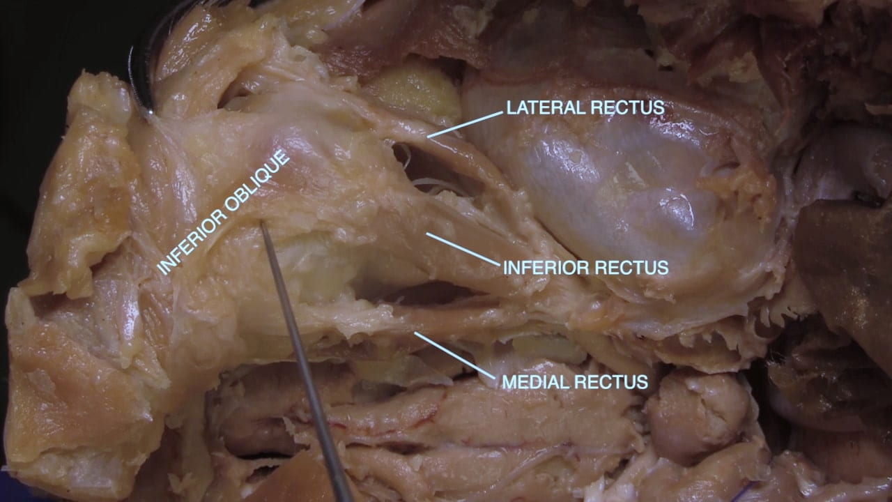
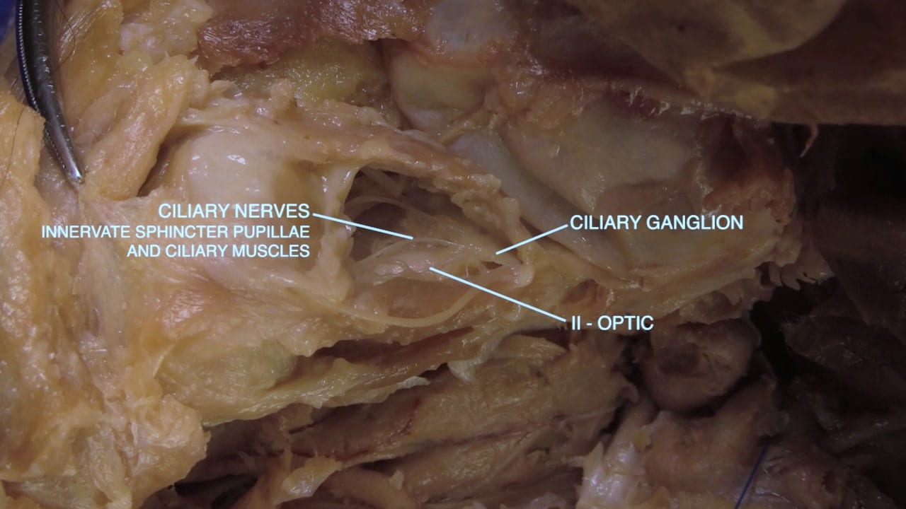
Advanced Dissection: Orbit 360°

View but do not perform this technically difficult exposure.

Question 3: What is the functional importance of the ciliary ganglion?

Superior View of Orbit

Inferior View of Orbit

Review Quiz