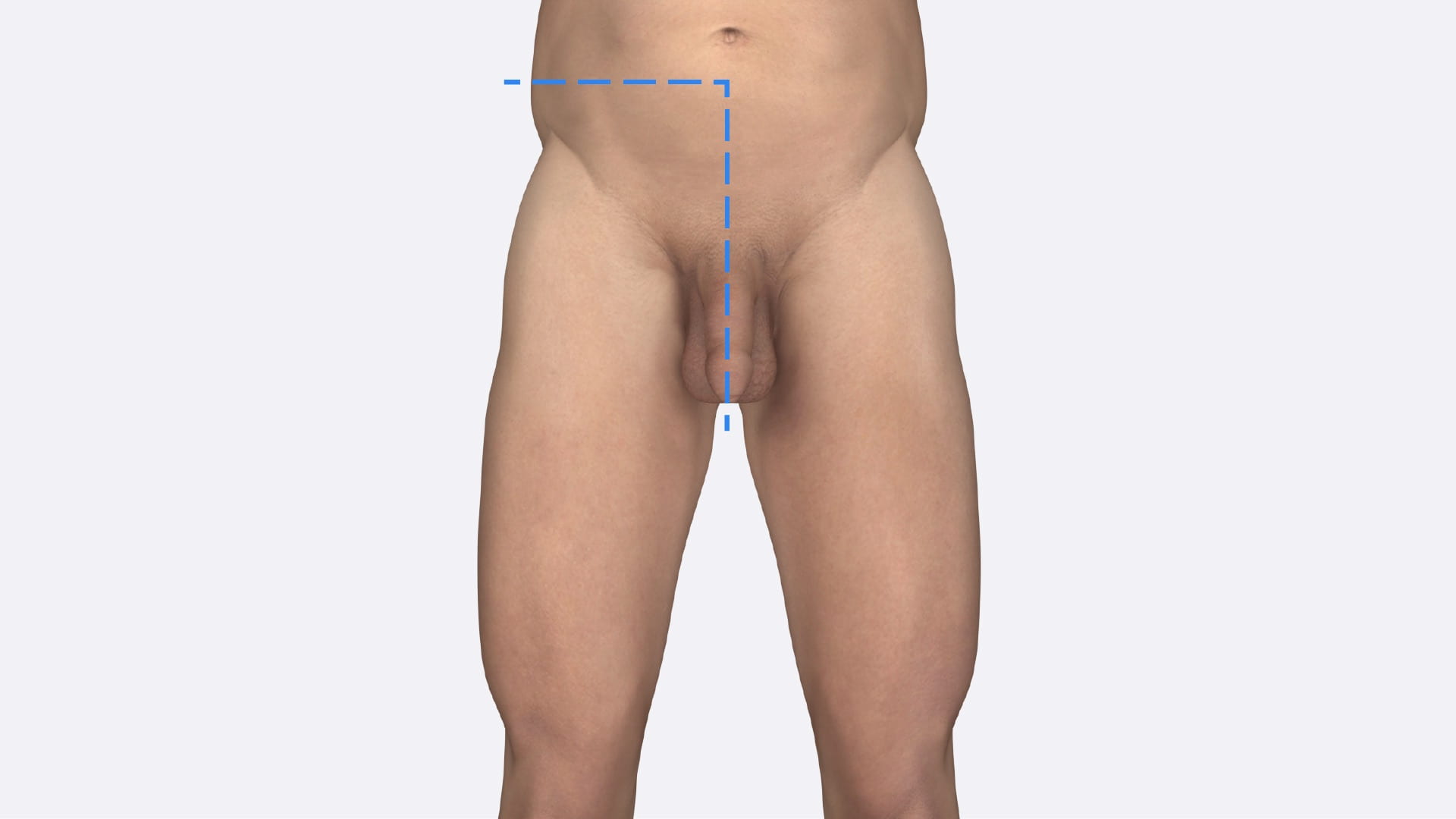
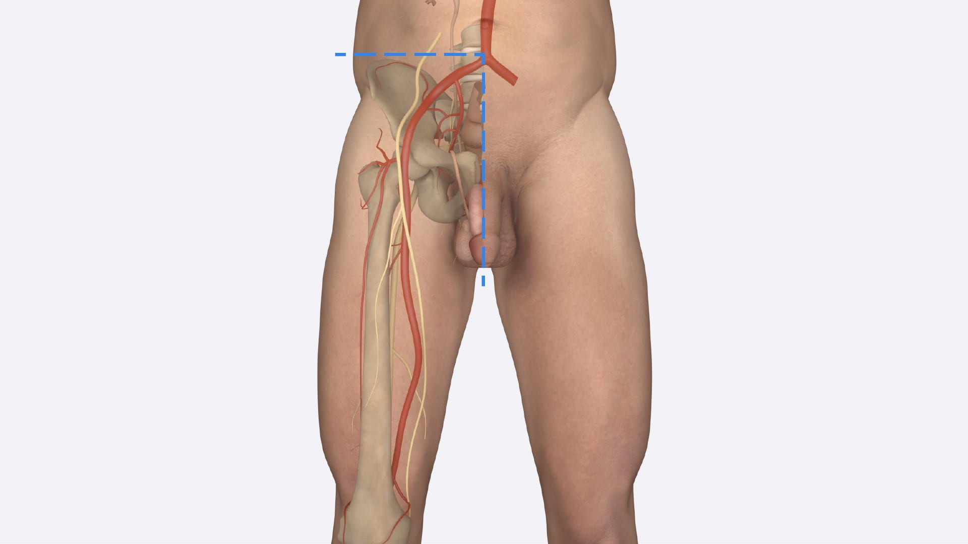
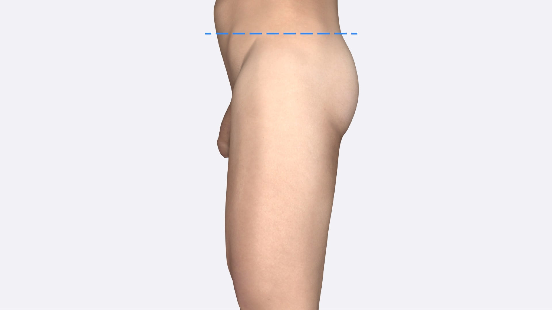
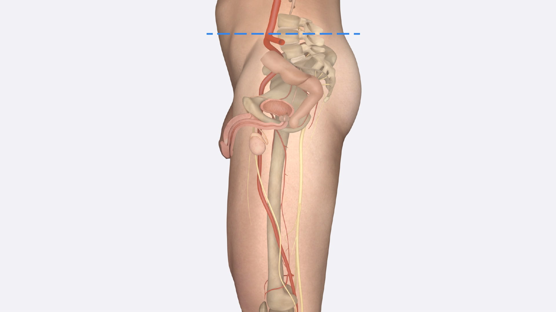
Assigned Male Pelvis

Lab Summary

This lab includes a presentation of the inguinal ligament, inguinal canal and testis. The male pelvis and perineum are presented from external structures to pelvic contents. Additionally, the male pelvis and perineal structures are shown from a midline sagittal perspective.

Lab Objectives

  1. Describe the erectile tissue of penis including location of penile urethra.
  2. Describe position of prostate gland and its relation to rectum, bladder and urogenital diaphragm. 
  3. Describe the pathway of sperm from testis to ejaculatory duct.
  4. Describe root of penis.
  5. Describe and differentiate between direct and indirect inguinal hernias.

Lecture List

Inguinal Ligament and Testis, Male Perineum, Male Pelvis, Male Sagittal Pelvis

Inguinal Ligament and Testis

Inguinal Ligament

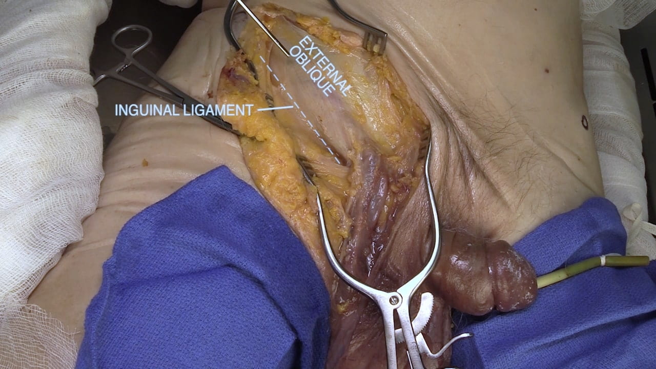
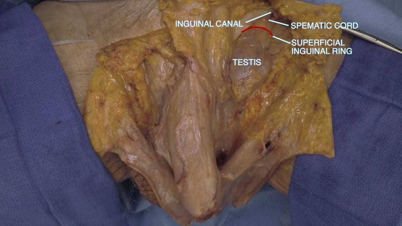
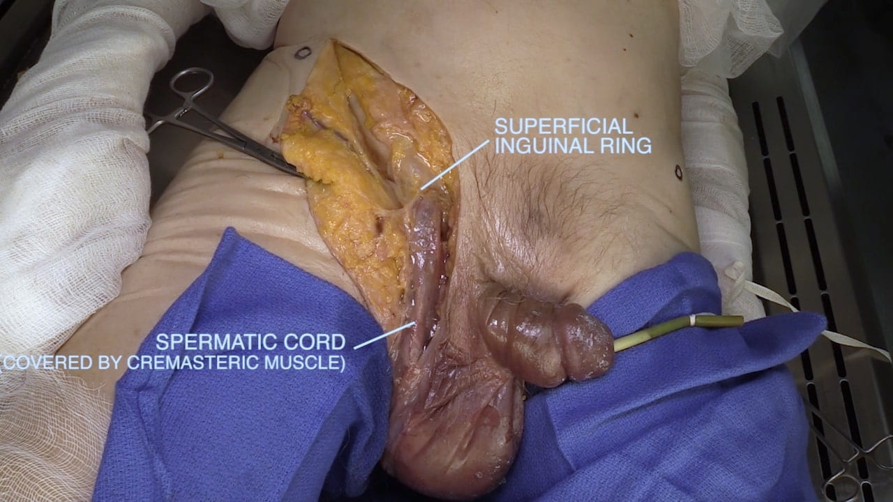
Make an incision superior and parallel to the inguinal ligament and extend the incision into the lateral scrotum.

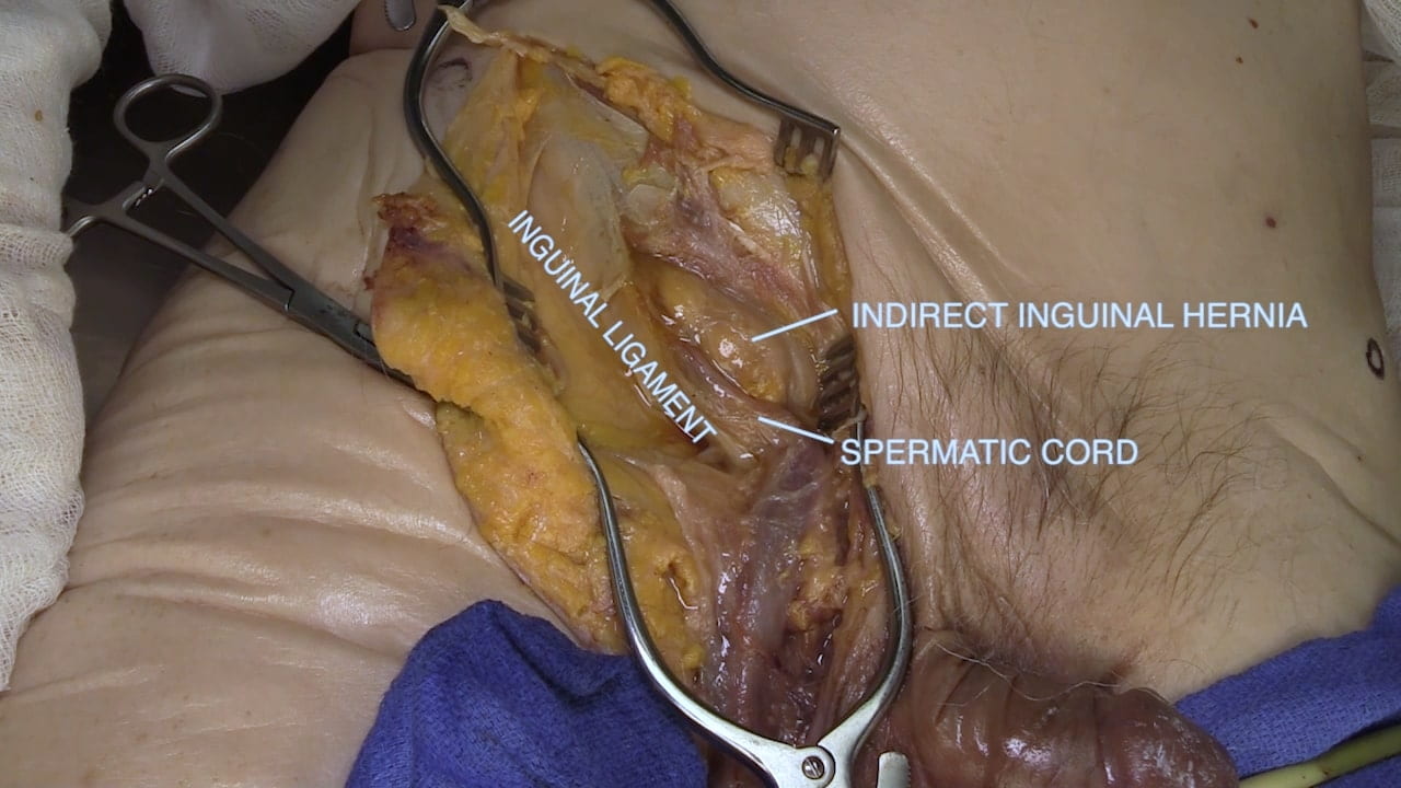
Identify the spermatic cord and expose its course into scrotum. Follow the spermatic cord superiorly to superficial inguinal ring. Open superficial ring and follow inguinal canal proximally. Does the donor have a hernia?

Note the donor in the video has an indirect inguinal hernia.

Spermatic Cord

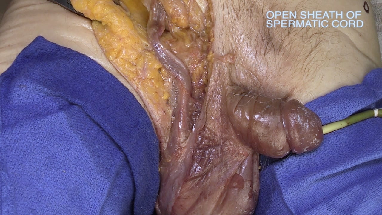
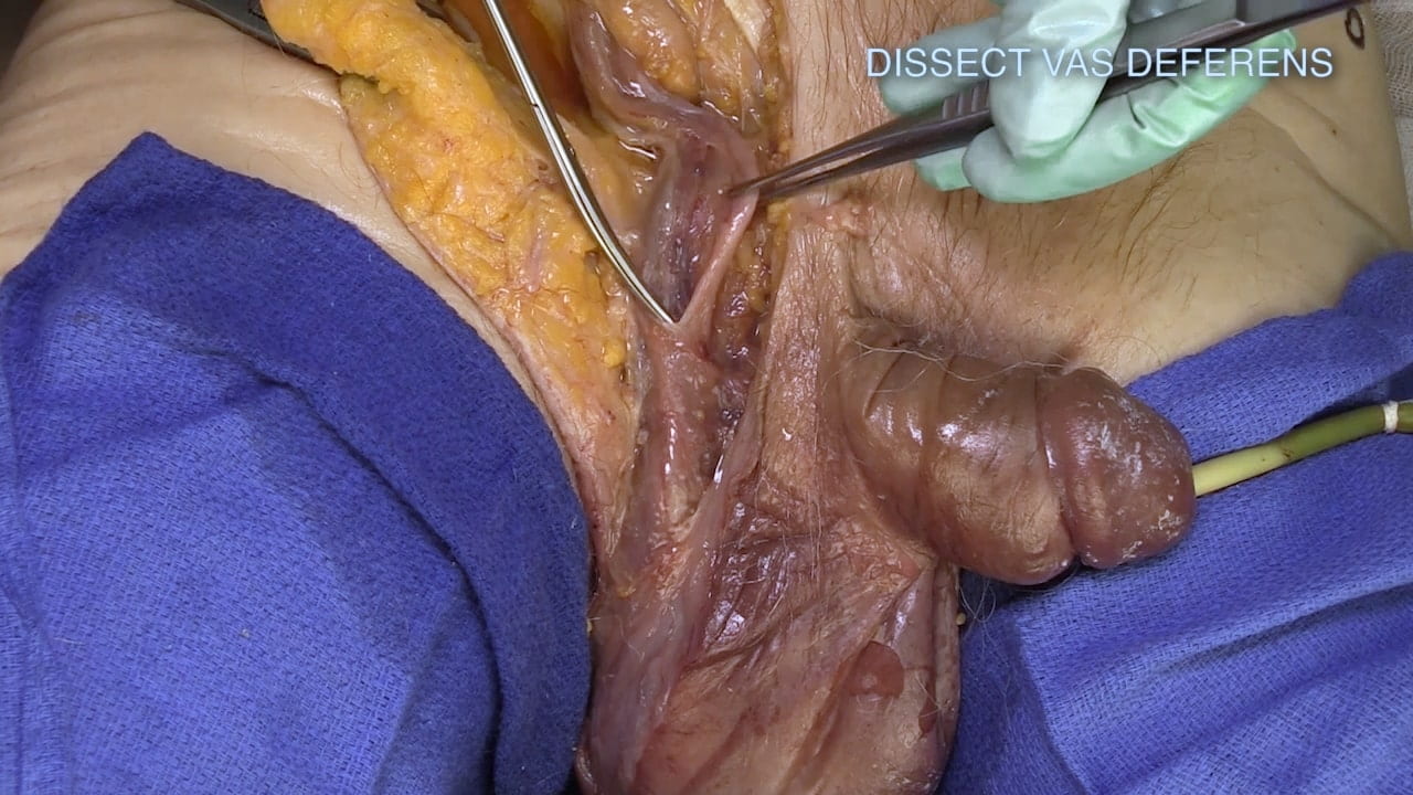
Incise the fascial coverings of the spermatic cord.

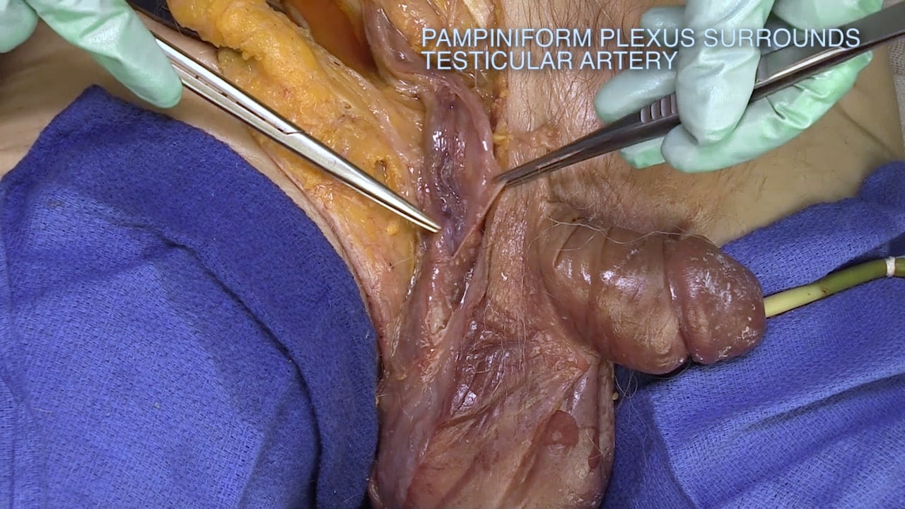
Identify vas deferens and plexus of veins (pampiniform plexus) surrounding testicular artery. The testicular artery will be difficult to identify.

Testis

Follow the spermatic cord to the testis. Mobilize the testis and free it from the scrotum.

Incise the dense fascial layer (tunica vaginalis) covering the testis. Identify it’s parietal and visceral layers.

Labels (top to bottom): Parietal tunica vaginalis, visceral tunica vaginalis, testis
1.3a) Tunica vaginalis

Epididymis

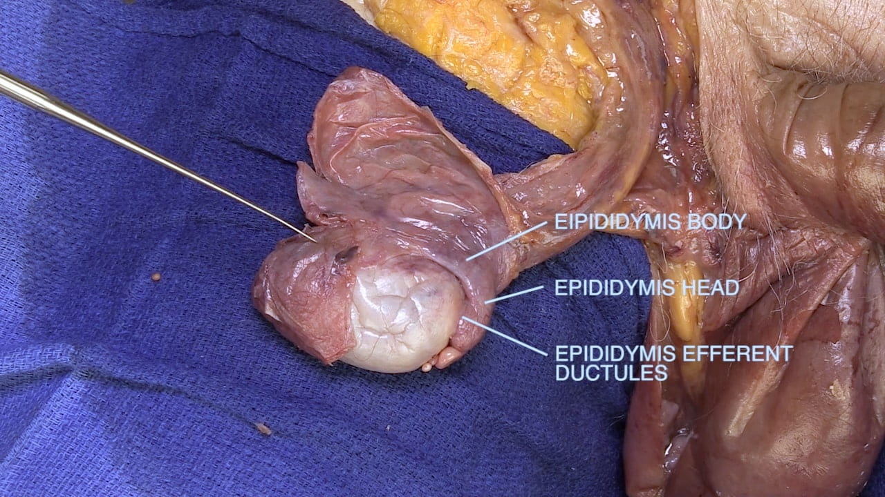
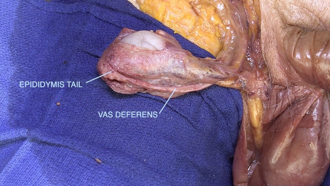
Locate the head of the epididymis and the efferent ductules.  Identify the body and tail of the epididymis and the vas deferens. 

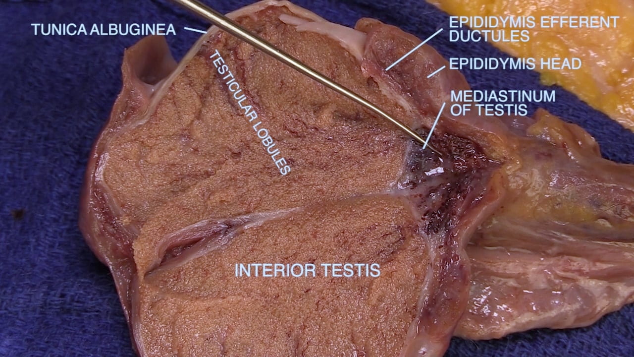
Sharply divide testis along the head of epididymis. Examine testicular lobules, tunica albuginea, ductules and head of epididymis and mediastinum testis.

Male Perineum

External Genitalia

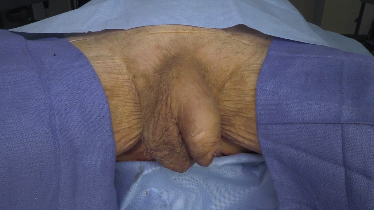
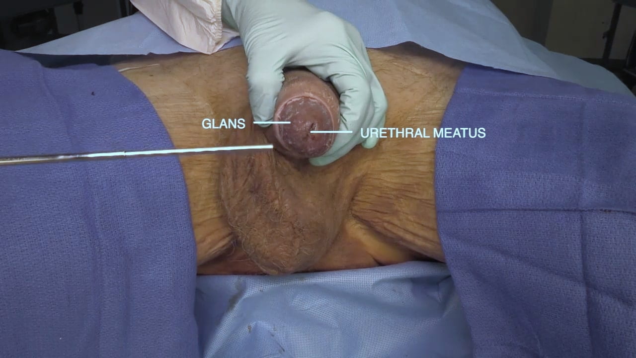
Examine the external genitalia.

Note the glans of the penis, urethral meatus, scrotum and testicles. Is your donor circumcised?

Penis and Scrotal Contents

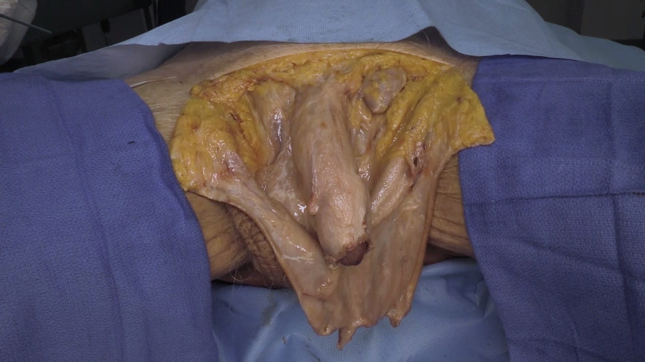
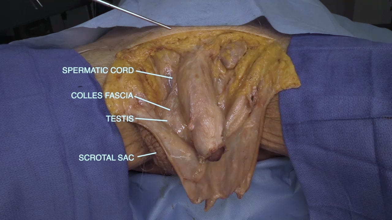
Make a horizontal incision from midpoint to midpoint of the inguinal ligament.

Make a second incision vertically along the dorsum of the penis to the glans. 

Remove the skin from the shaft of the penis.  Incise the scrotal sac on the contralateral side from previous dissection to show testis and spermatic cord.

Question 1: The video and Fig. 2.2c demonstrate an anomalous location of testis on left.  How might this occur?

Erectile Tissue

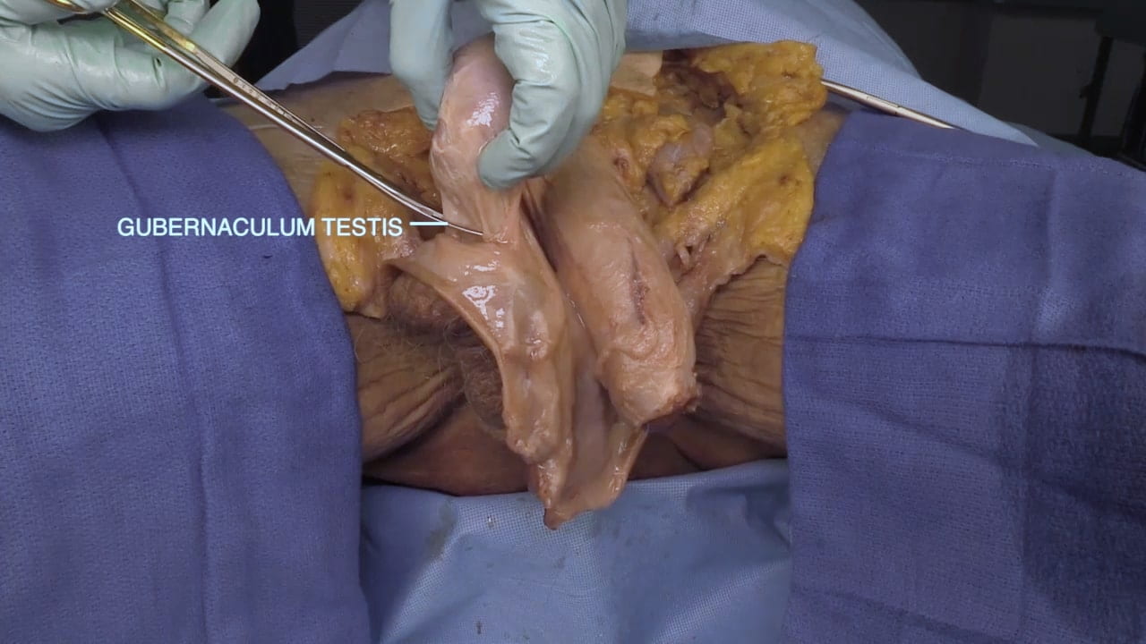
Elevate the testes and divide the gubernaculum testis; this structure attaches the testis to the scrotum.

Divide the scrotum in the midline and reflect the flaps laterally to expose the base of the penis.

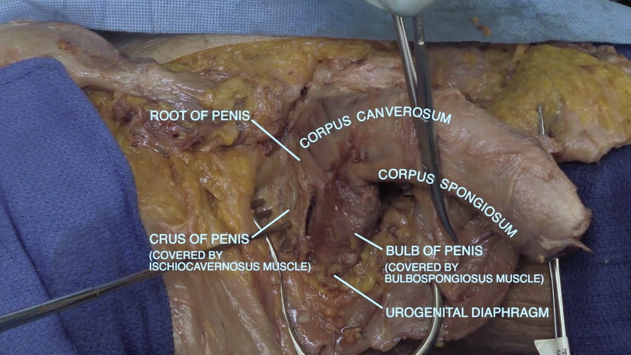
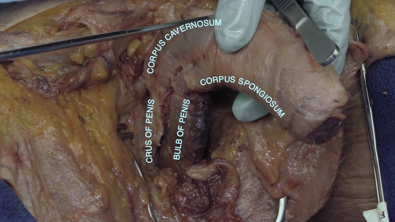
Identify the bulb, crus, corpus spongiosum and corpus cavernosum.

Dorsal Nerve of Penis

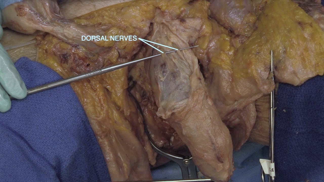
On the dorsum of penis, identify paired dorsal nerves of penis. These are terminal branches of pudendal nerve.

Remove bulbospongiosus muscle covering bulb of penis and confirm that the bulb is continuous with corpus spongiosum.

Similarly, remove ischiocavernosus muscle covering crus of penis and confirm that the crus is continuous with corpus cavernosum.

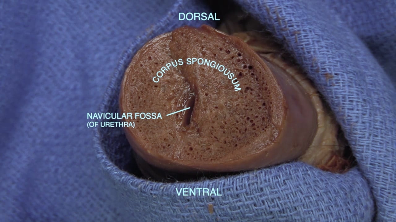
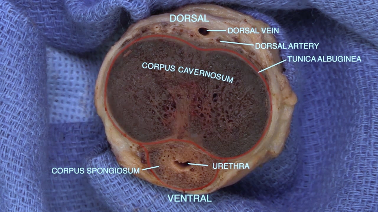
Cross Sections of Penis

View the next section of the video on the root of the penis and cross sections. Do not perform this dissection as these structures will be better demonstrated in sagittal section that you will perform later.

The erectile tissue of penis is supplied by the internal pudendal artery, a branch of internal iliac artery. Dilation of the arterioles of the corpus cavernosum and spongiousum and accompanying compression of the venous return by the ischiocavernosus and bulbospongiosus muscles causes an erection.

Male Pelvis

Internal Pelvic Structures

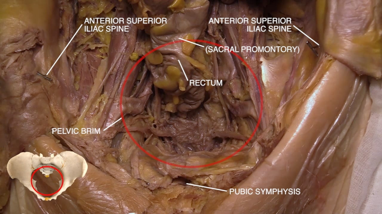
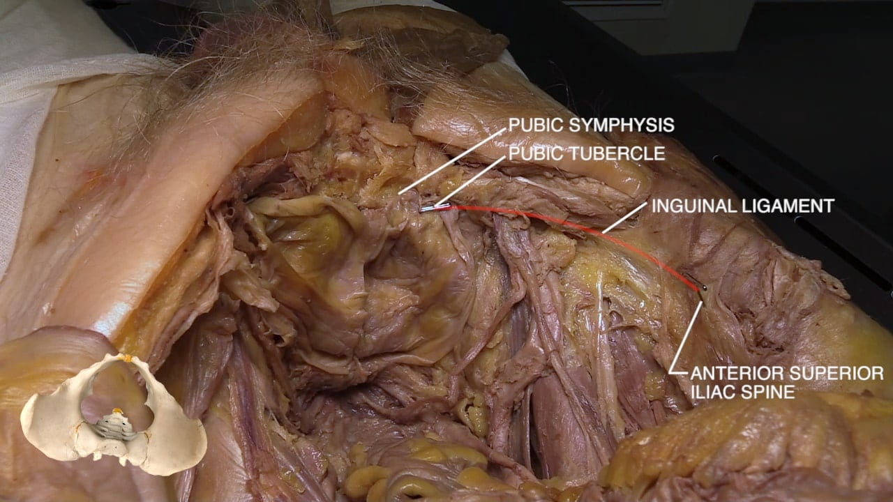
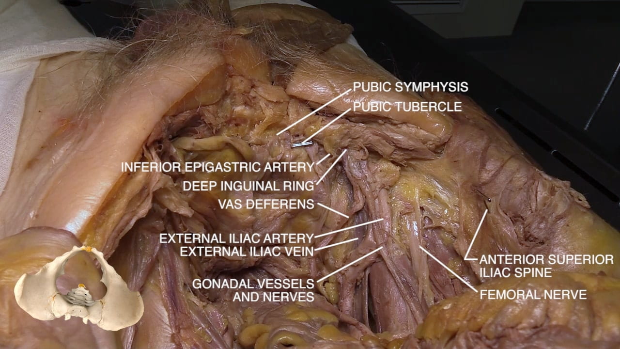
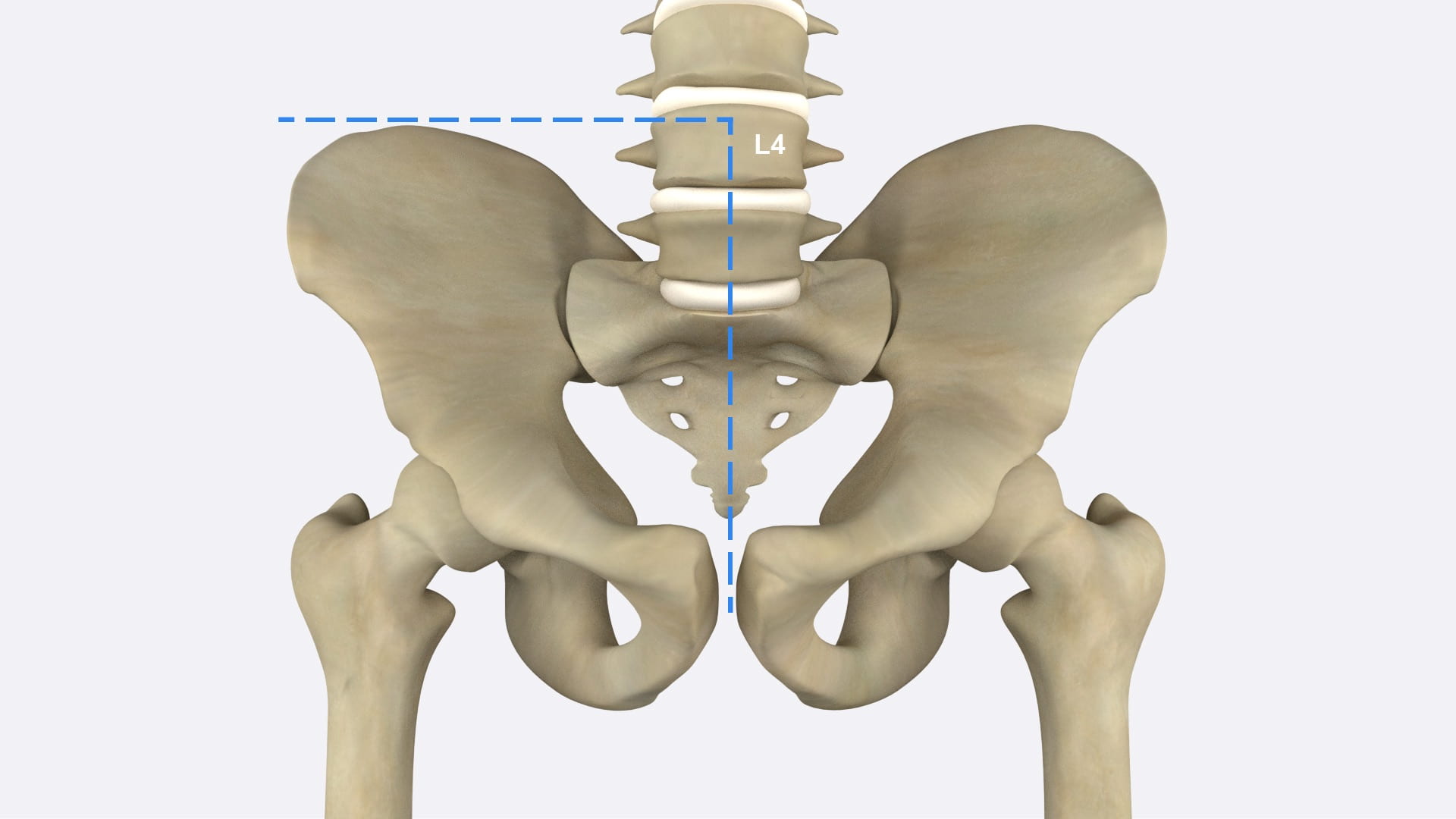
On a skeleton, examine the pelvis. Identify pubic symphysis, anterior superior iliac spine and sacral promontory. On donor locate the same structures.

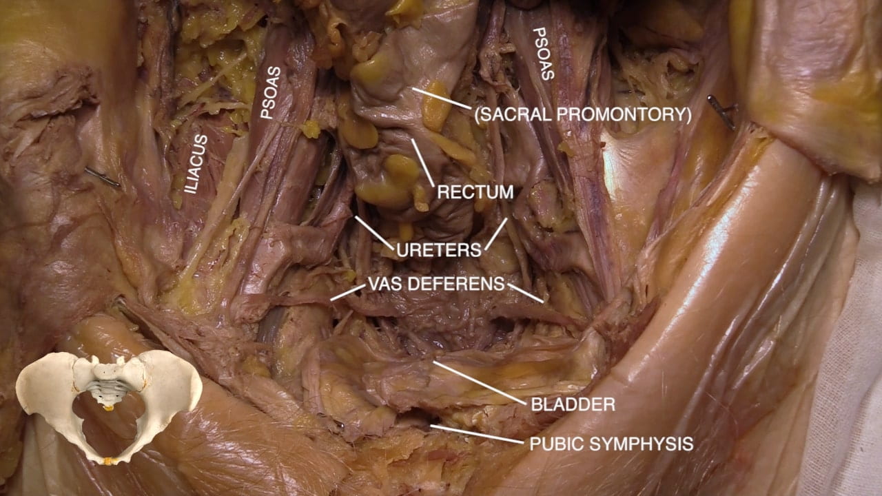
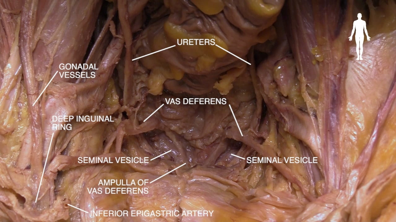
Locate psoas, bladder, ureters, vas deferens and rectum.

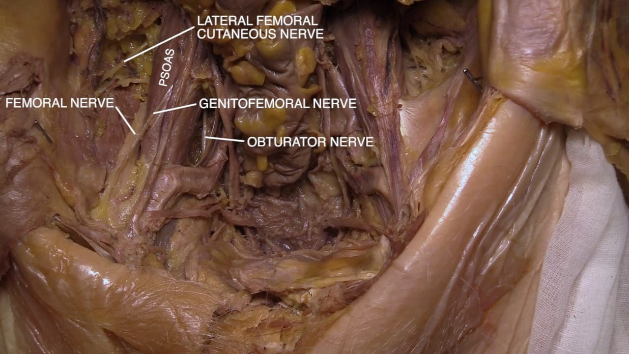
Using psoas as a landmark, locate femoral nerve lateral and obturator nerve medial to this muscle.

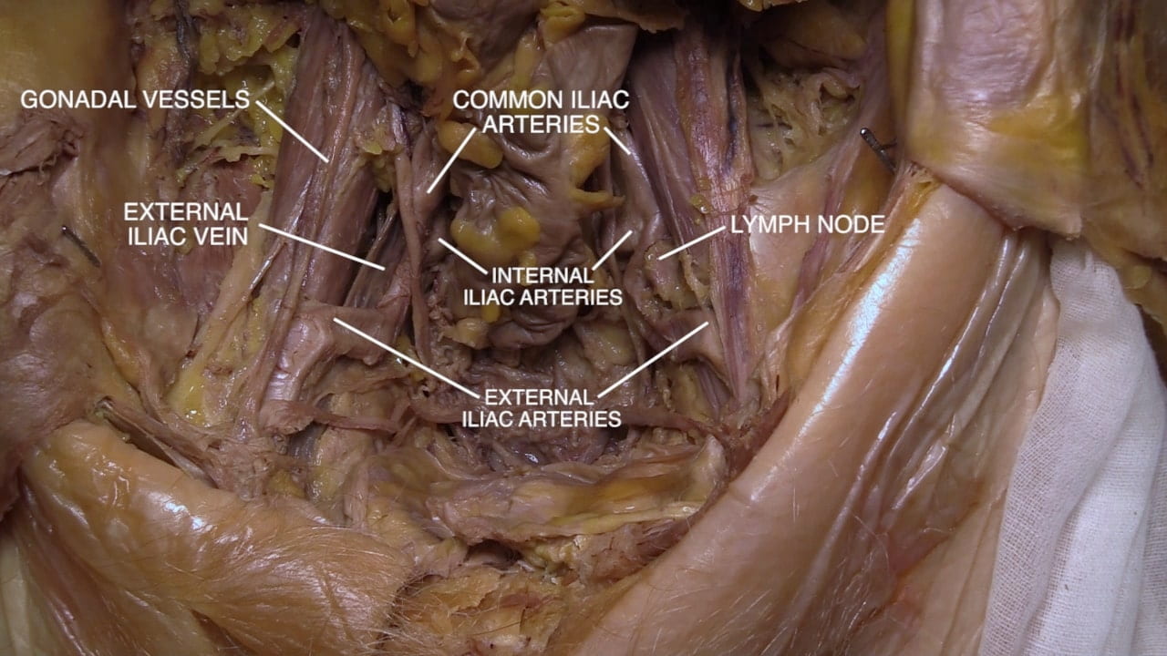
On the lateral wall of pelvis medial to the psoas muscle, identify common, internal and external iliac vessels.  Locate testicular (gonadal) vessels along psoas muscle.

Bladder

Follow the ureter to posterior surface of bladder base.

Follow vas deferens from deep inguinal ring to posterior surface of bladder (base). Identify seminal vesicles near vas deferens entry into posterior prostate.

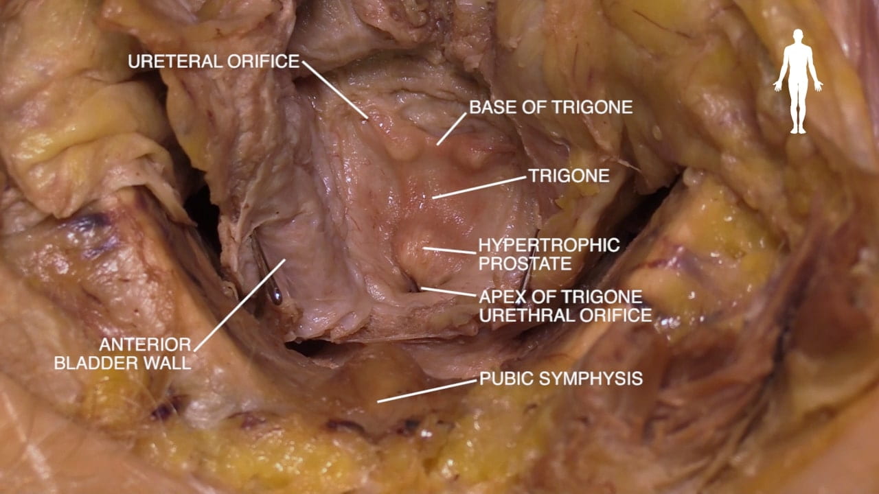
Incise superior and anterior walls of bladder.  Locate trigone, a smooth region on the interior of base.  Locate the urethral orifice and openings of ureters into bladder at base of trigone. The prostate surrounds this opening.

Note trabeculations on the remainder of the bladder wall.

Deep Inguinal Ring

Examine the deep inguinal ring from its pelvic surface. Along the inguinal ligament, find the deep inguinal ring, vas deferens and testicular vessels.  Note that inferior epigastric vessels are at the medial border of the deep inguinal ring. Indirect inguinal hernias enter the inguinal canal lateral to inferior epigastric vessels and direct inguinal hernias are medial to these vessels.

Key Sagittal Pelvic Dissection Notes

While splitting the pelvis provides the best view of the pelvis and perineum, we appreciate that some may not be comfortable with this process and suggest the following alternatives:

  1. Split pelvis
  2. For AMAB spend time on inguinal canal, testis and try to visualize prostate – laparoscope may be helpful – and maybe even a prostatectomy with Urology.
  3. For all, examine pelvic vasculature, course of ureter and lumbosacral plexus.

As you think through these options and discuss with your partners, please feel free to ask us questions and voice concerns.

Sagittal Dissection Preparation Gallery

In preparation for sagittal dissection of the pelvis, please review the above images.

Sagittal Dissection Preparation

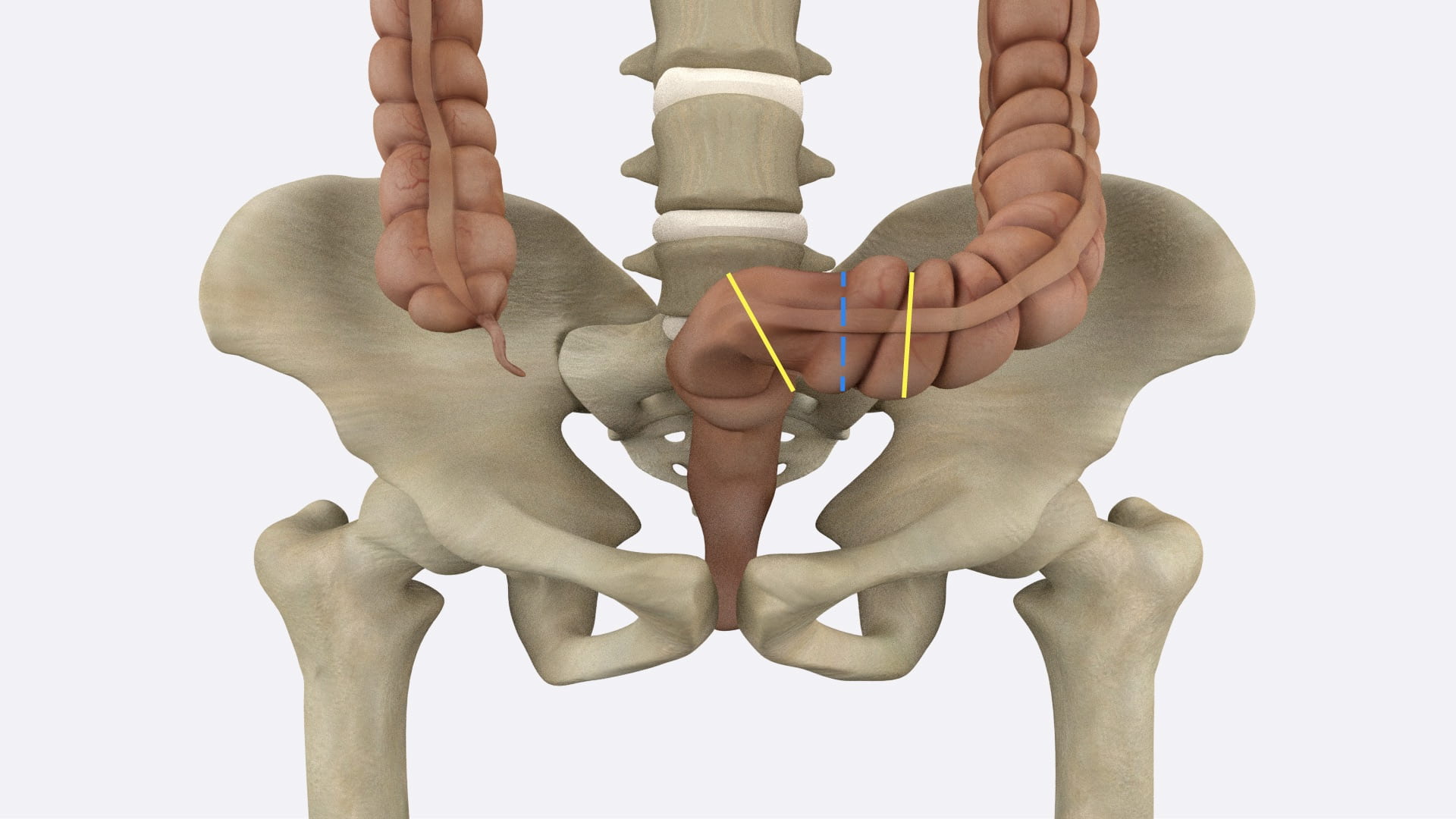
Fig. 4.2a: Place clamps several inches apart at yellow lines. Divide bowel between these clamps. Place ties on each side of bowel well away from bowel division.

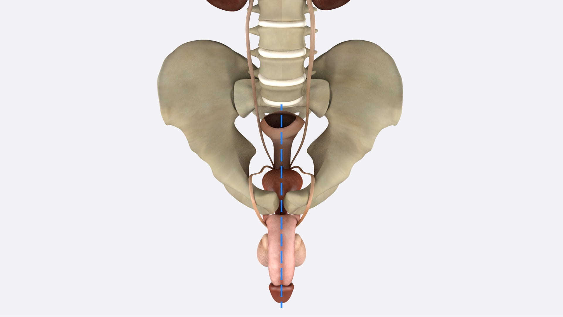
 Fig. 4.2b: Divide soft tissue including pubic symphysis, bladder, bowel and penis in the midline.

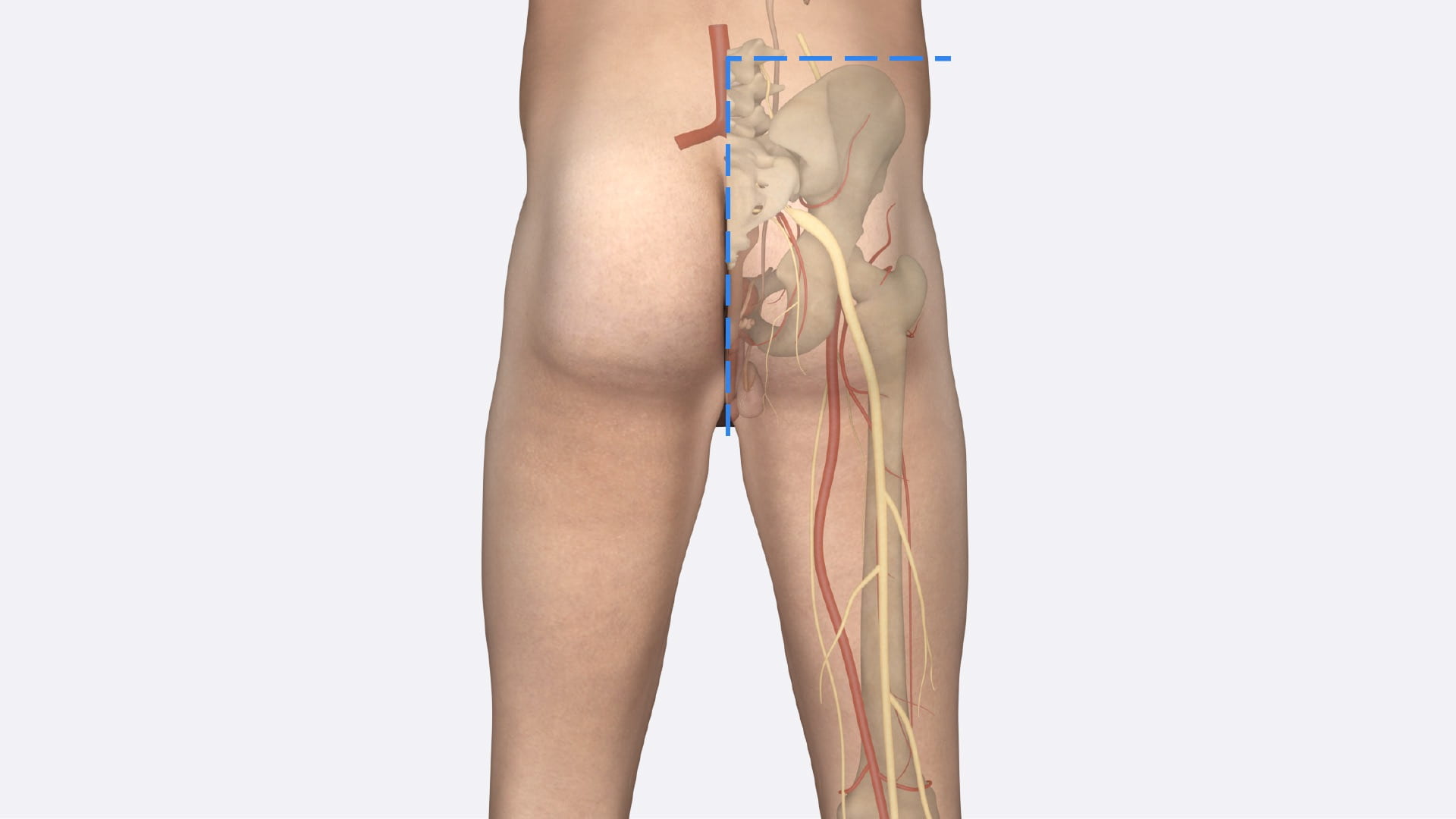
Fig. 4.2c: Divide lateral soft tissue and midline bony structures. Lateral positioning may be helpful. Be sure to retract large and small intestines away from the cuts.

Male Sagittal Pelvis

Pelvic Hemisection

Place ligatures at the rectosigmoid junction and divide the bowel. Elevate the colon and small intestine away from the saw cuts noted below. 

With a long knife, divide the penis in the midline and divide the soft tissues of the perineum and pelvis.

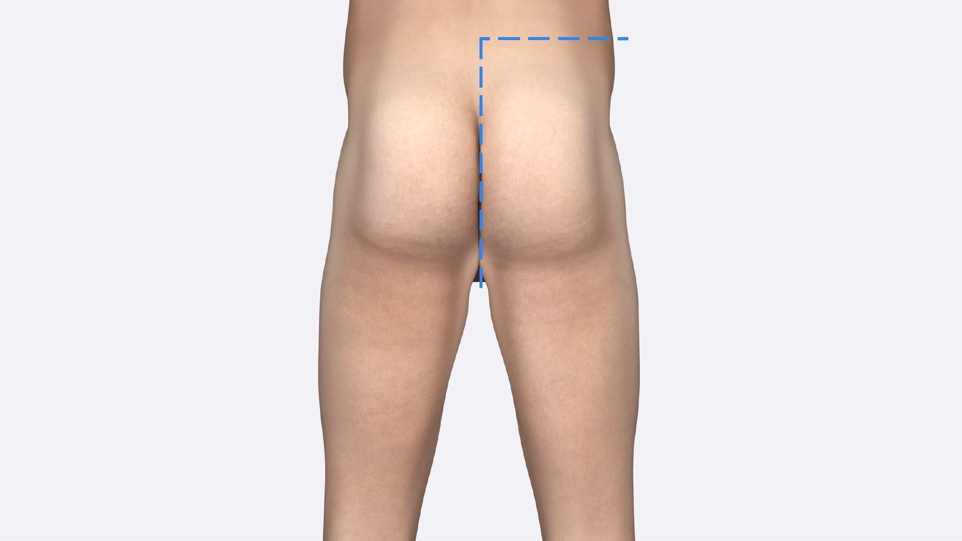
Using a hand saw, divide the pubic symphysis, sacrum, and the lumbar spine as far superiorly as L3. Stay in the midline.

Make a transverse cut across the trunk just superior to the iliac crest to the midline. Remove the attached lower limb and pelvis to view the sagittal pelvis.

Hemisection of Pelvis

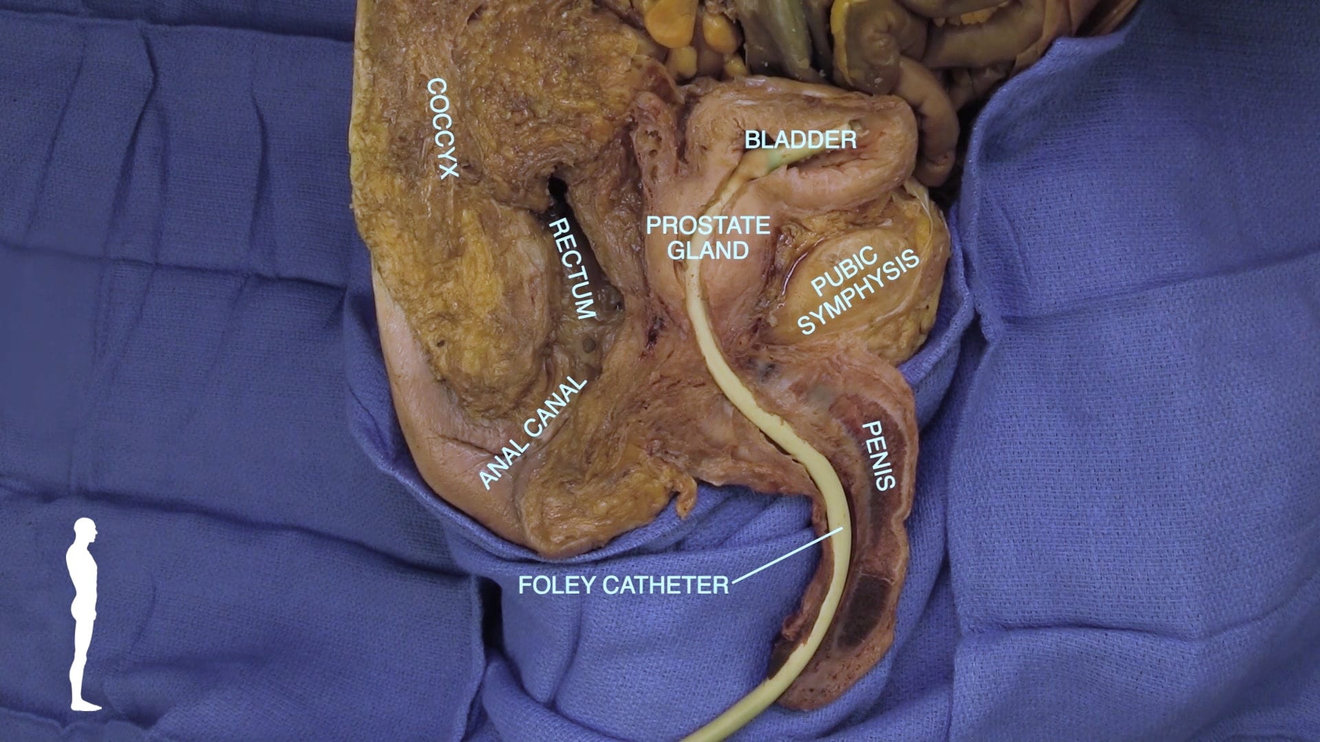
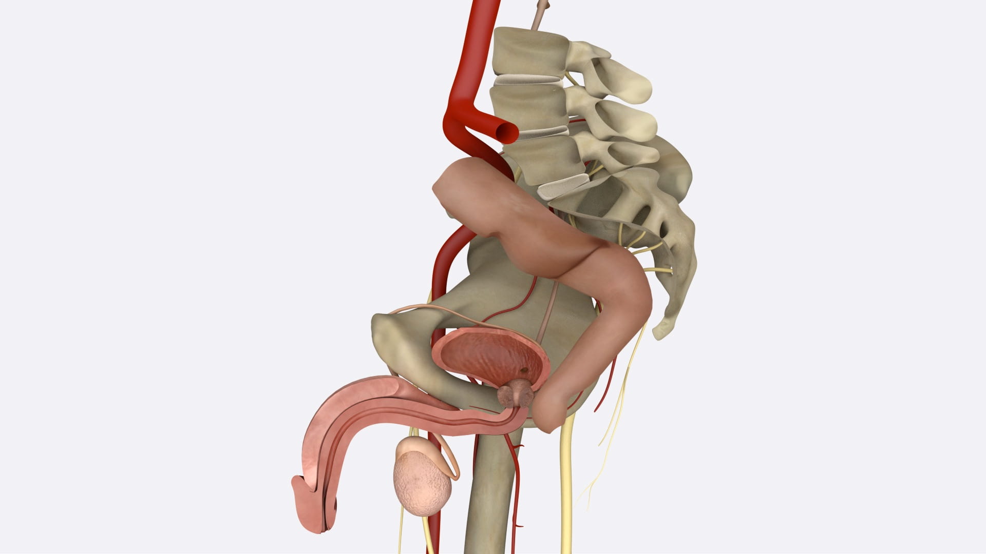
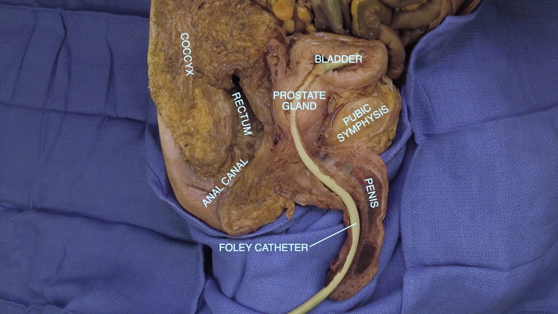
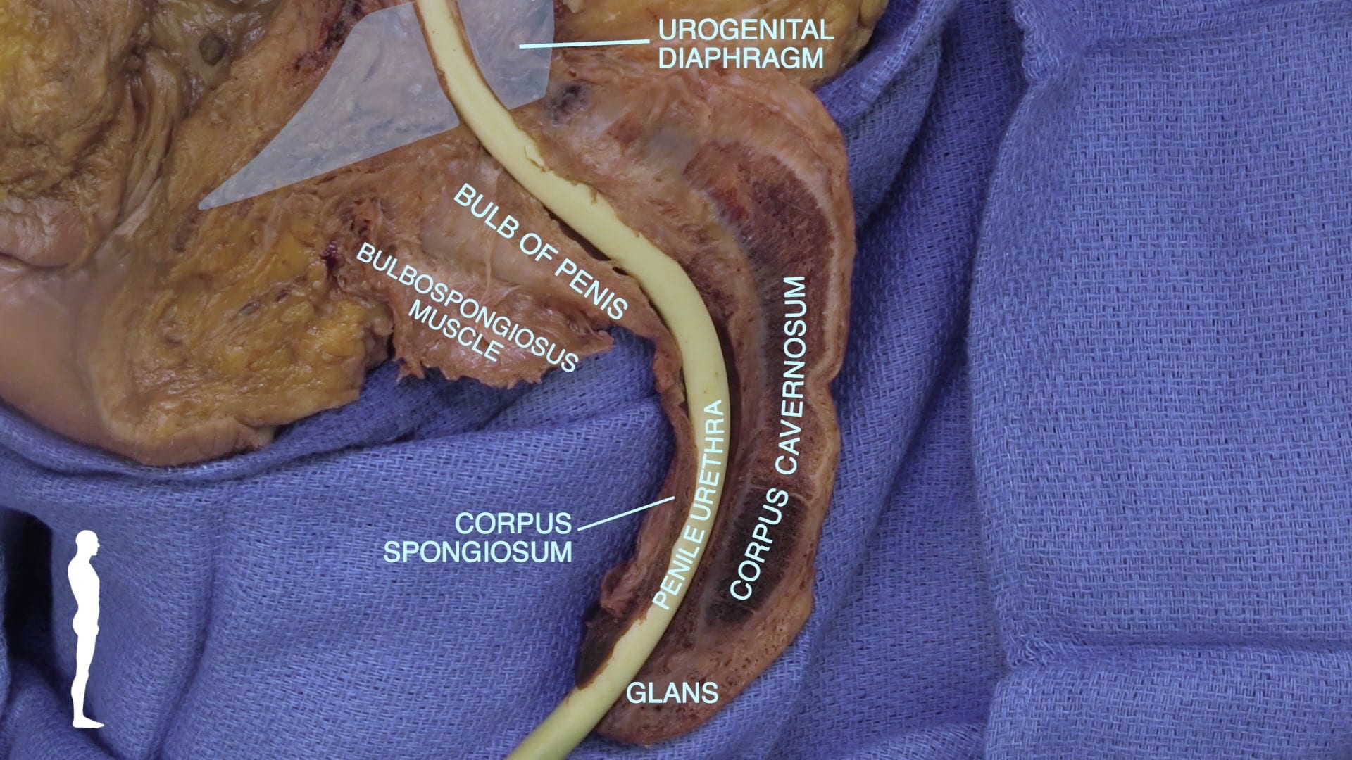
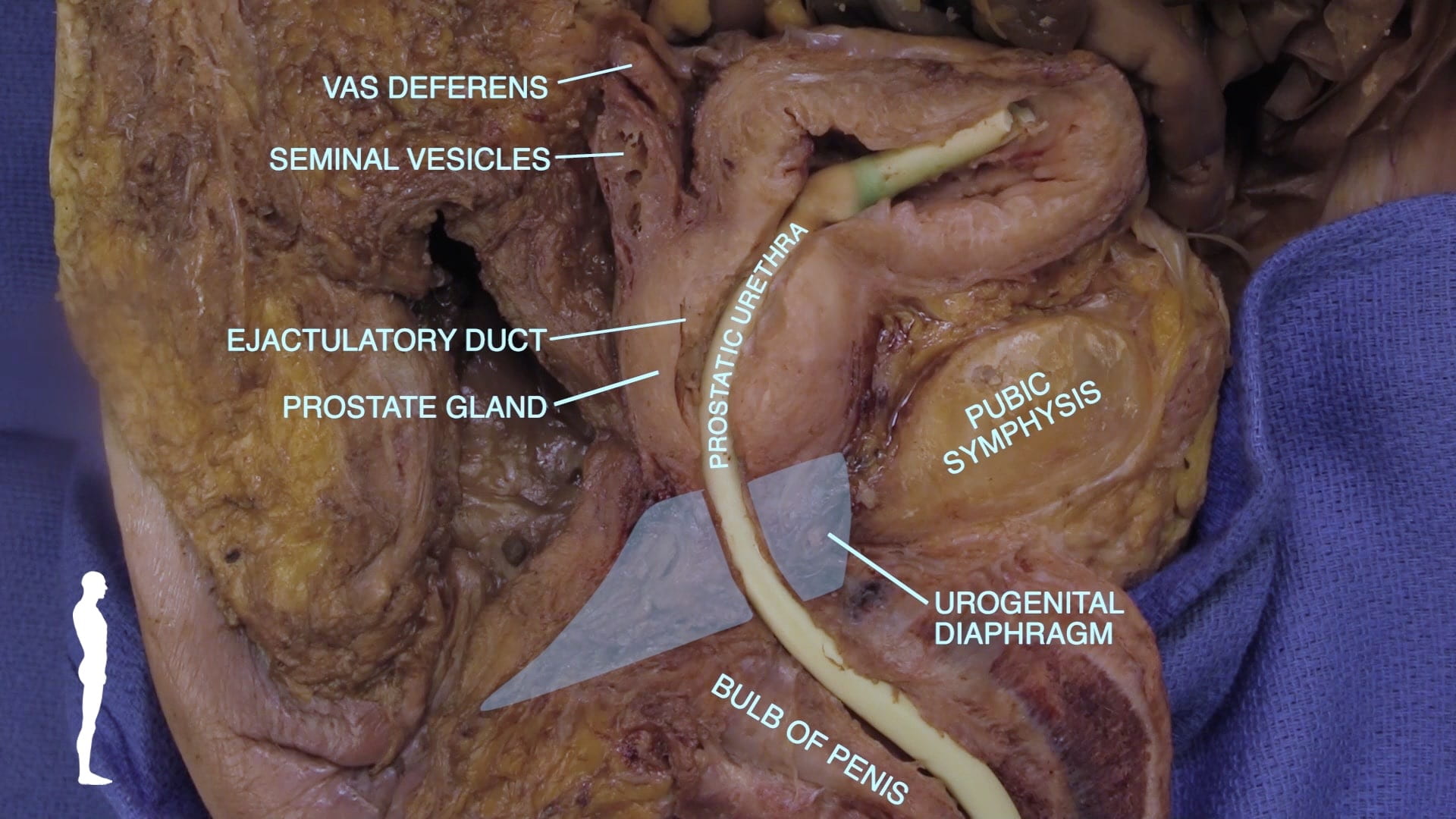
Identify the pubic symphysis, coccyx and sacral promontory in the sagittal plane. Identify penile urethra, prostate gland, bladder, rectum and anal canal.

On the penis, locate the corpus spongiosum, corpus cavernosum, penile urethra and bulb of the penis. Identify the prostatic urethra, seminal vesicles and vas deferens. The prostate rests on the urogenital diaphragm that reinforces the pelvic floor.
Prostate

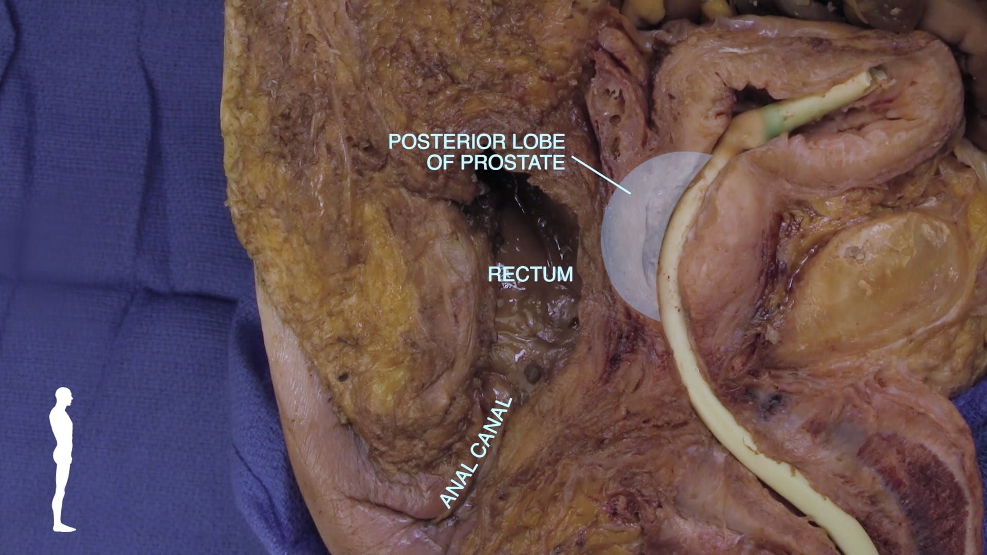
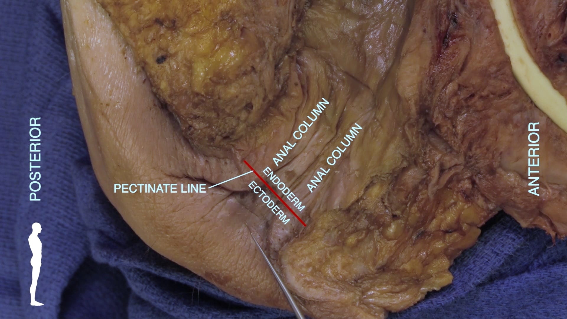
Locate rectum and anal canal. Note the close relationship between rectum and posterior lobe of prostate. In the anal canal, identify anal columns and pectinate line.

Male Cystoscopy

Male Bladder Drainage

Review Quiz