Assigned Female Pelvis

Lab Summary

The structure of the female pelvis and the perineum along with their contents are taught. The female pelvis and perineum are presented from external structures to pelvic contents. Additionally, the female pelvis is shown from a midline sagittal perspective. The course of retroperitoneal vessels and nerves are demonstrated.

Lab Objectives

  1. Name structures in broad ligament.
  2. Describe relationship between uterine artery and ureter.
  3. Describe positions of glans and crus of clitoris and vestibular bulb.
  4. Name principal structures in midline pelvis of female.
  5. Describe A-P dimension of pelvic inlet and how it is estimated.
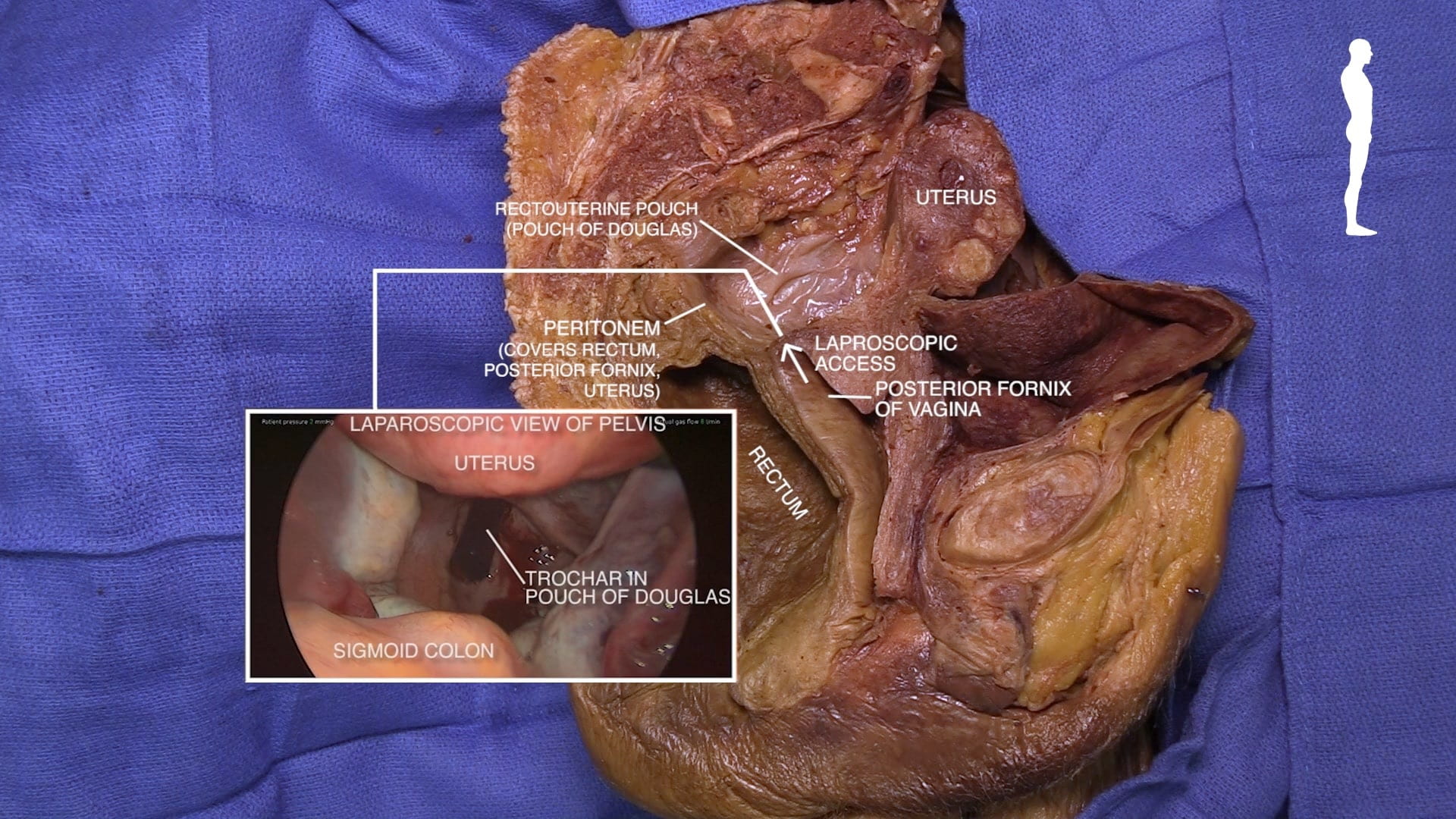
  6. Explain clinical importance of posterior fornix of vagina.
  7. Describe diagonal conjugate.
  8. Describe the erectile tissue of clitoris and vestibular bulb.

Lecture List

Female Perineum, Female Pelvis, Female Sagittal Pelvis, Pelvic Vessels and Nerves

Female Perineum

External Genitalia

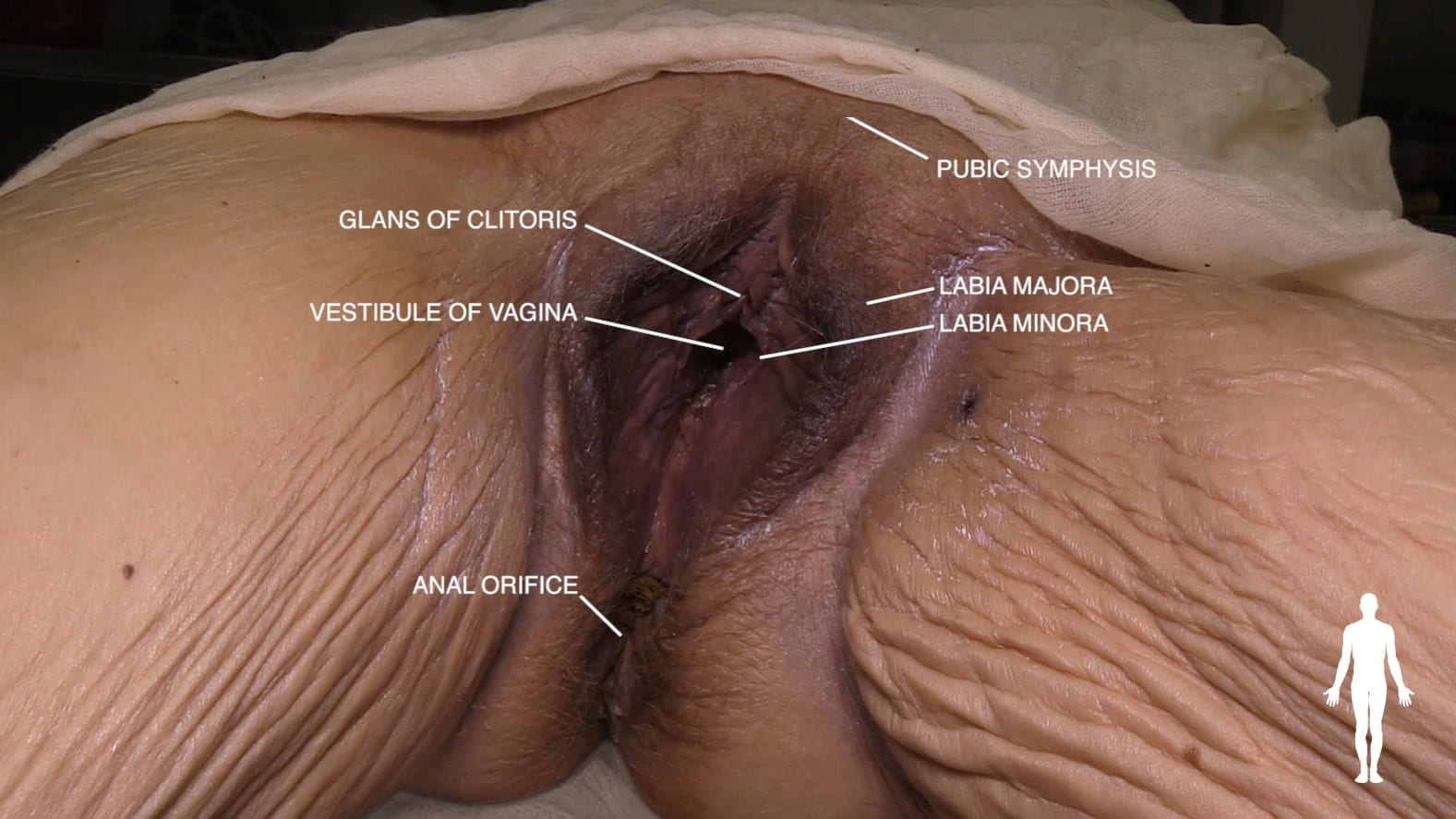
Examine the perineum. Identify pubic symphysis, anal aperture, vestibule of vagina, glans of clitoris, labia majora and labia minora.

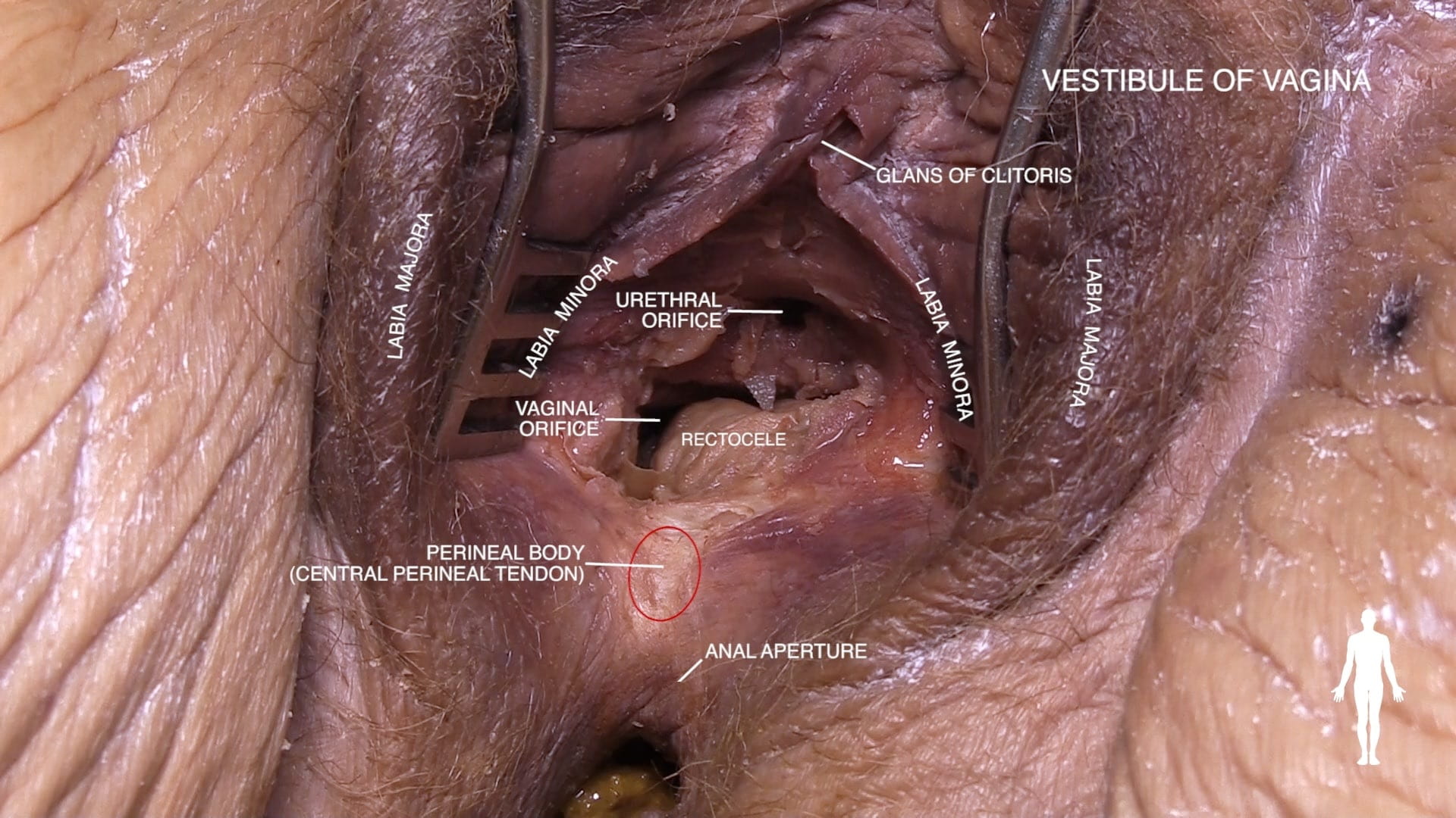
Identify the urethral orifice in the vestibule of vagina.  Posterior to vaginal orifice locate the bulge of the perineal body (central perineal tendon).

Erectile Tissue

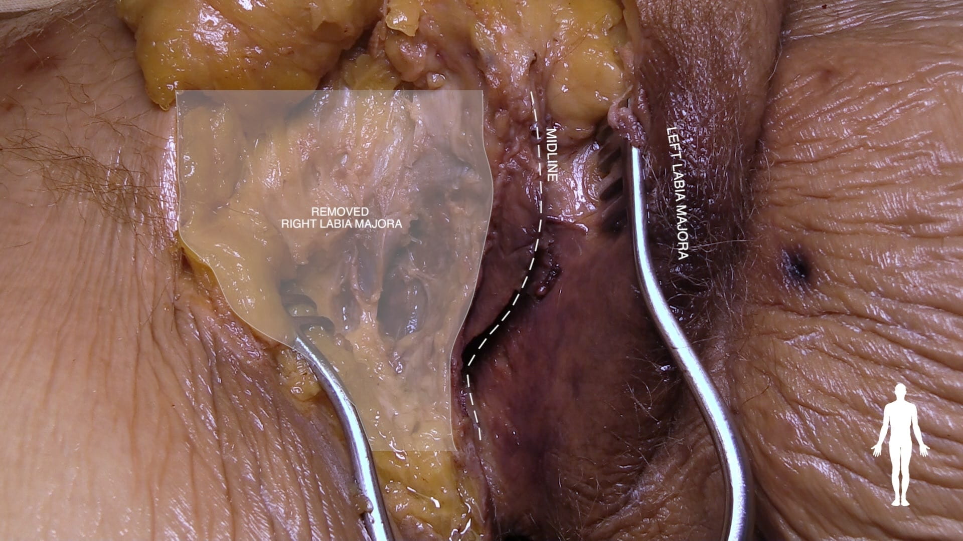
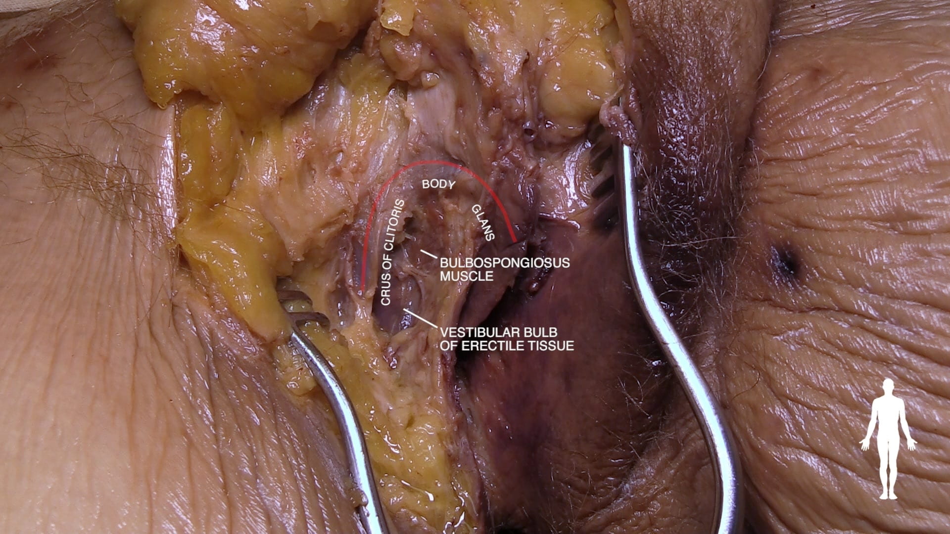
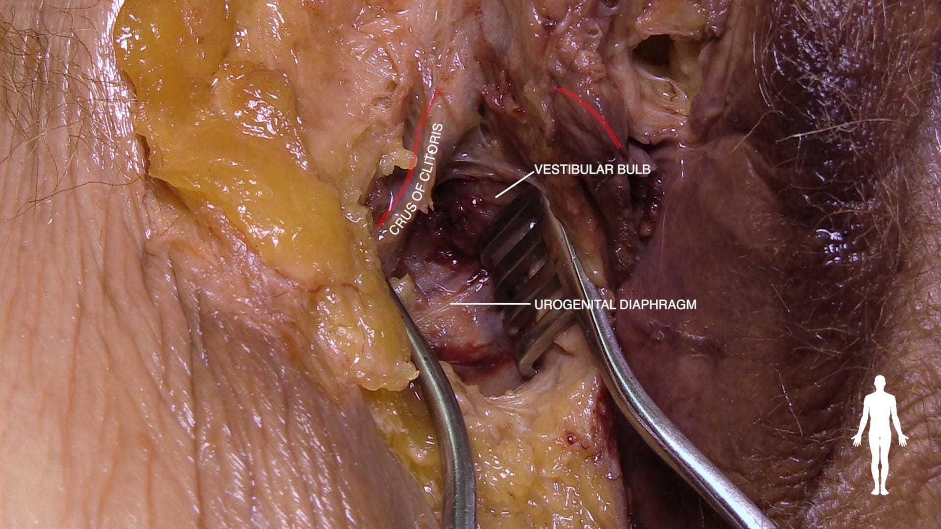
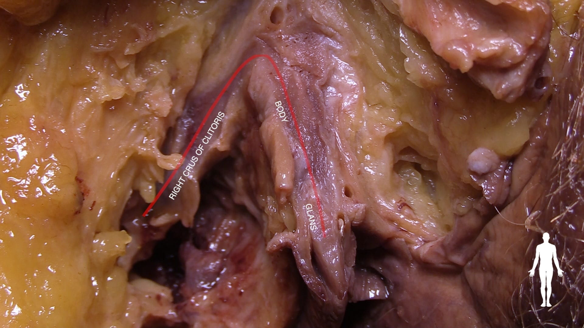
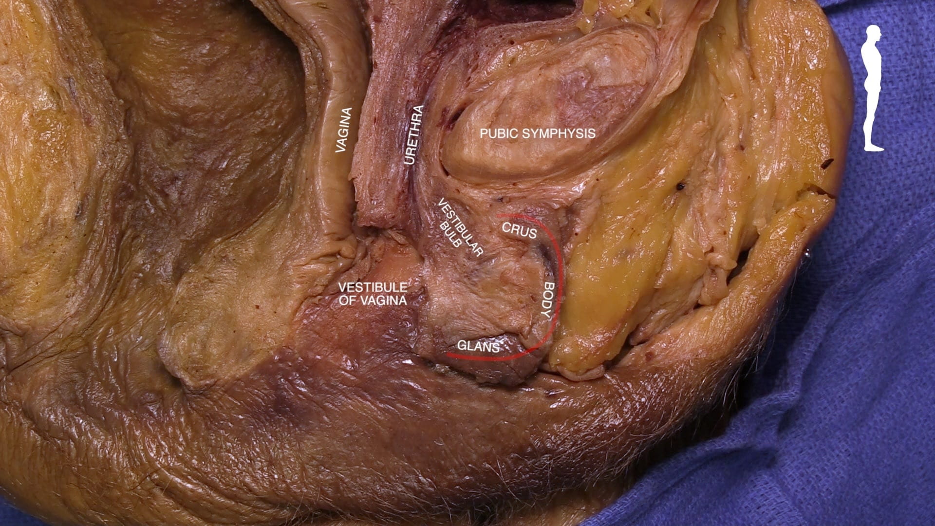
Remove the skin and the superficial fascia of the labia majora.  Locate the glans, body and crus of the clitoris.

Identify the vestibular bulb of erectile tissue, which surrounds the vestibule of the vagina.

Place a retractor between the crus of the clitoris and the vestibular bulb to expose the urogenital diaphragm.

Remove the outer portion of the glans, body and crus of the clitoris. Note the appearance of this erectile tissue.

Female Pelvis

Uterus

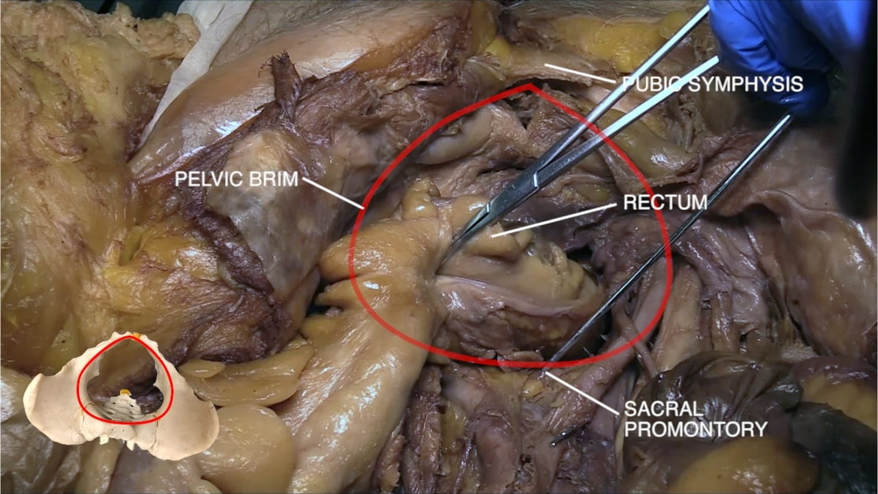
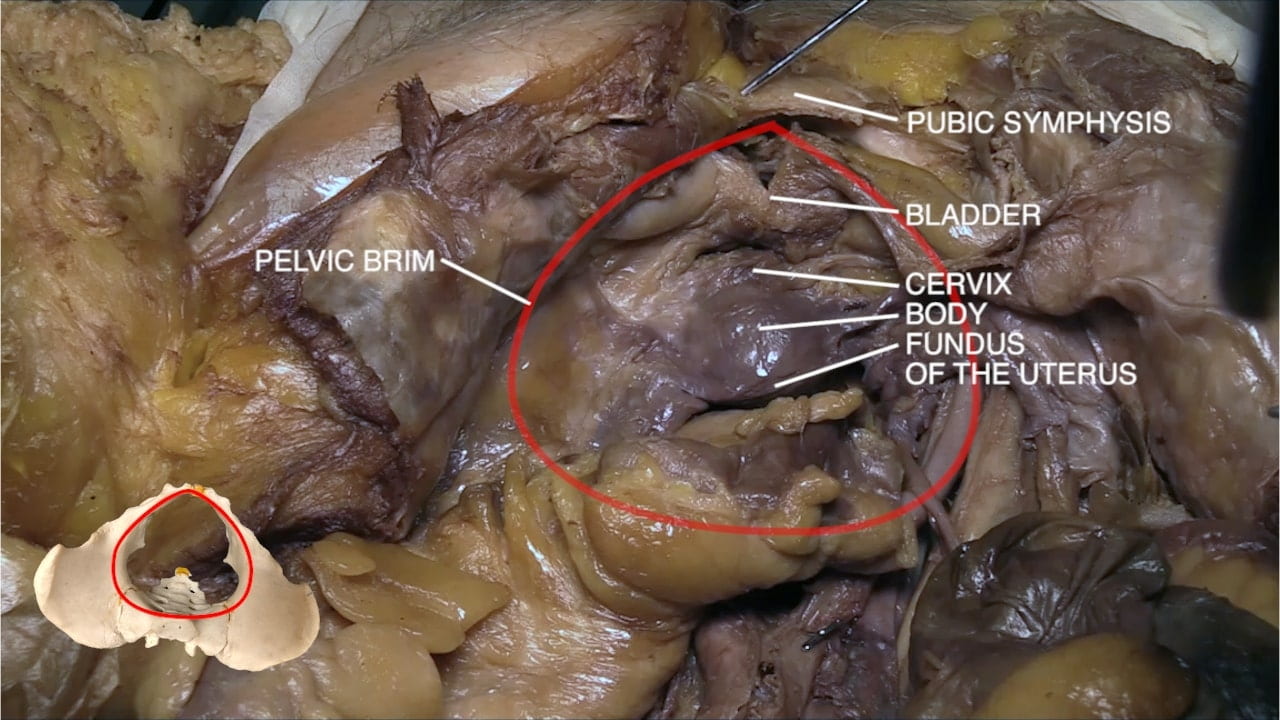
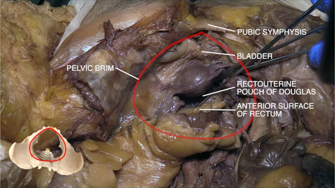
Examine the interior of the pelvis. Orient by locating the pubic symphysis and sacral promontory. Identify the pelvis brim.

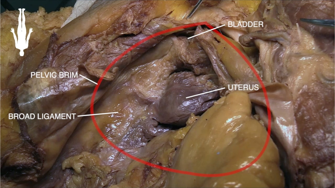
Locate the bladder, uterus and rectum.

Identify the fundus, body and cervix of the uterus.  Note that the donor in the video has a retroverted uterus. Locate the rectouterine pouch of Douglas between the uterus and rectum.

Uterine Adnexa

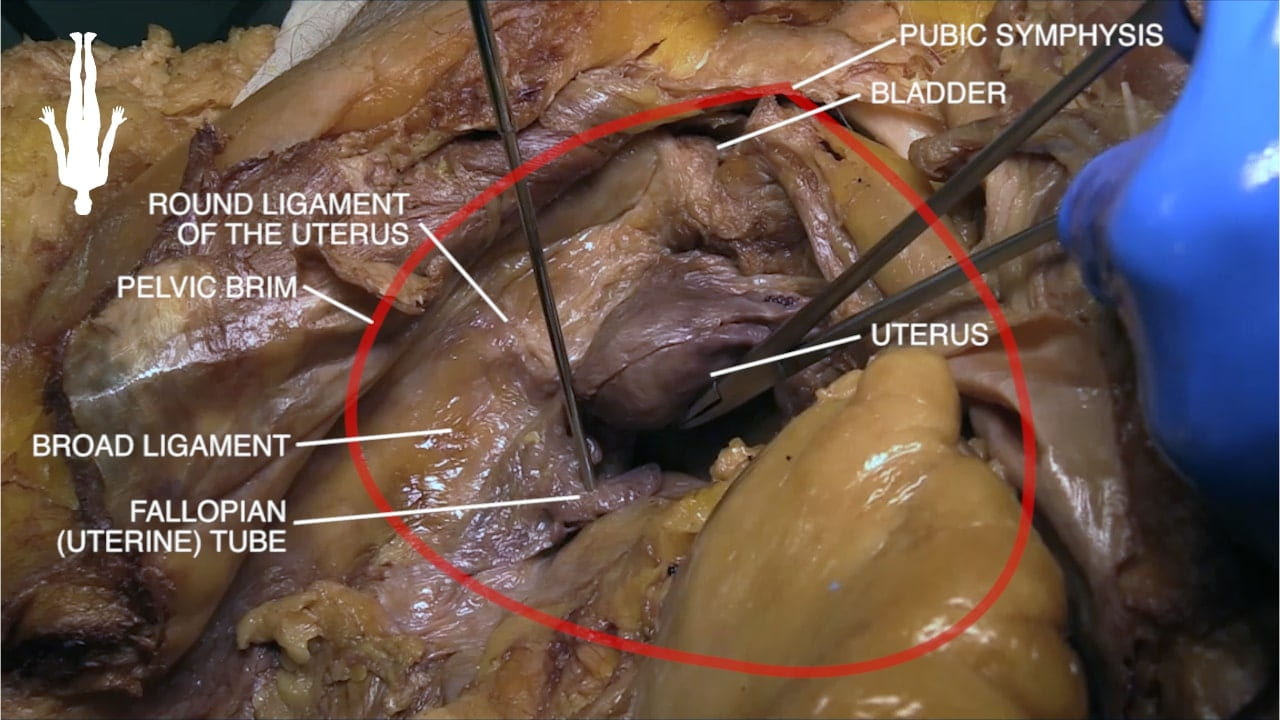
Identify the broad ligament that connects the uterus to the pelvic wall on each side.

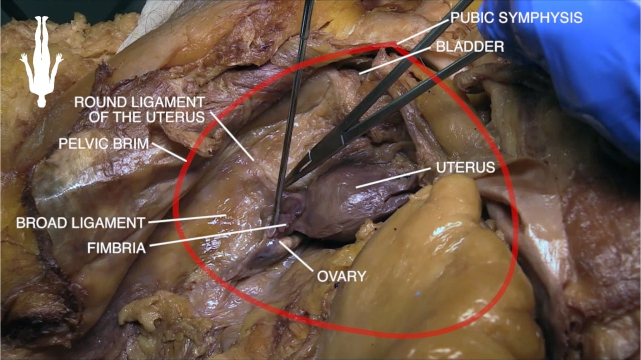
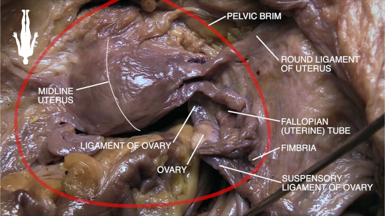
In the free border of the broad ligament, locate the fallopian tube (uterine tube). Fold the broad ligament anteriorly to expose the ovary.

Dissect the broad ligament to expose the round ligament of the uterus, uterine tube and ovary. Identify the suspensory ligament of the ovary which contains the ovarian vessels.

Iliac Vessels and Femoral Canal

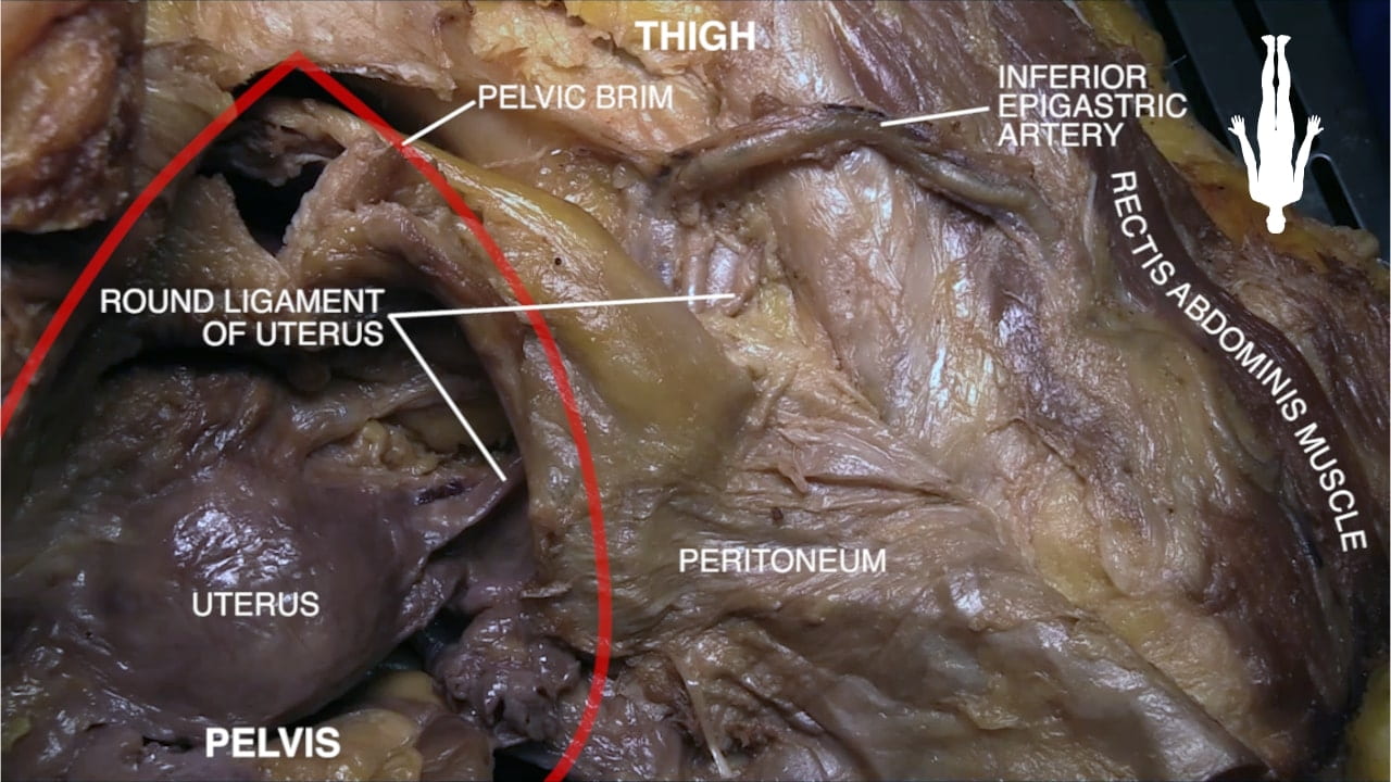
Trace the round ligament of the uterus anterolaterally. Reflect the peritoneum and identify the deep inguinal ring, where the round ligament enters the inguinal canal. Medial to the deep inguinal ring, locate the inferior epigastric vessels.

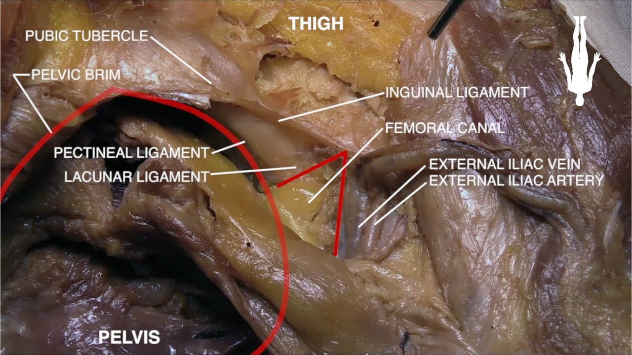
Locate the external iliac vessels. Medial to the vessels, locate the femoral canal. The femoral canal is the site of femoral hernias, which pass inferior to the inguinal ligament and are more common in women.

Ureter and Uterine Artery

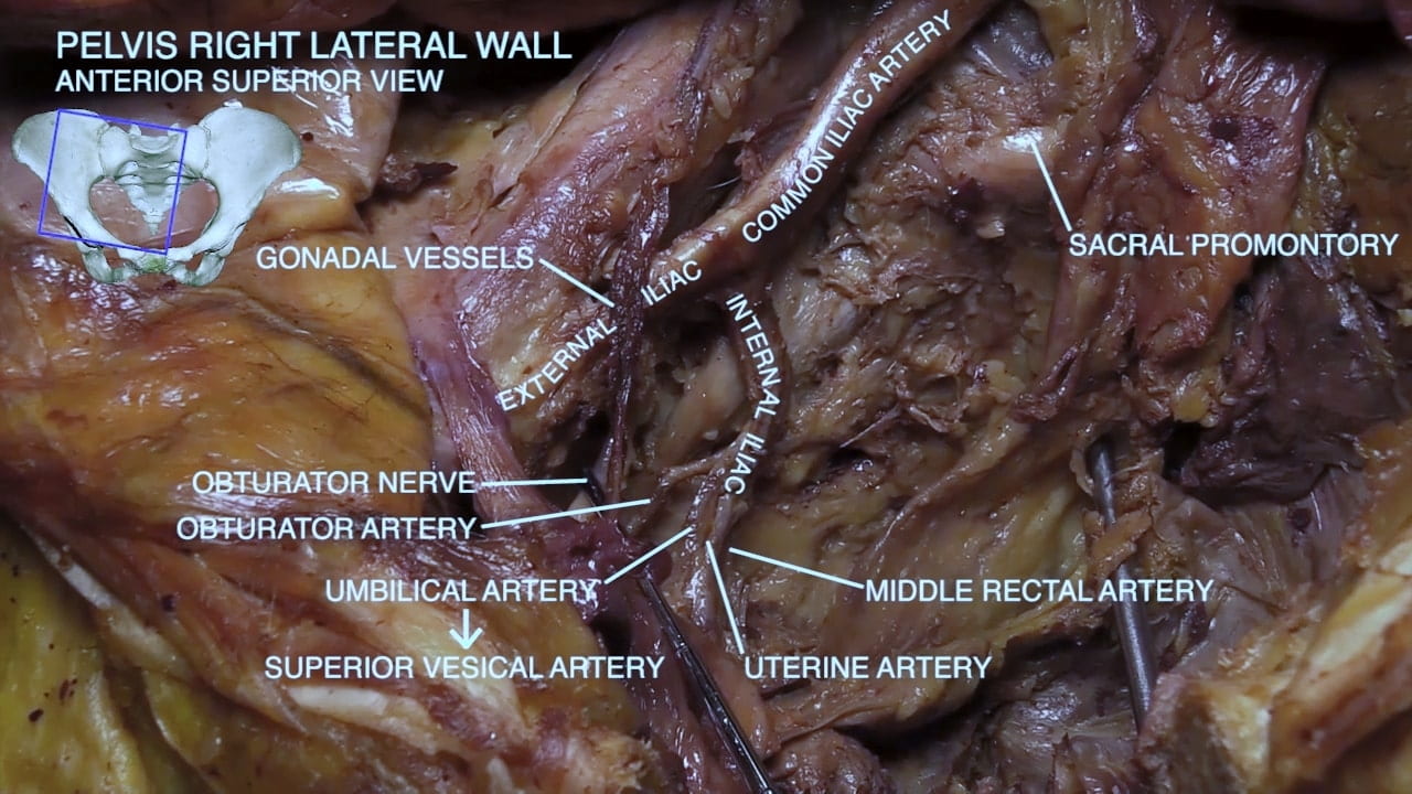
Identify the ureter. Trace it into the pelvis and follow it anteriorly to the bladder. Note the close relationship of the uterine artery to the ureter lateral to the uterus.

Labels (left to right): Pelvic brim, ureter, uterine artery, external iliac vessels
2.4a) Ureter and uterine artery relationship

Bladder

Incise the superior and anterior walls of the bladder.  Identify the trigone, a smooth region on the interior of the base.  Locate the urethral orifice and openings of the ureters into the bladder.

Examine the remaining interior surface of the bladder for its rough appearance from the detrusor muscle.

Labels (top to bottom): Ureteral orifice, base of trigone, trigone, hypertrophic prostate, apex of trigone urethral orifice, anterior bladder wall, pubic symphysis
2.5a) Trigone of bladder (image from male donor)

Key Sagittal Pelvic Dissection Notes

While splitting the pelvis provides the best view of the pelvis and perineum, we appreciate that some may not be comfortable with this process and suggest the following alternatives:

  1. Split pelvis
  2. If no split, perineum in AFAB is difficult – try separating legs progressively with blocks to hold them apart.
  3. For AFAB with uterus, try a hysterectomy with Ob-Gyn – laparoscope may be helpful for visualizing deep structures. Note relationship of uterine artery and ureter.
  4. For all, examine pelvic vasculature, course of ureter and lumbosacral plexus.

As you think through these options and discuss with your partners, please feel free to ask us questions and voice concerns.

Sagittal Dissection Preparation Gallery

In preparation for sagittal dissection of the pelvis, please review the above images.

Sagittal Dissection Preparation

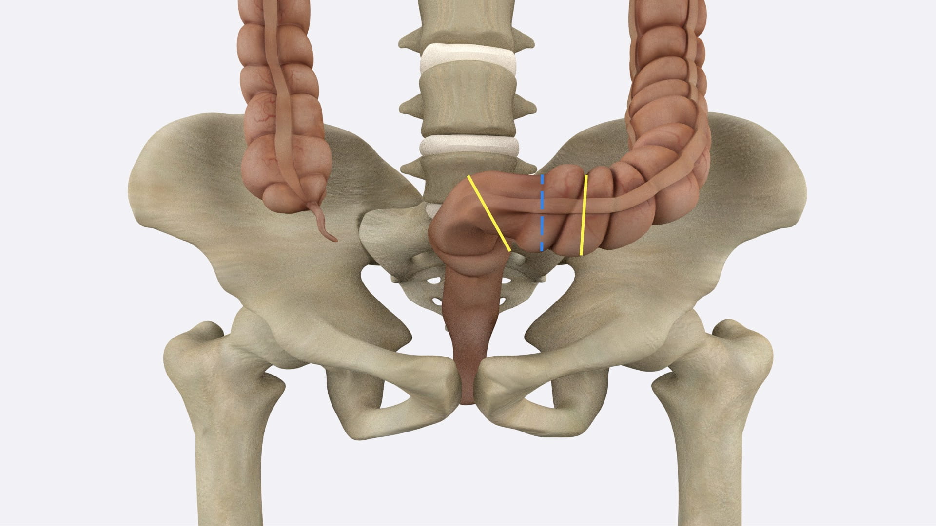
Fig. 3.2a: Place clamps several inches apart at yellow lines. Divide bowel between these clamps. Place ties on each side of bowel well away from bowel division.

 Fig. 3.2b: Divide soft tissue including pubic symphysis, bladder, bowel, uterus and vagina in the midline.

Fig. 3.2c: Divide lateral soft tissue and midline bony structures. Lateral positioning may be helpful. Be sure to retract large and small intestines away from the cuts.

Female Sagittal Pelvis

Pelvic Hemisection

Place ligatures at the rectosigmoid junction and divide the bowel. Elevate the colon and small intestine away from the saw cuts noted below. 

With a long knife, divide the soft tissues of the perineum and pelvis in the midline.

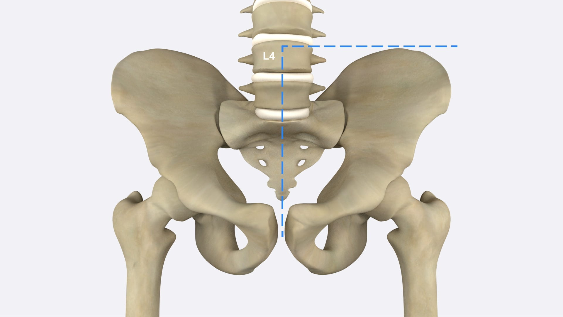
Using a combination of hand saw and long knife divide the pubic symphysis, remaining soft tissue, sacrum and the lumbar spine as far superiorly as L3. Stay in the midline.

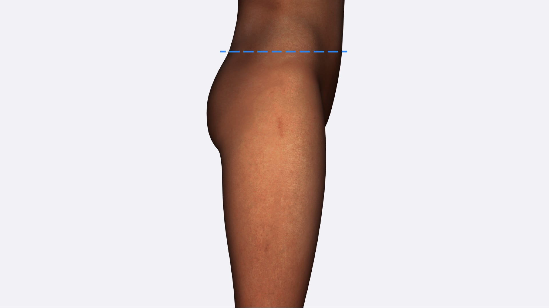
Make a transverse cut across the trunk just superior to the iliac crest to the midline. Remove the attached lower limb and pelvis to view the sagittal pelvis.

4.1a) Planes for pelvic section

Hemisection of Pelvis

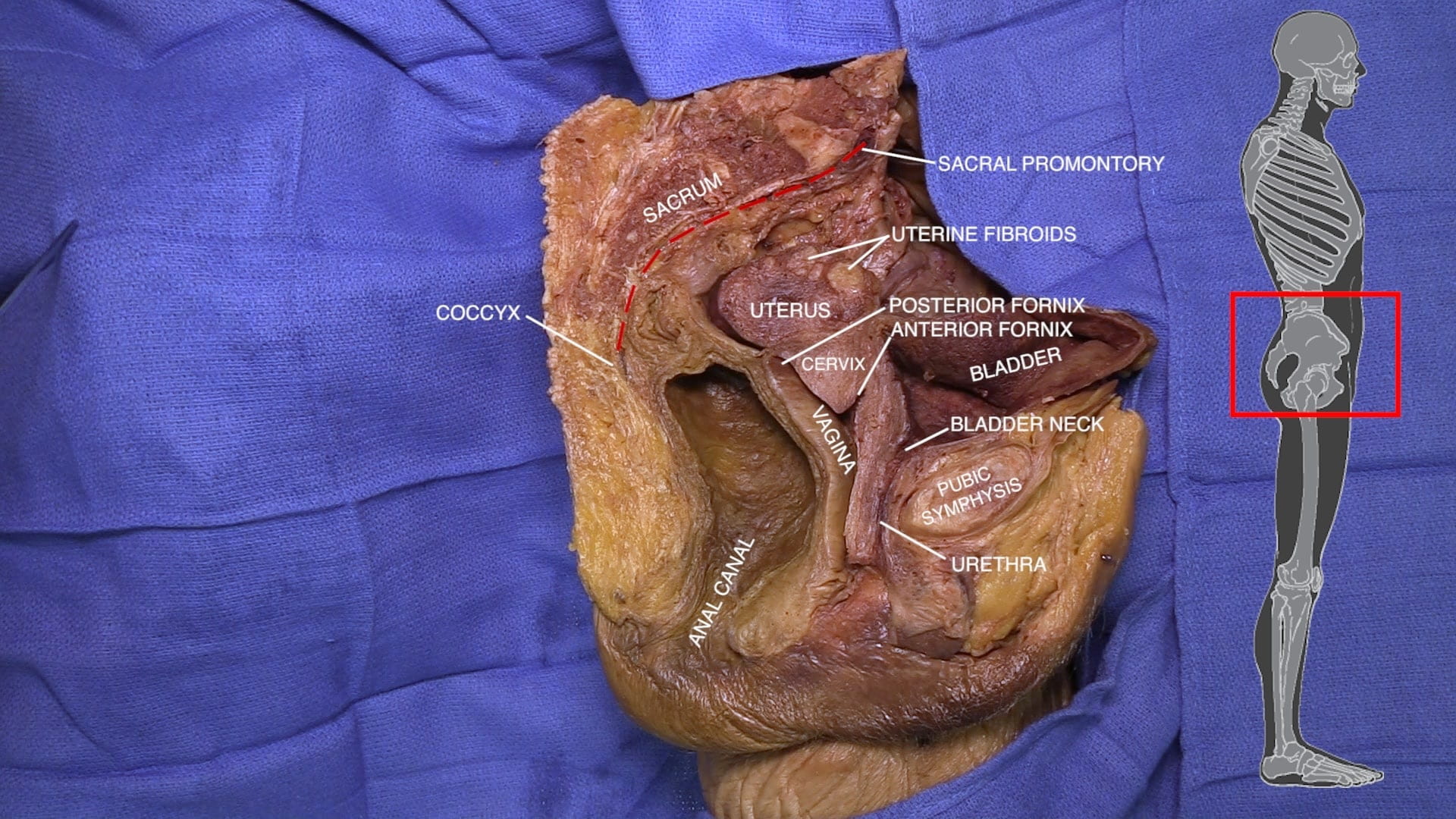
Identify the pubic symphysis, sacral promontory and coccyx.

Locate the bladder, urethra, rectum and anal canal.

Identify the labia majora and minora, glans, body and crus of clitoris, vestibular bulb and vestibule of vagina.

Identify the vagina anterior and posterior fornices of vagina, cervix and uterus. What is the position of your donor’s uterus: anteverted, retroverted, or midposed?  If your donor’s uterus has been surgically removed, examine a nearby donor.

Rectouterine Pouch

Locate the rectouterine pouch of Douglas and note its close relationship with the posterior fornix of vagina. The rectouterine pouch of Douglas is an important access point to the peritoneum in gynecologic surgery.

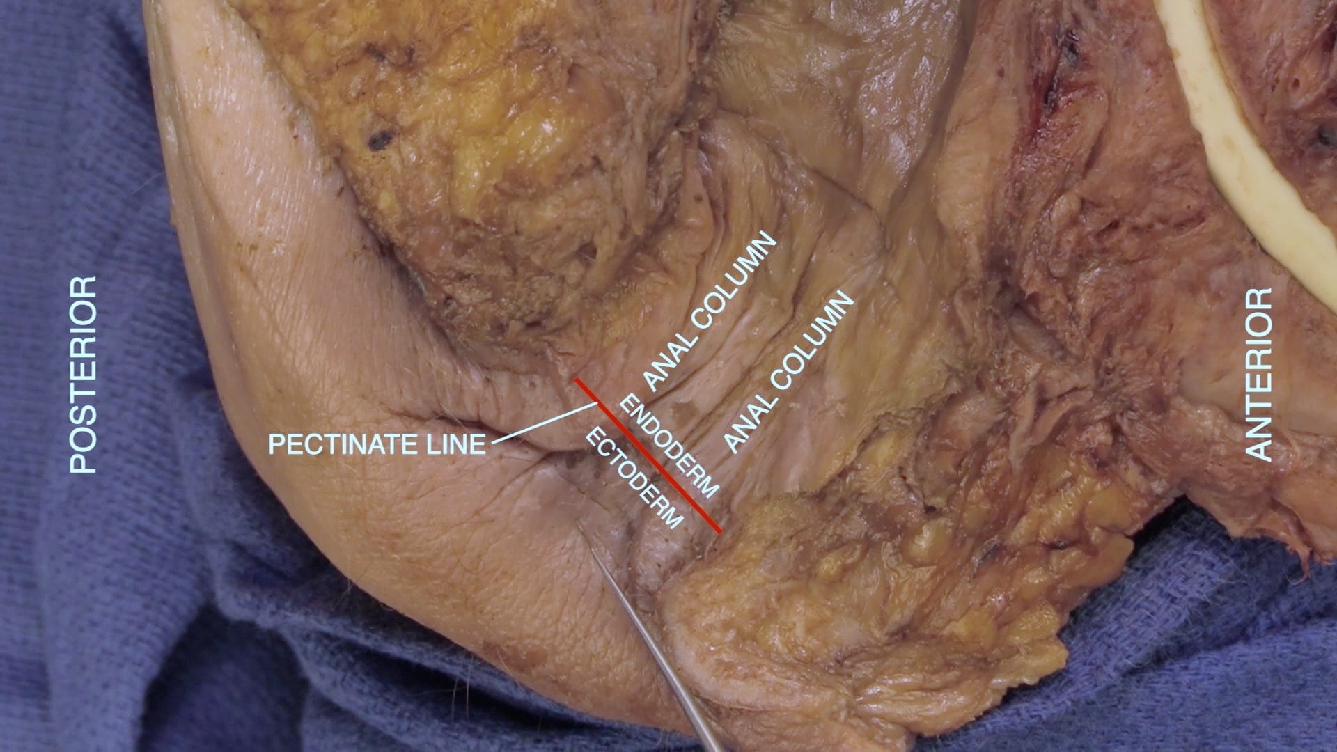
Relocate the rectum and the anal canal.  In the anal canal, identify the anal columns and the pectinate line.

Pelvic Vessels and Nerves

Pelvic Vessels and Nerves

Reflect the rectum from the anterior surface of the sacrum. Develop a retroperitoneal (extraperitoneal) plane along the lateral surface of the pelvis.

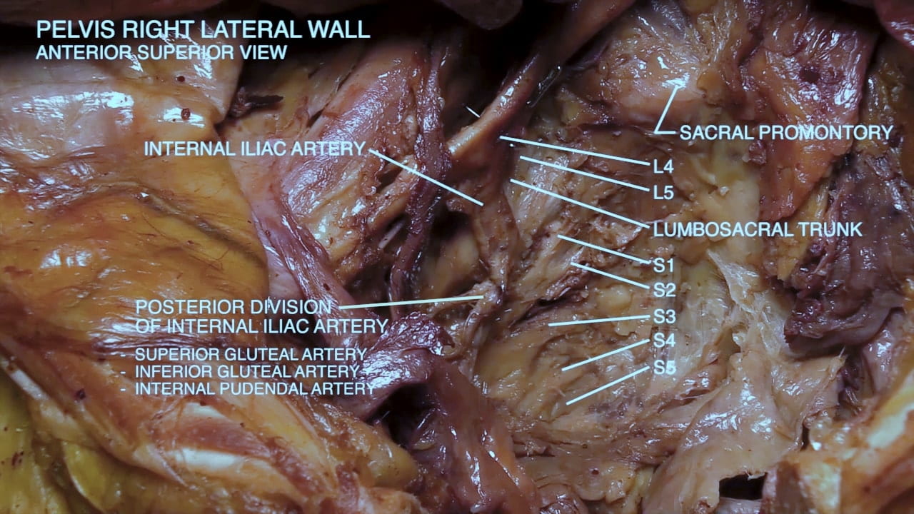
Identify the common iliac artery and its external and internal branches. Follow the internal iliac into the pelvis. Identify the anterior branch and its uterine branch. Identify the posterior branch which gives rise to the superior and inferior gluteal arteries and internal pudendal artery. 

Posterior to the iliac vessels identify L4, L5, comprising the lumbosacral trunk. Follow the sacral roots, S1-S5, as they exit from the sacrum.

These sacral roots, along with L4 and L5, combine to form the lumbosacral plexus whose major branches include the sciatic, pudendal and superior and inferior gluteal nerves. These nerves are further explored during gluteal dissection.

Female Cystoscopy

Bladder Drainage Devices

Review Quiz