Proximal Lower Extremity

Lab Summary

The structures of the proximal lower extremity are considered from anterior, femoral triangle, medial, lateral and gluteal perspectives. The neurovascular structures of the femoral triangle are key in a vast array of clinical scenarios. The hip joint is discussed in terms of surrounding structures, anatomic and surgical approaches and its basic configuration. Vasculature and innervation of the lower extremity are taught with emphasis on their courses and functionality. Considerable time is spent on the gluteal region with its connections to pelvis and perineum.

Lab Objectives

  1. Describe the boundaries and contents of the femoral triangle.
  2. Describe the major muscle groups of the thigh and their innervation.
  3. Describe the vascular supply to the lower extremity from the inguinal ligament to popliteal fossa.
  4. Describe the superficial and deep venous drainages of the lower extremity.
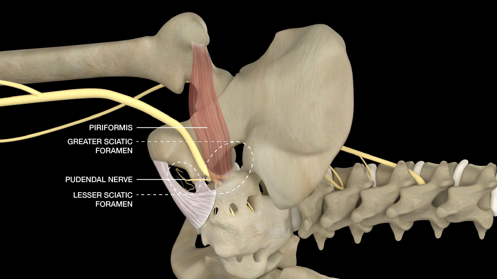
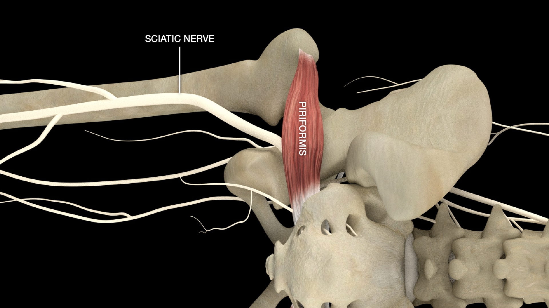
  5. Describe the relationship of the piriformis muscle to the sciatic nerve and the gluteal nerves and vessels.
  6. Describe the course of the pudendal nerve from sacrum to perineum.
  7. Name the functions of the pudendal nerve.

Lecture List

Anterior Thigh, Femoral Triangle, Medial Thigh, Lateral Thigh, Gluteal Region Part I, Gluteal Region Part II

Anterior Thigh

Exposing the Anterior Thigh

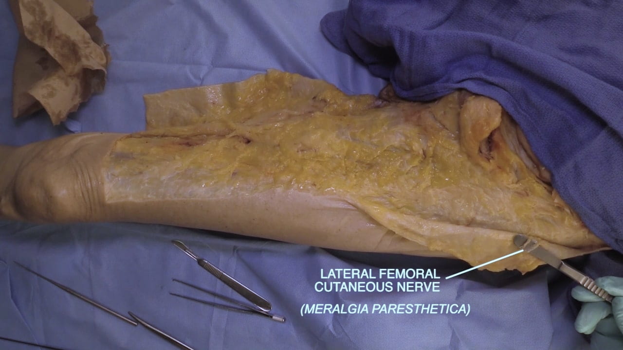
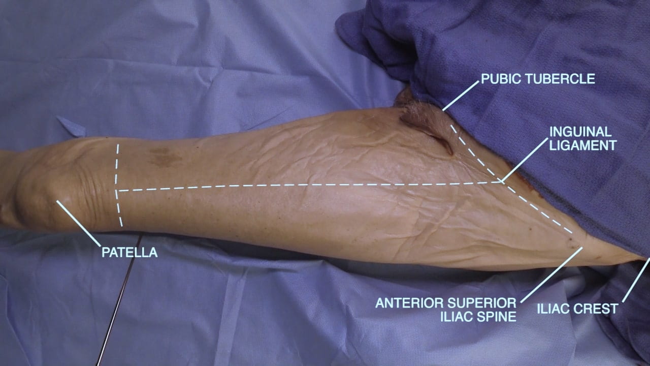
Incise the skin along the inguinal ligament. Make a vertical incision through the skin and superficial fascia on the anterior thigh to just superior to the patella and a distal perpendicular incision across thigh.

Dissect in the plane of the superficial fascia to reflect the skin flaps. Look for the lateral femoral cutaneous nerve medial and inferior to the anterior superior iliac spine. This nerve has a variable position and can difficult to locate.

Superficial View Anterior Thigh

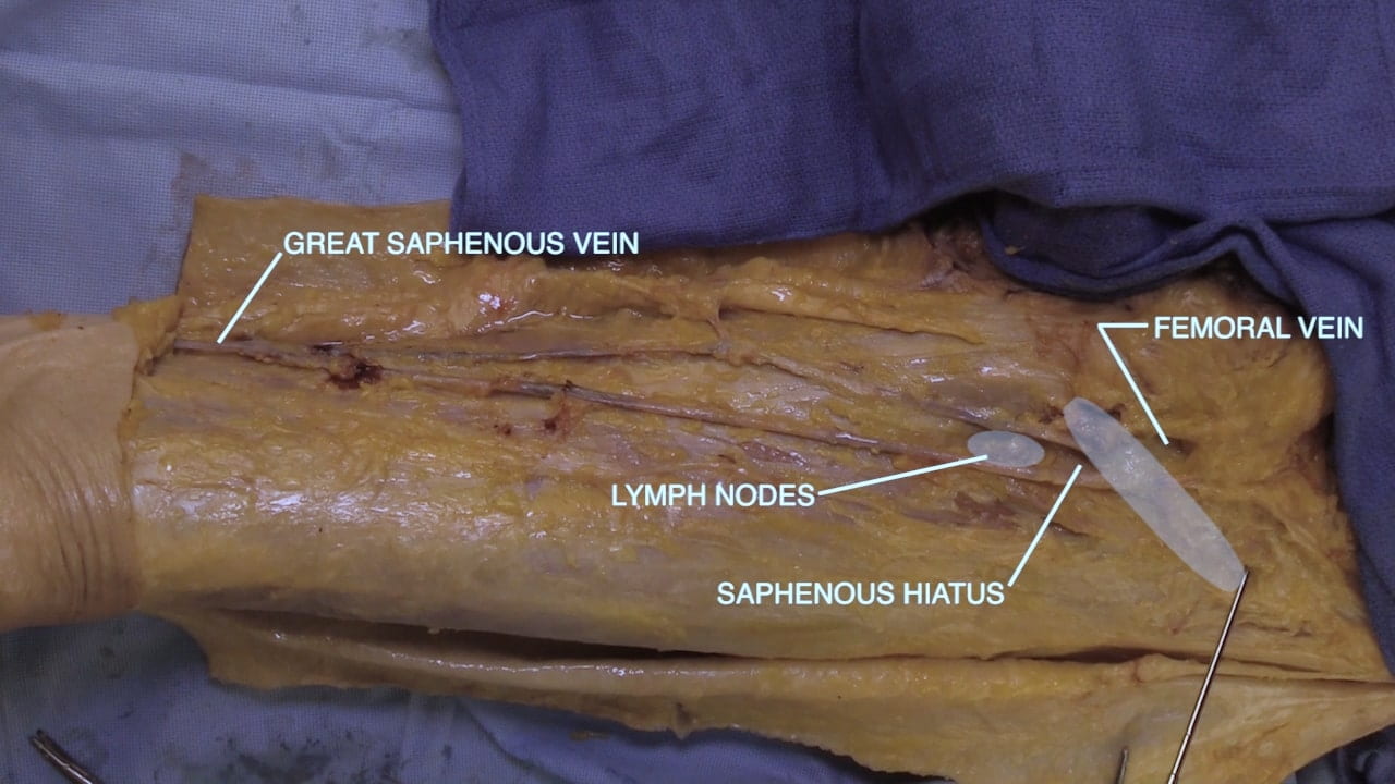
Dissect the superficial fascia on the medial thigh to expose the great saphenous vein from the lower thigh to the saphenous hiatus where it drains into the femoral vein.

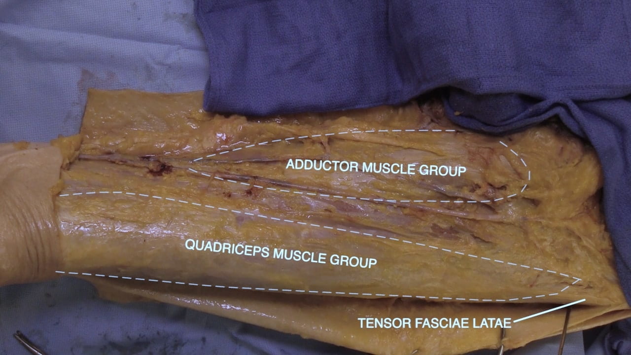
From medial to lateral, identify the adductor and quadriceps muscle groups as well as the tensor fascia latae.

Femoral Triangle

Femoral Triangle

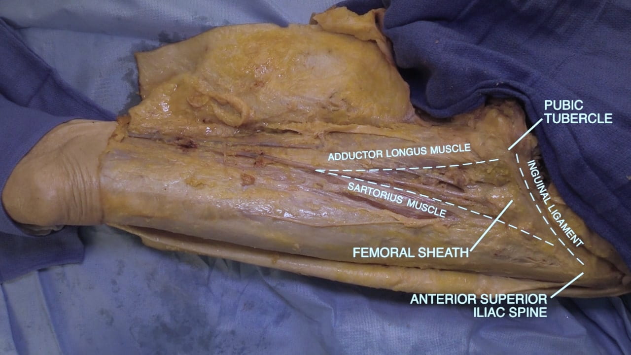
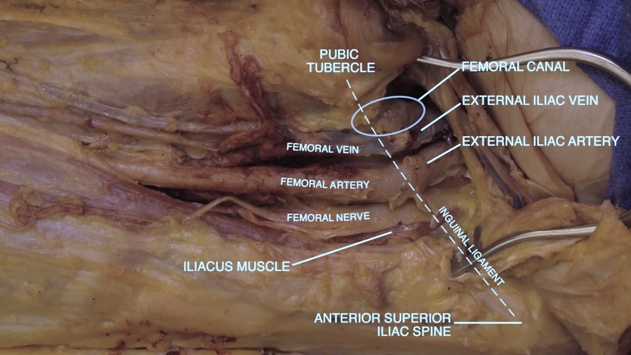
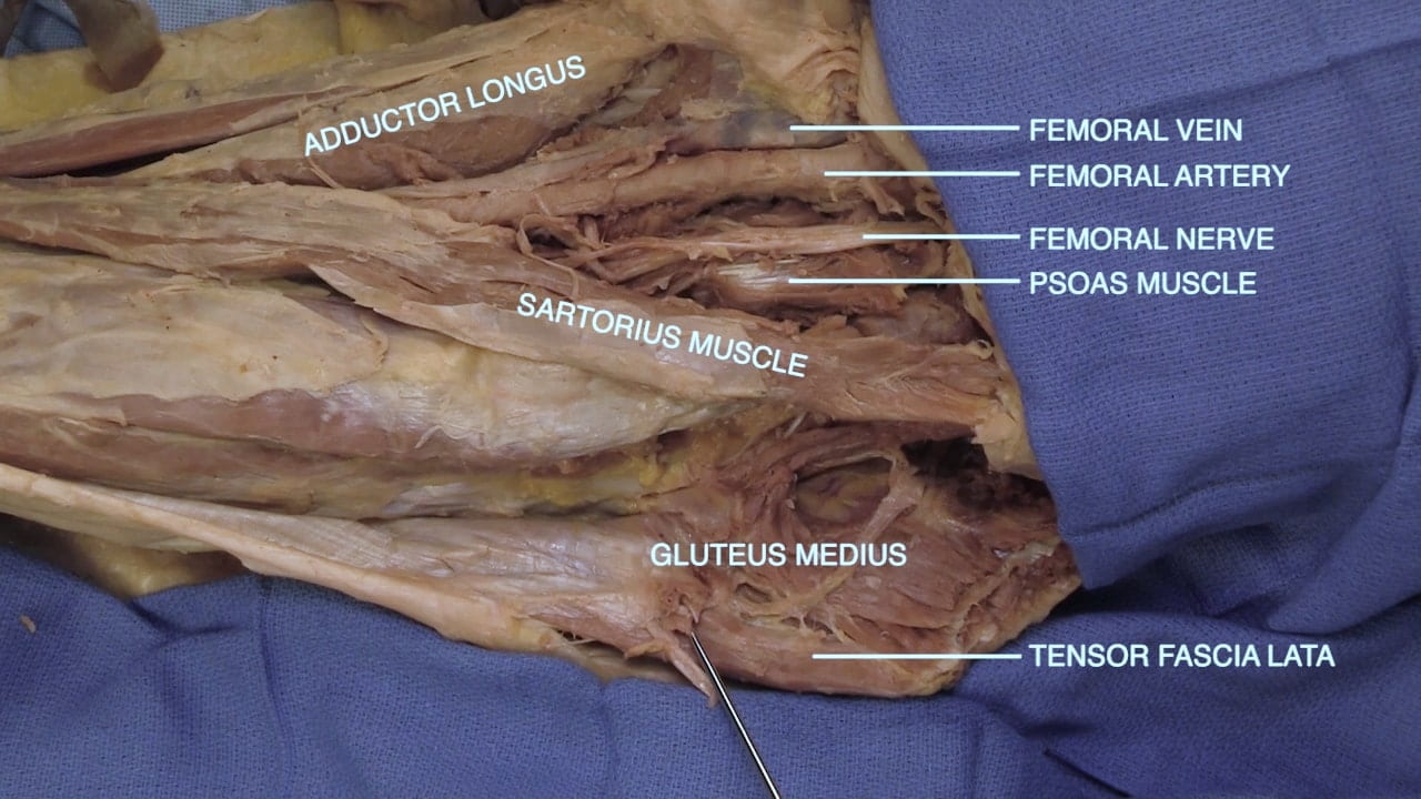
On the anterior thigh, expose the femoral triangle, which is bounded by the inguinal ligament superiorly, adductor longus medially and sartorius laterally.

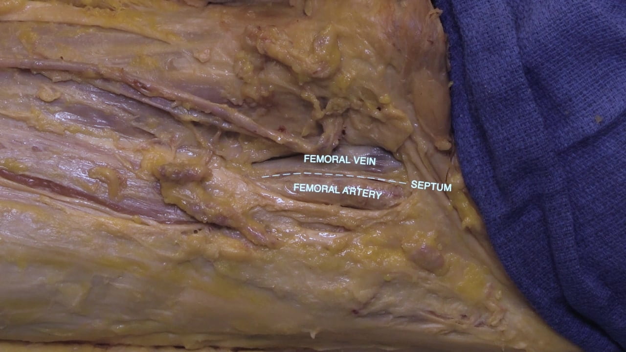
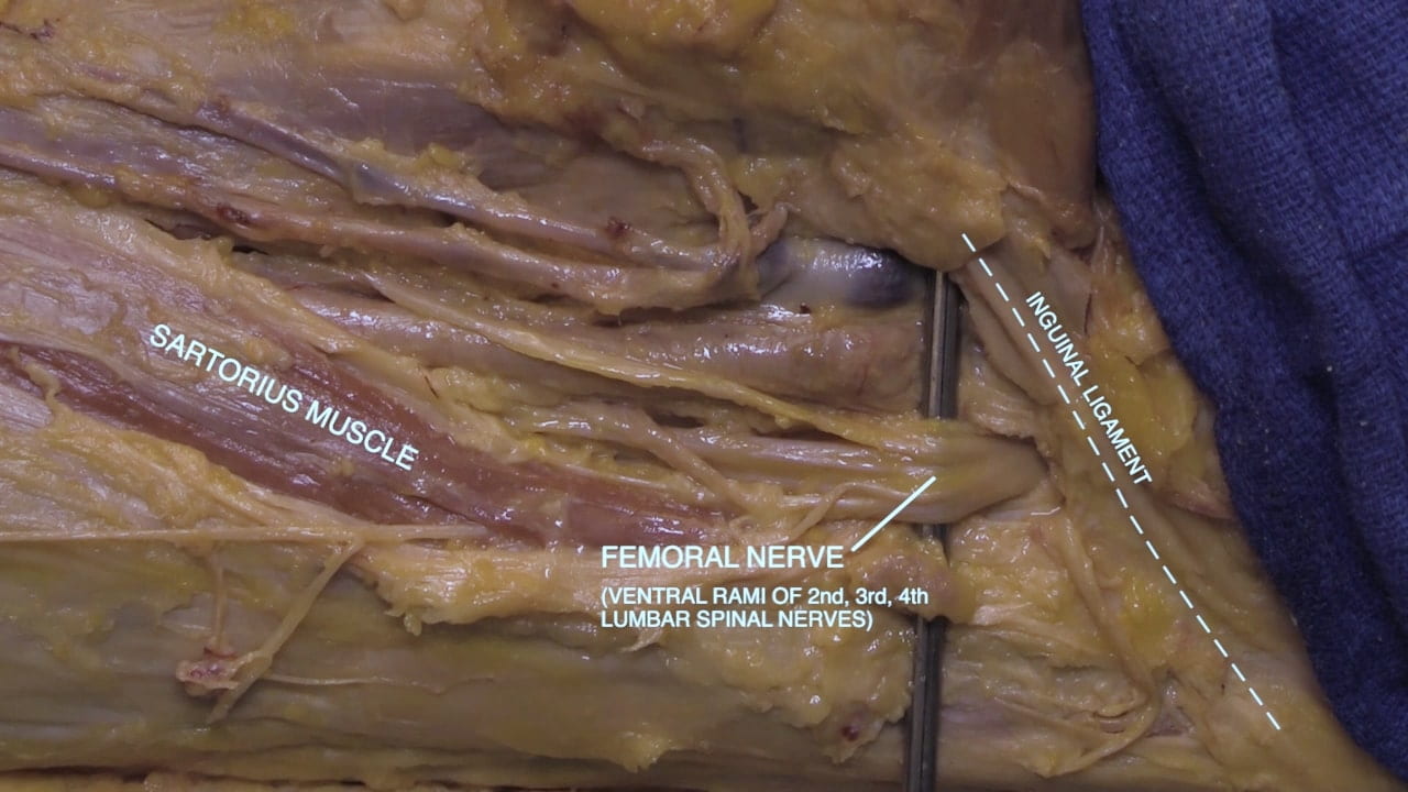
Incise the femoral sheath to expose the femoral artery and vein.  Expose the femoral nerve lateral to the femoral artery.

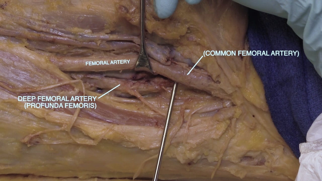
Retract the femoral artery medially to expose the deep femoral (profunda femoris) artery.  Look for the medial and lateral circumflex branches of deep femoral artery.

Femoral Canal

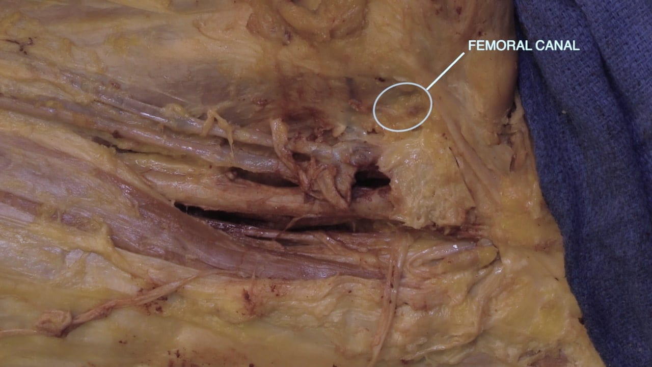
Locate the femoral canal medial to the femoral vein and inferior to the inguinal ligament. The femoral canal is the location of femoral hernias.

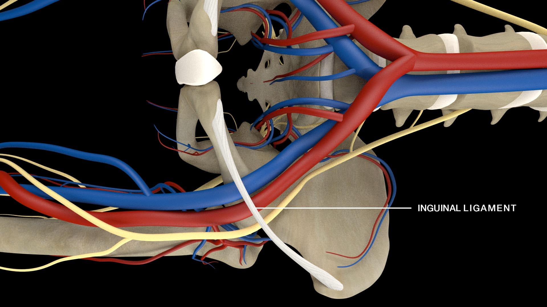
Divide and reflect the inguinal ligament to show the passage of vessels and nerve from the abdomen to the thigh. The external iliac vessels become the femoral vessels as they pass inferior to the inguinal ligament.

Medial Thigh

Obturator Nerve

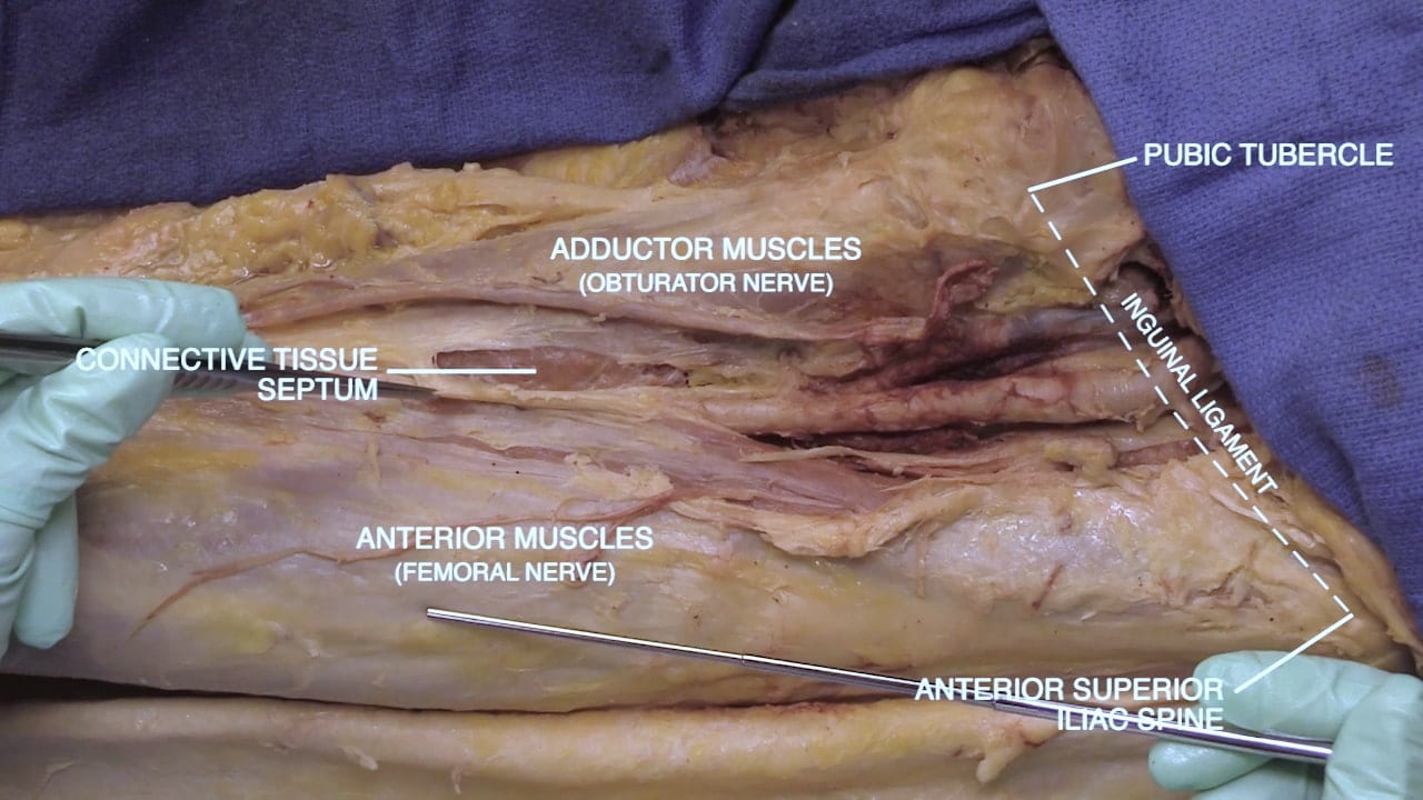
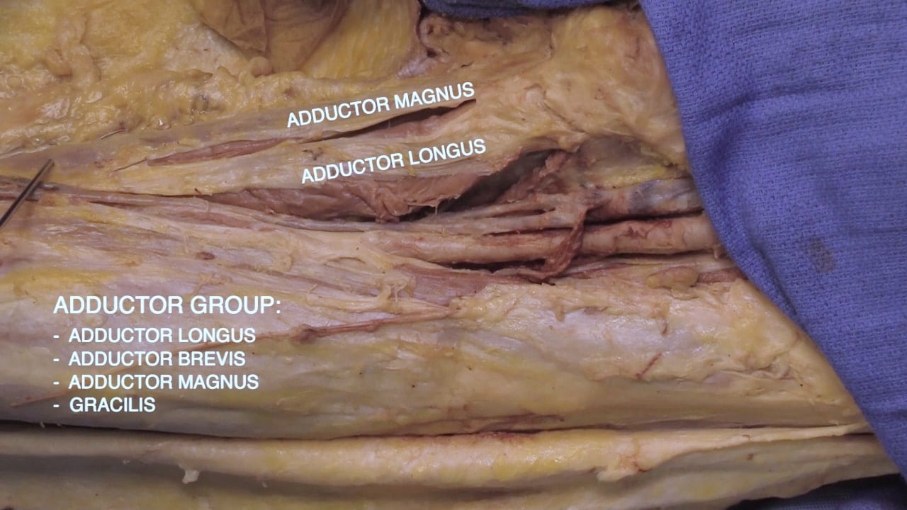
Expose the muscles of the medial thigh by incising the deep fascia covering the medial compartment just medial to the femoral triangle. Within this medial compartment, identify the adductor longus and magnus muscles.
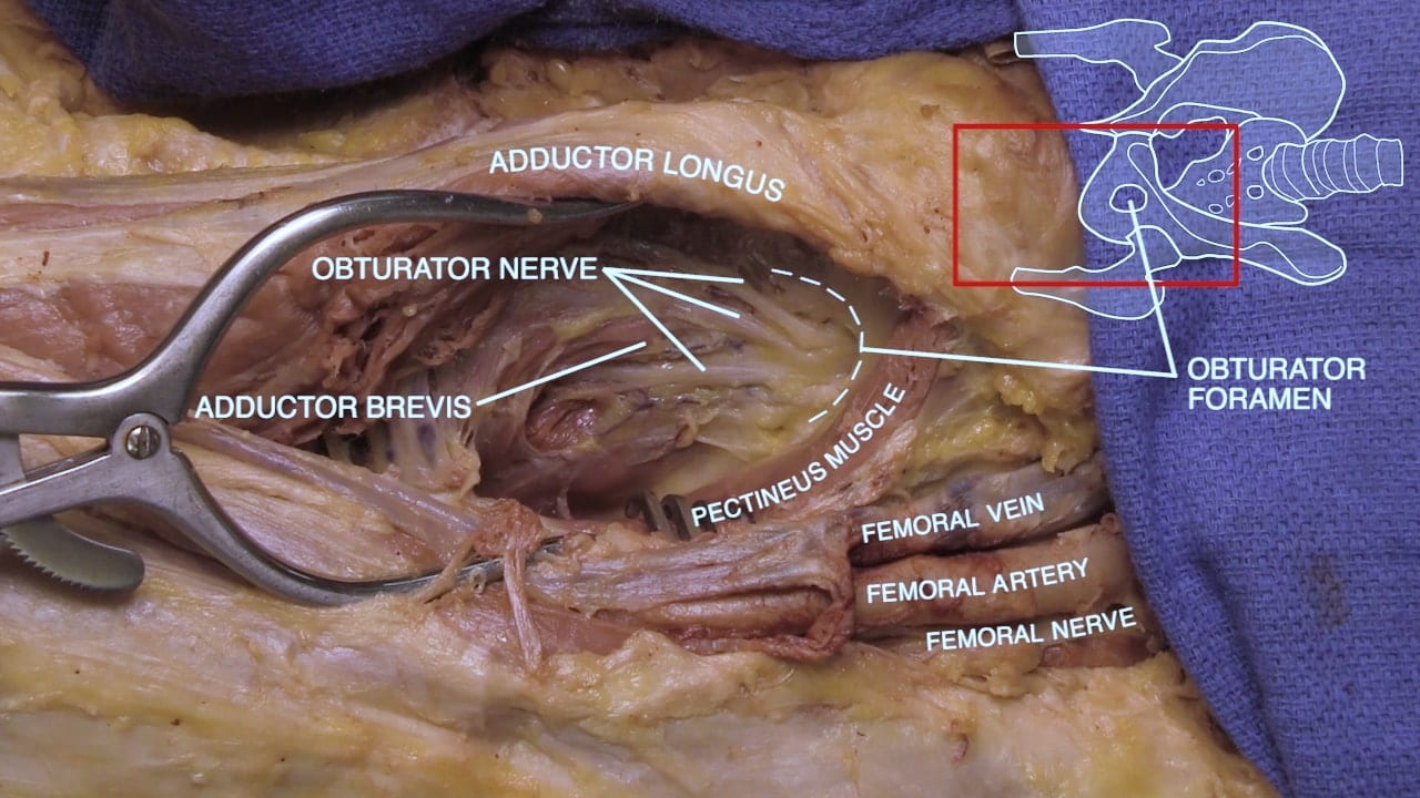
Reflect the adductor longus medially to locate the obturator nerve on the surface of the adductor brevis. The obturator nerve passes through the obturator foramen to supply the adductor muscles of the medial thigh. The obturator foramen is the site of obturator hernias.

Adductor Hiatus

Separate the sartorius from surrounding tissue proximally to distally. Retract the sartorius medially to identify the femoral vessels.

Locate the adductor hiatus where the femoral vessels pass into the popliteal fossa. This route moves the femoral vessels from medial to posterior in the thigh.

Labels (left and right): Saphenous nerve, popliteal vessels, adductor hiatus, femoral artery, sartorius muscle
3.2a) Adductor hiatus

Lateral Thigh and Hip

Exposing the Lateral Thigh

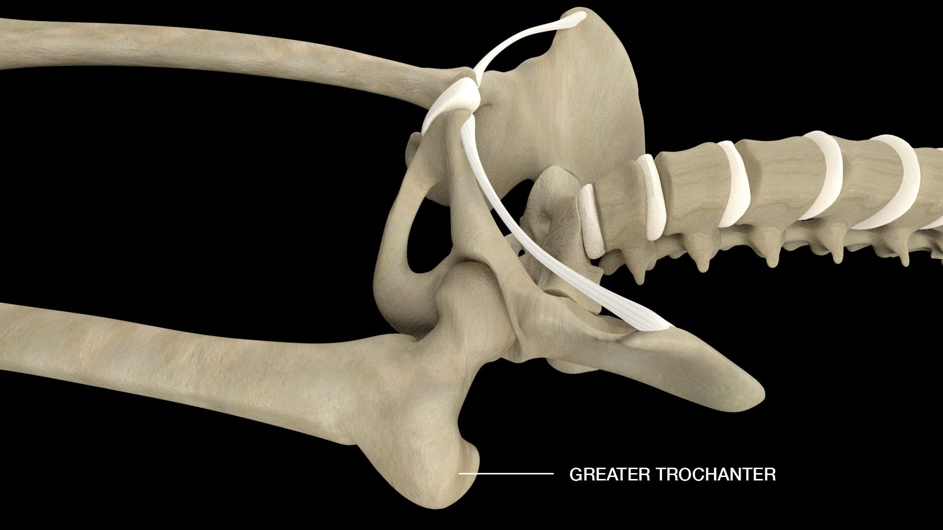
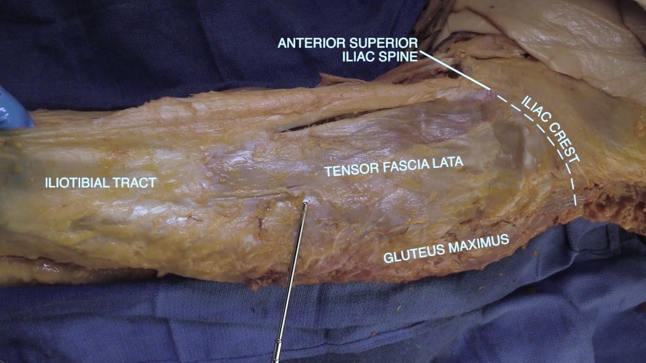
On the lateral thigh, identify the iliac crest and anterior superior iliac spine. Locate the greater trochanter by palpation of the lateral thigh. Examine a skeleton to assist with landmarks.

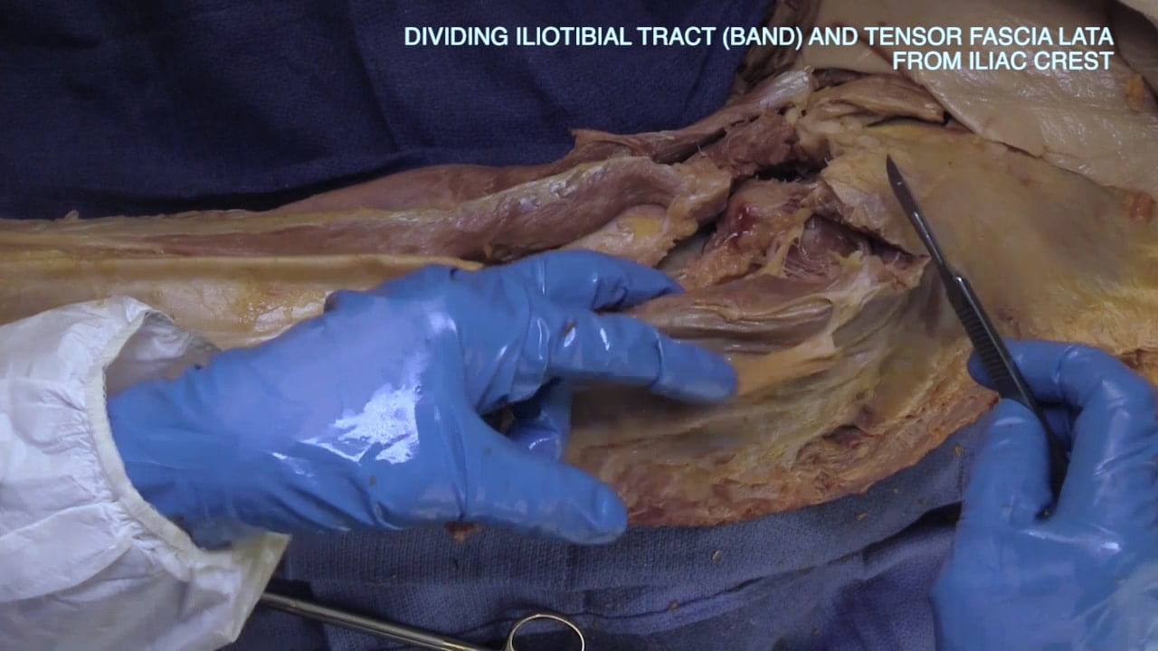
Incise along the lateral border of the iliac crest to divide the proximal attachments of the tensor fascia latae (and iliotibial tract) and gluteus medius. Reflect these muscles posteriorly.

Superior Gluteal Nerves and Vessels

Locate the superior gluteal vessels and nerve along the deep surface of the gluteus medius as it runs between the gluteus medius and minimus.

All muscles of the lateral thigh are innervated by the superior gluteal nerve.

Labels (left to right): Tesnro fascia lata, superior gluteal nerve, gluteus medius, gluteus minimus, superior gluteal vessels and nerves, iliac crest
4.2a) Superior gluteal nerve

Exposure of Hip Joint

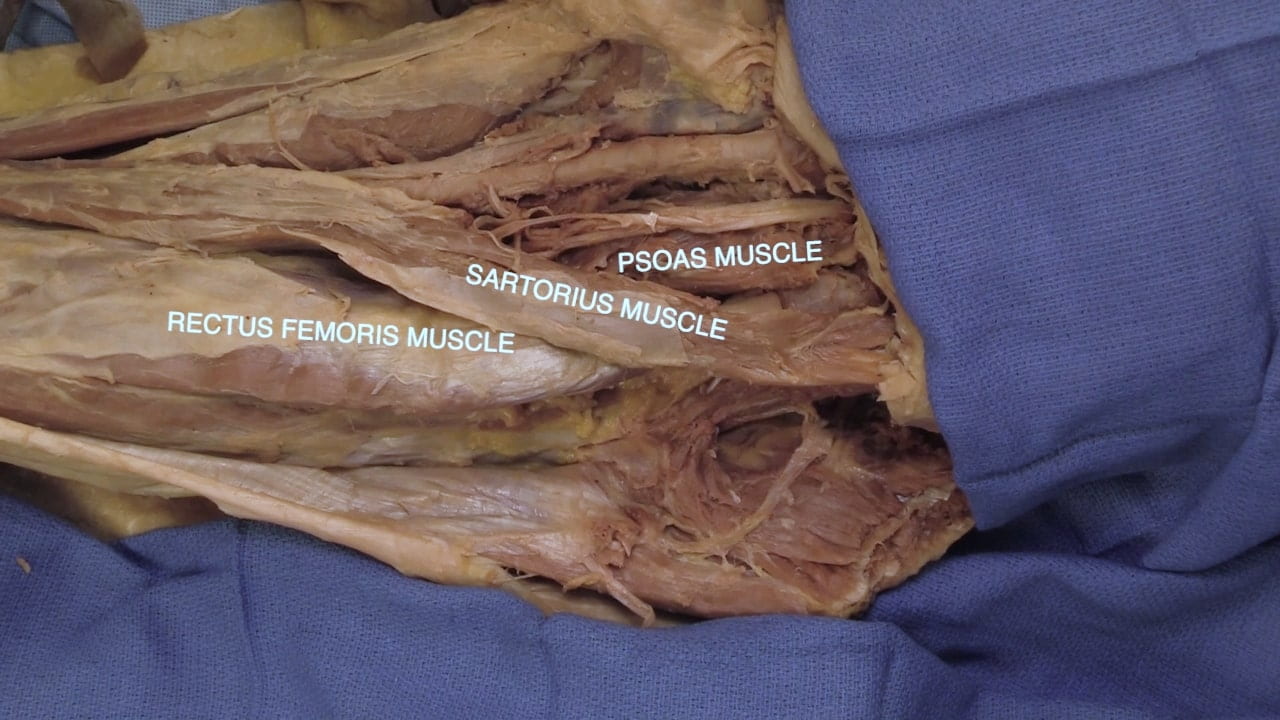
To expose the hip joint, identify the muscles of the anterior thigh which lie anterior to the joint. These are the sartorius, rectus femoris and psoas muscles.

Muscles of the anterior thigh are innervated by the femoral nerve.

Divide the sartorius and underlying rectus femoris as proximal as possible to facilitate hip exposure.

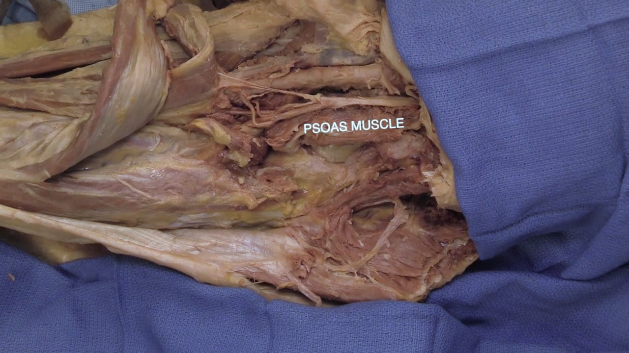
Palpate the hip joint through the psoas muscle. This is facilitated by rotating the lower extremity. Divide the psoas overlying the joint and reflect the muscle superiorly and inferiorly to expose the joint capsule.

Hip Joint

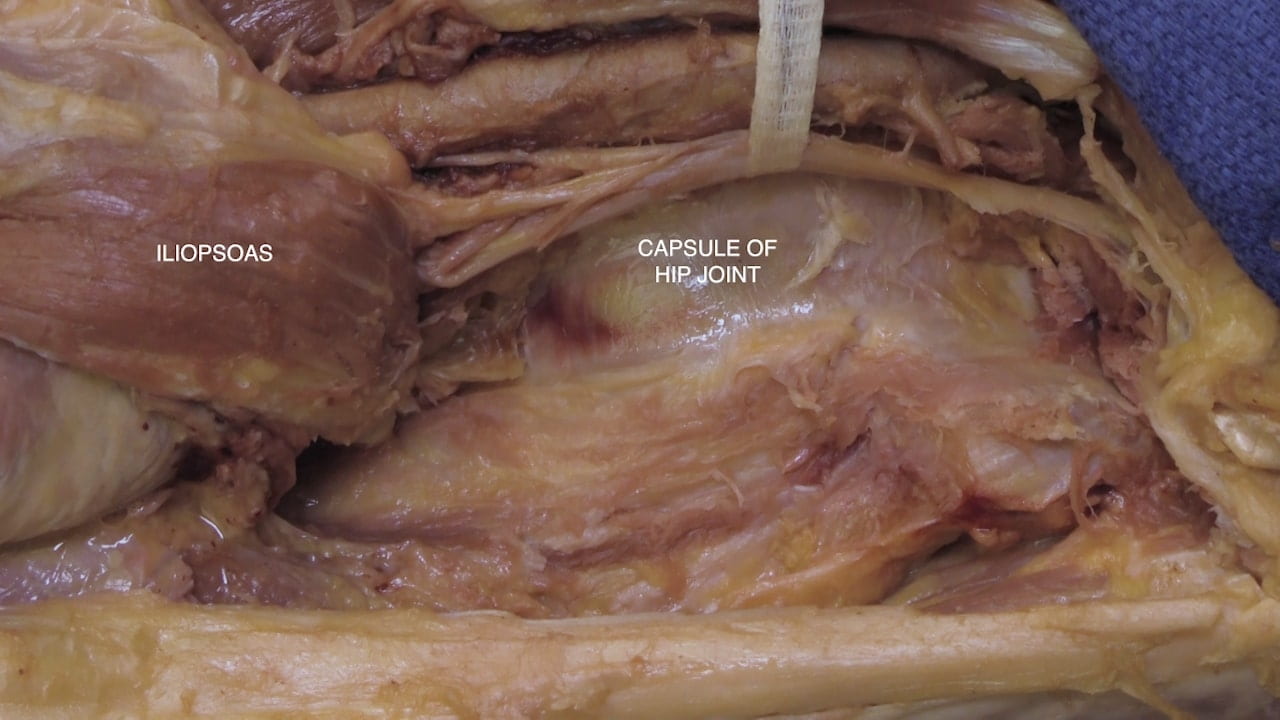
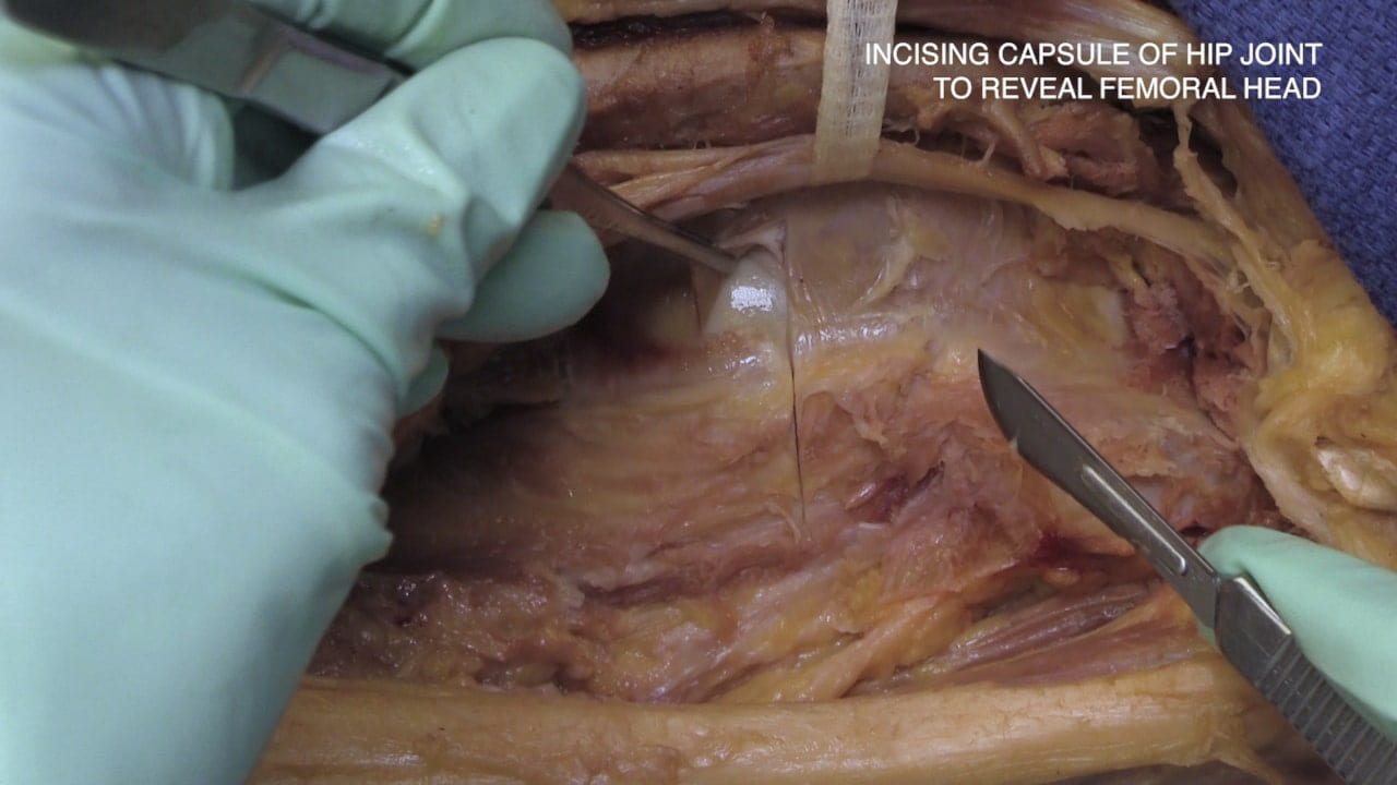
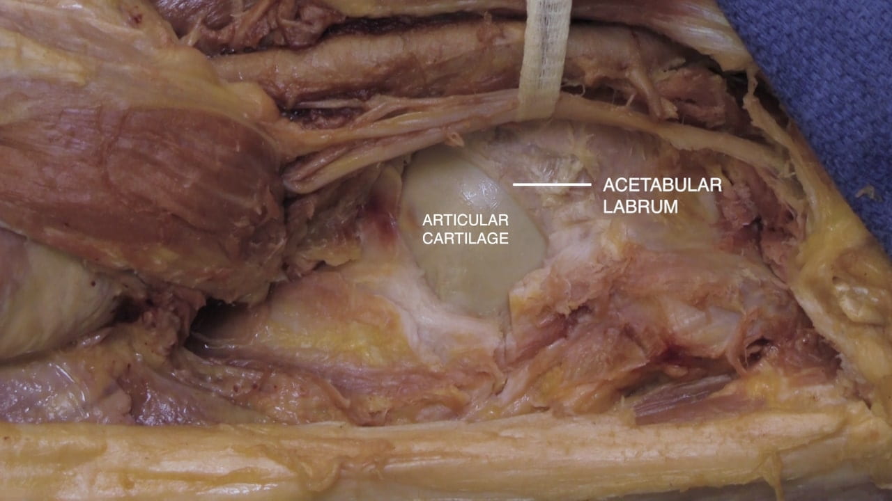
Open the dense capsule of the hip joint using a cruciate incision to reveal the femoral head, labrum and acetabulum.

Dislocate the hip by adducting and externally rotating the lower extremity to expose the head of the femur and acetabulum. This will require considerable force. A portion of the acetabulum may need to be resected to dislocate the hip.

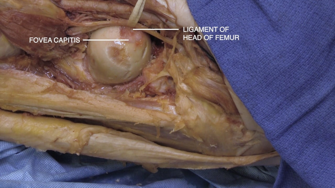
Identify the ligament of head of femur (ligamentum teres) which provides some of its blood supply.

Proximal Femur

A common surgical approach to the hip is through the interval between the anterior and lateral compartments since they have separate innervation. This interval is termed the inter-nervous plane.

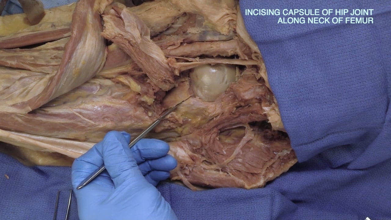
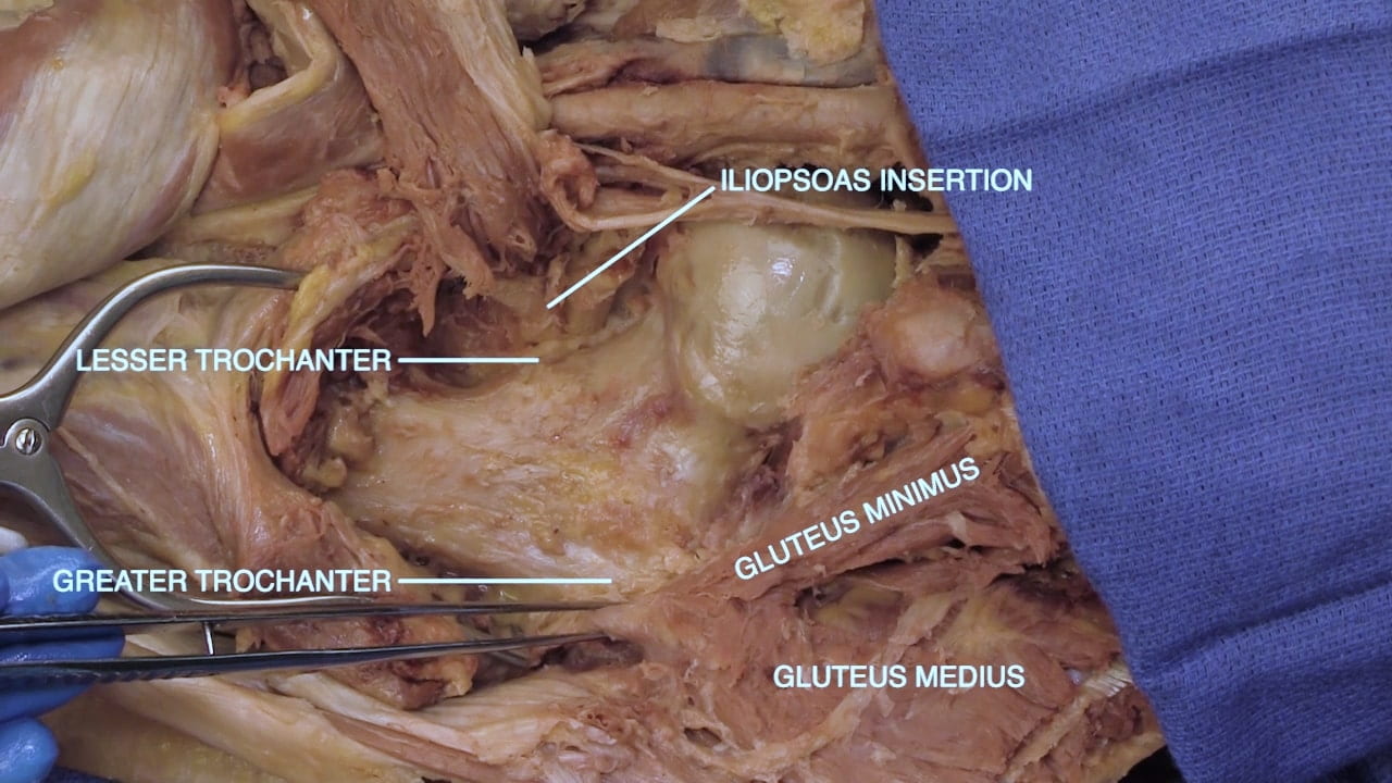
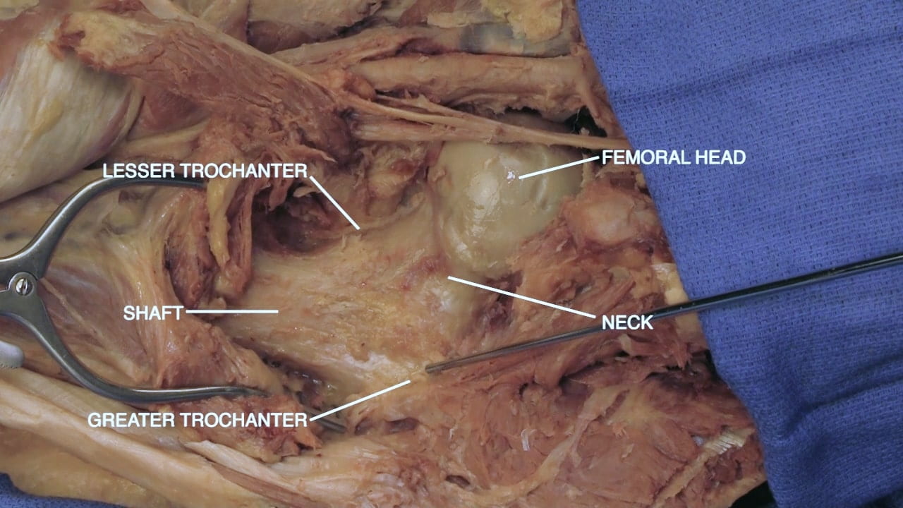
Continue the incision of the capsule of the hip joint along the neck of the femur to reveal the neck and shaft. Identify the lesser and greater trochanters of the proximal femur.

Identify the attachment of the iliopsoas to the lesser trochanter and the attachment of the gluteus medius and minimus to the greater trochanter.

Gluteal Region Part I

Exposure of the Gluteal Region

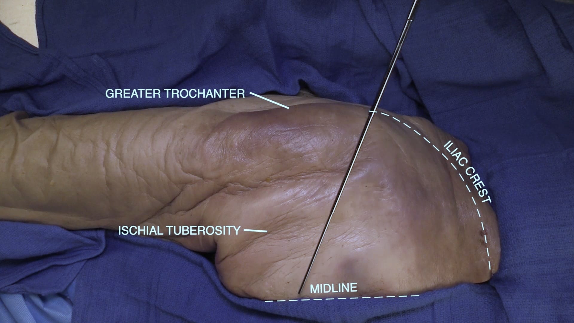
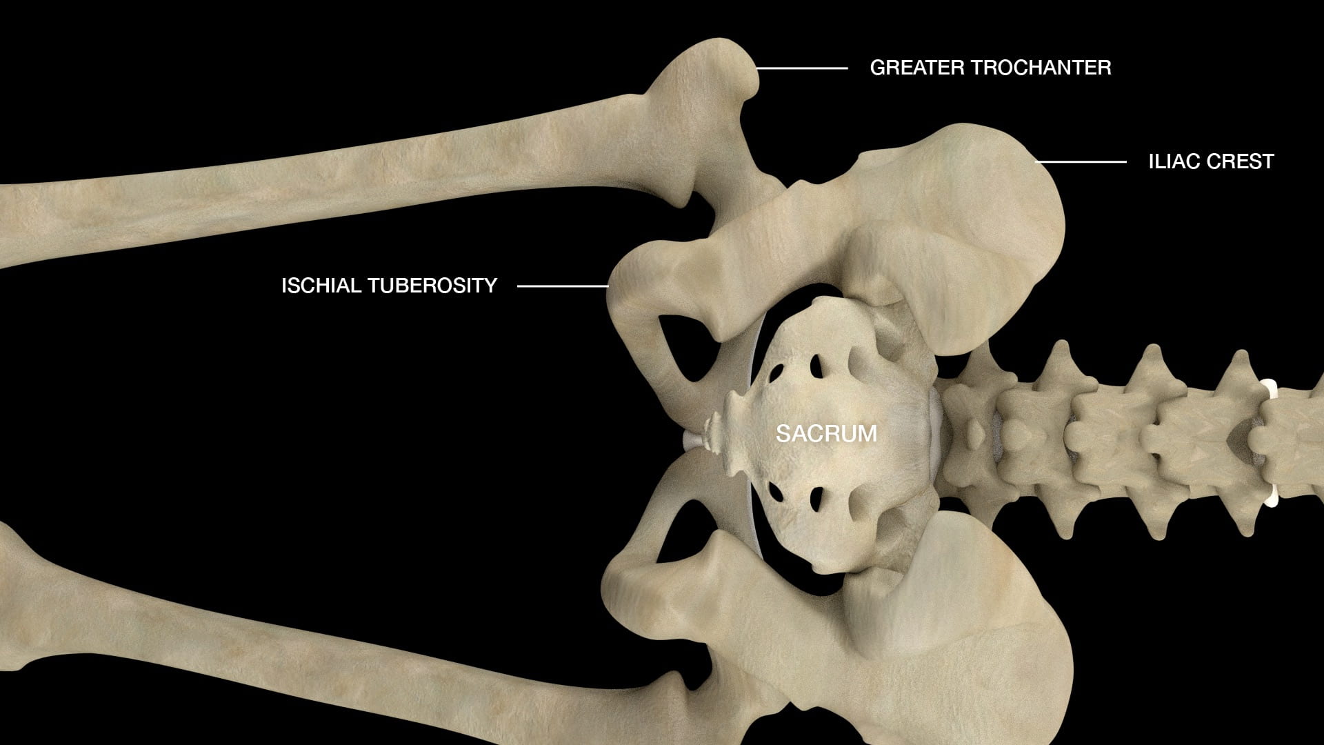
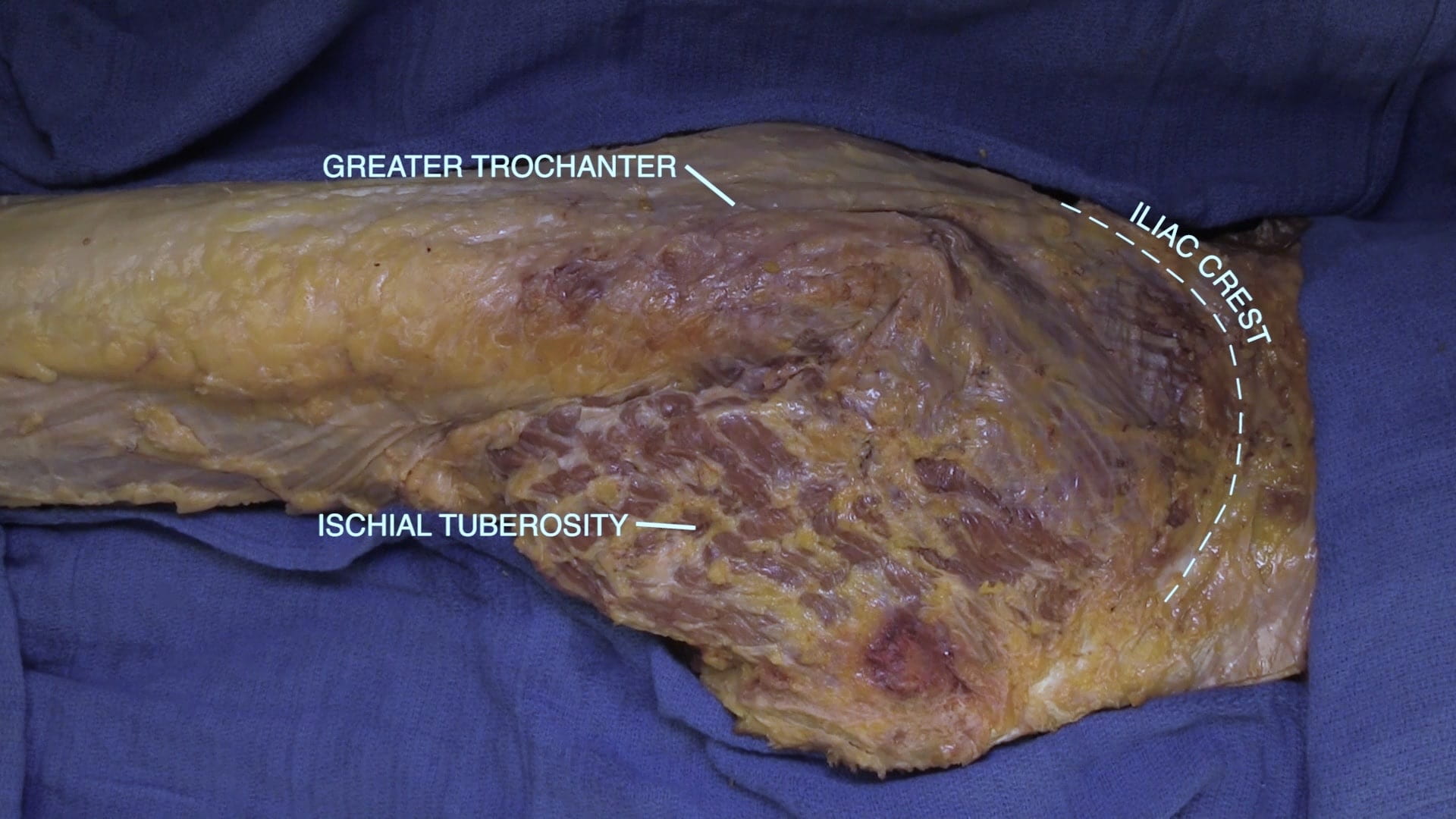
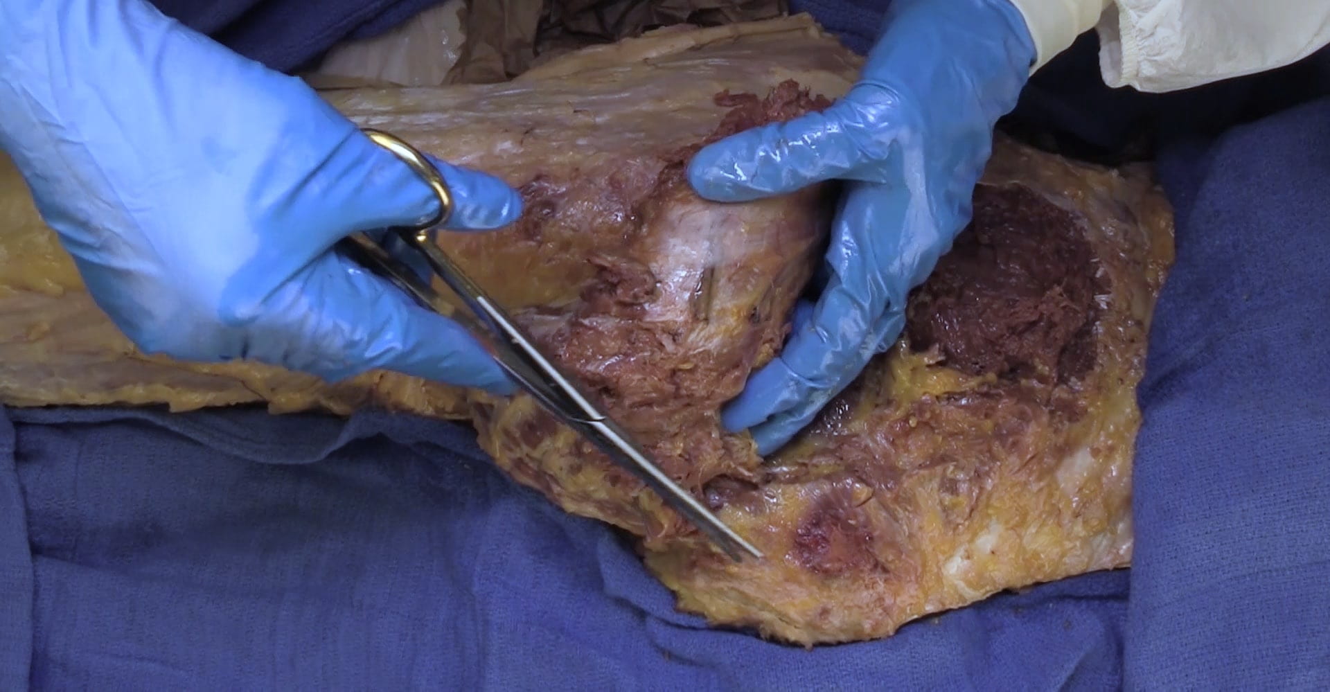
With the donor prone, identify the positions of the iliac crest, greater trochanter, ischial tuberosity and sacrum. Confirm landmarks by examining a skeleton.

Remove the skin and superficial fascia by incising along the iliac crest and the midline of the sacrum. Connect these incisions to the previous anterior and lateral thigh flaps.

Re-identify the positions of the iliac crest, greater trochanter, ischial tuberosity and sacrum.

Gluteus Maximus Dissection

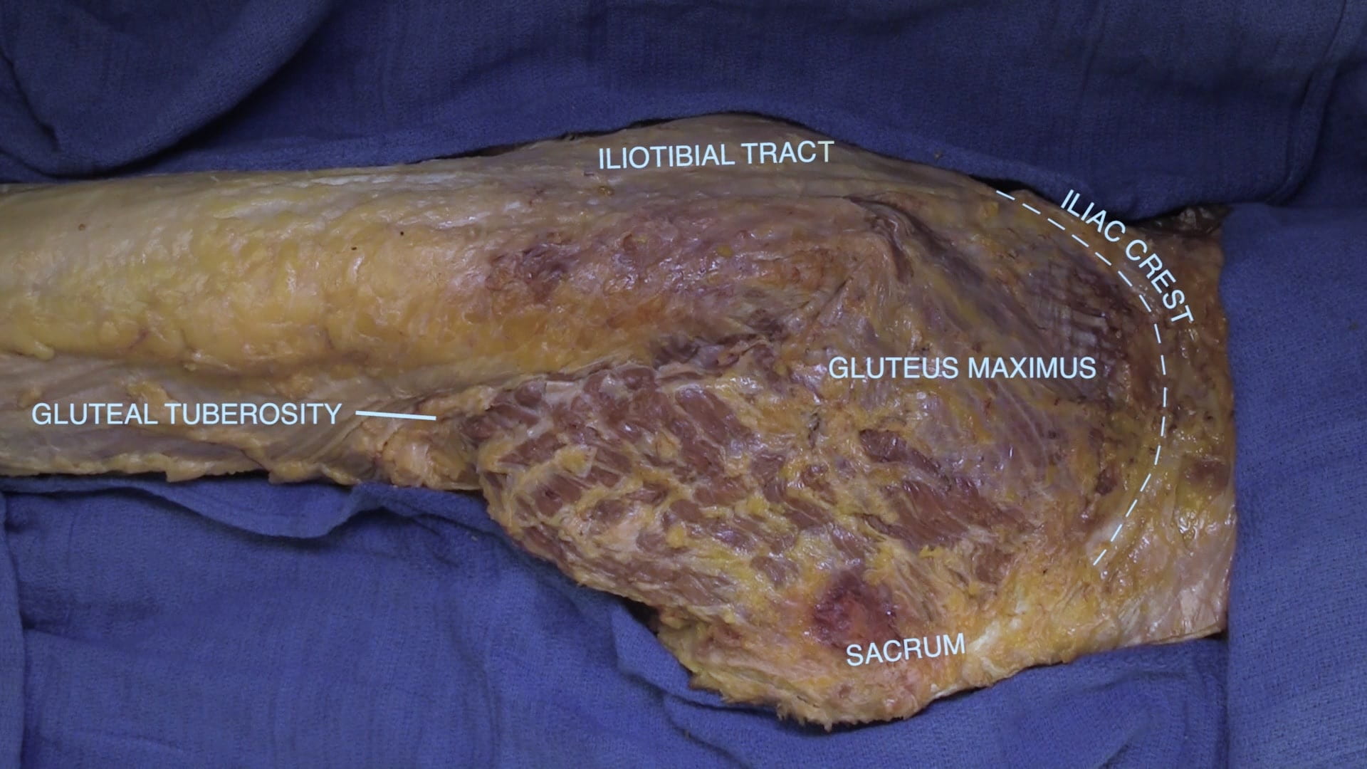
Identify the gluteus maximus and its iliac crest, sacrum and iliotibial tract attachments.

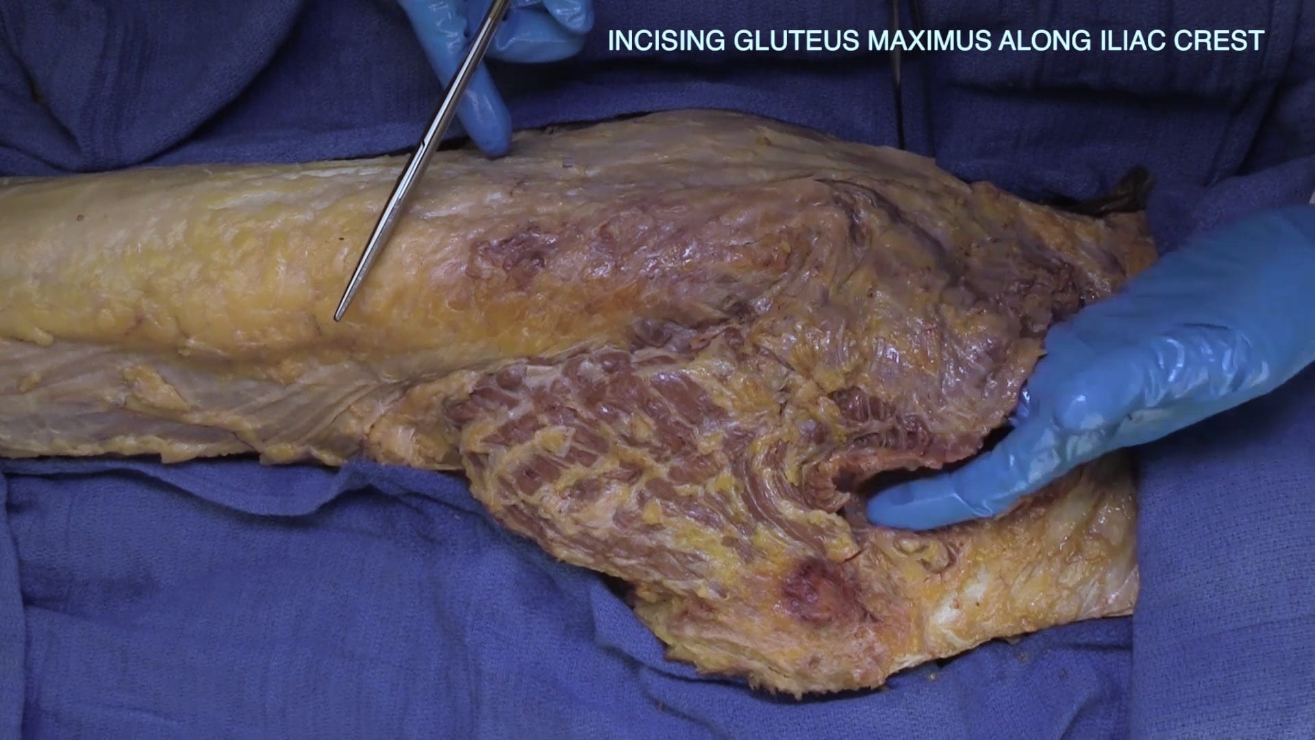
Divide the gluteus maximus along the iliac crest.

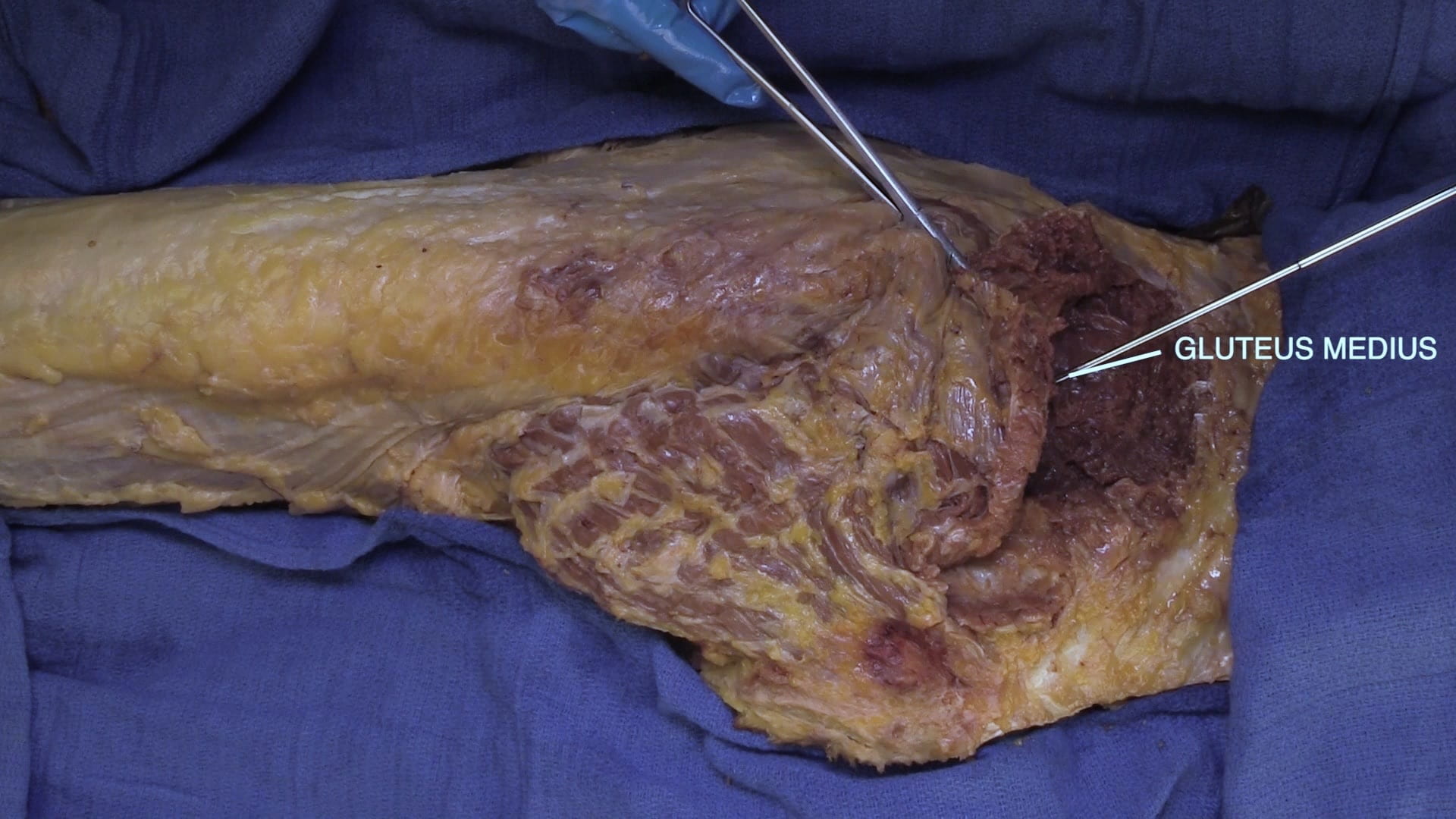
Locate the fascial plane between the gluteus maximus and medius. Once this plane is identified divide the gluteus maximus along its sacral attachment.

Piriformis

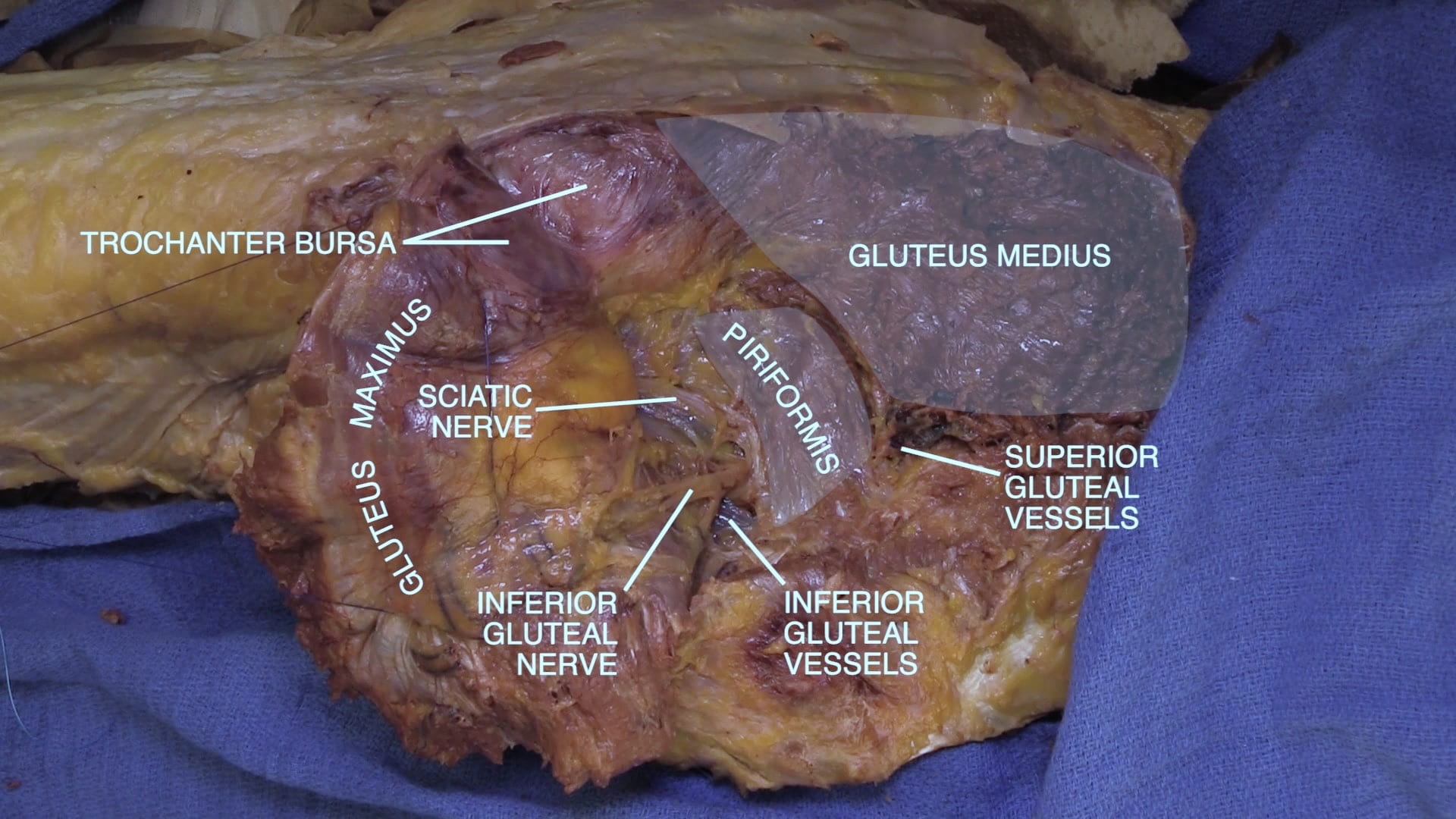
Reflect the gluteus maximus inferiorly and locate the gluteus medius deep to the gluteus maximus.

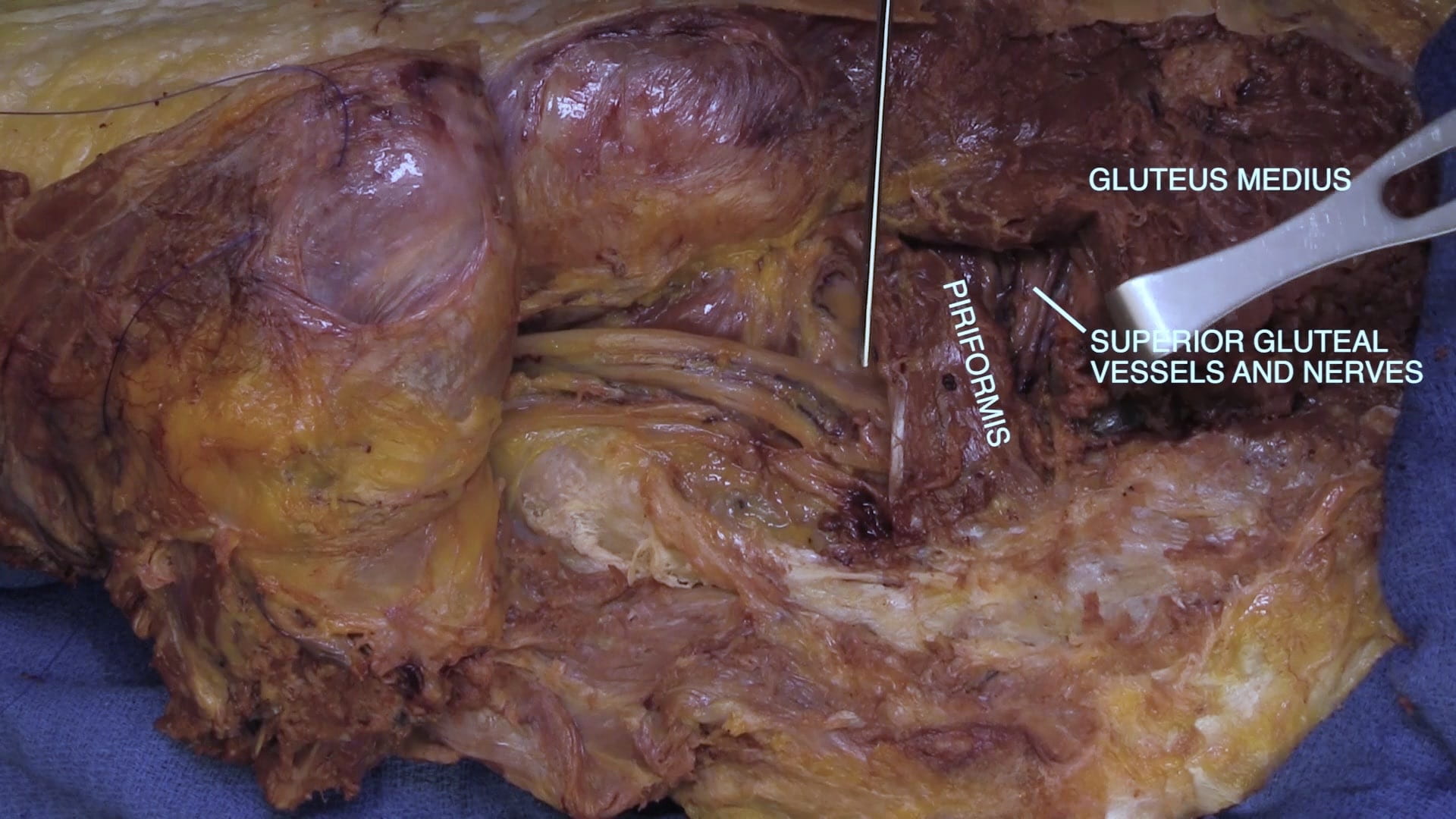
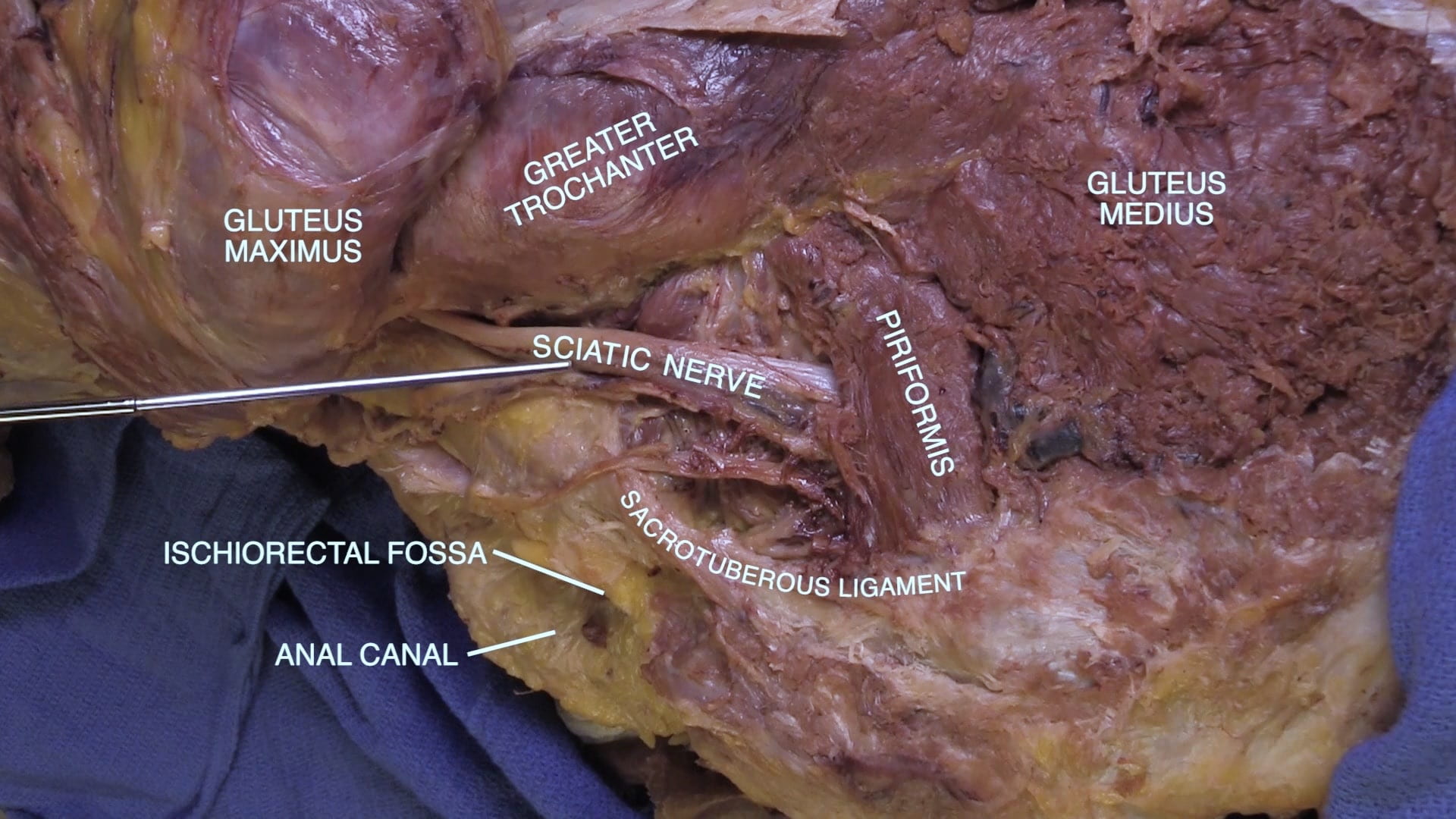
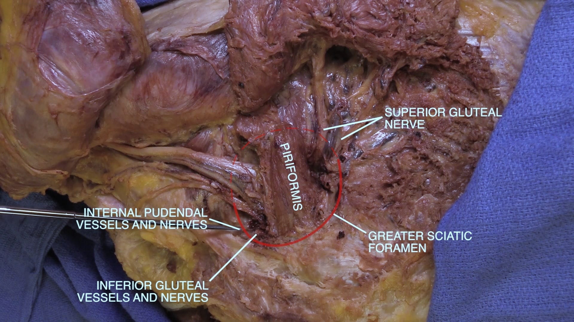
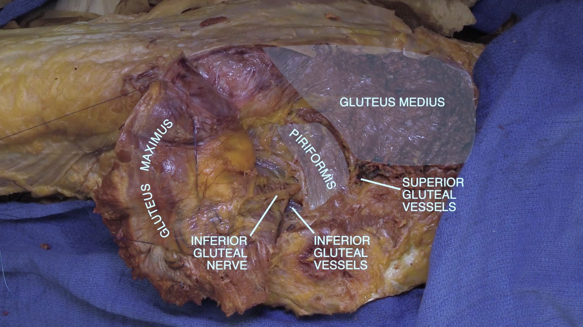
Locate the piriformis inferior to the gluteus medius. The piriformis is a landmark since the sciatic nerve emerges inferiorly and the superior and inferior gluteal nerves emerge above and below, respectively.

Sciatic Nerve

Divide the inferior gluteal vessels and nerves to further reflect the gluteus maximus thereby exposing more of the sciatic nerve.

Identify the superior gluteal nerves and vessels above the piriformis. Retract the gluteus medius laterally to follow the vessels and nerve between the gluteus medius and minimus.

  • Superior gluteal nerve – gluteus medius and minimus and tensor fascia lata
  • Inferior gluteal nerve – gluteus maximus

Gluteal Region Part II

Pudendal Vessels and Nerve

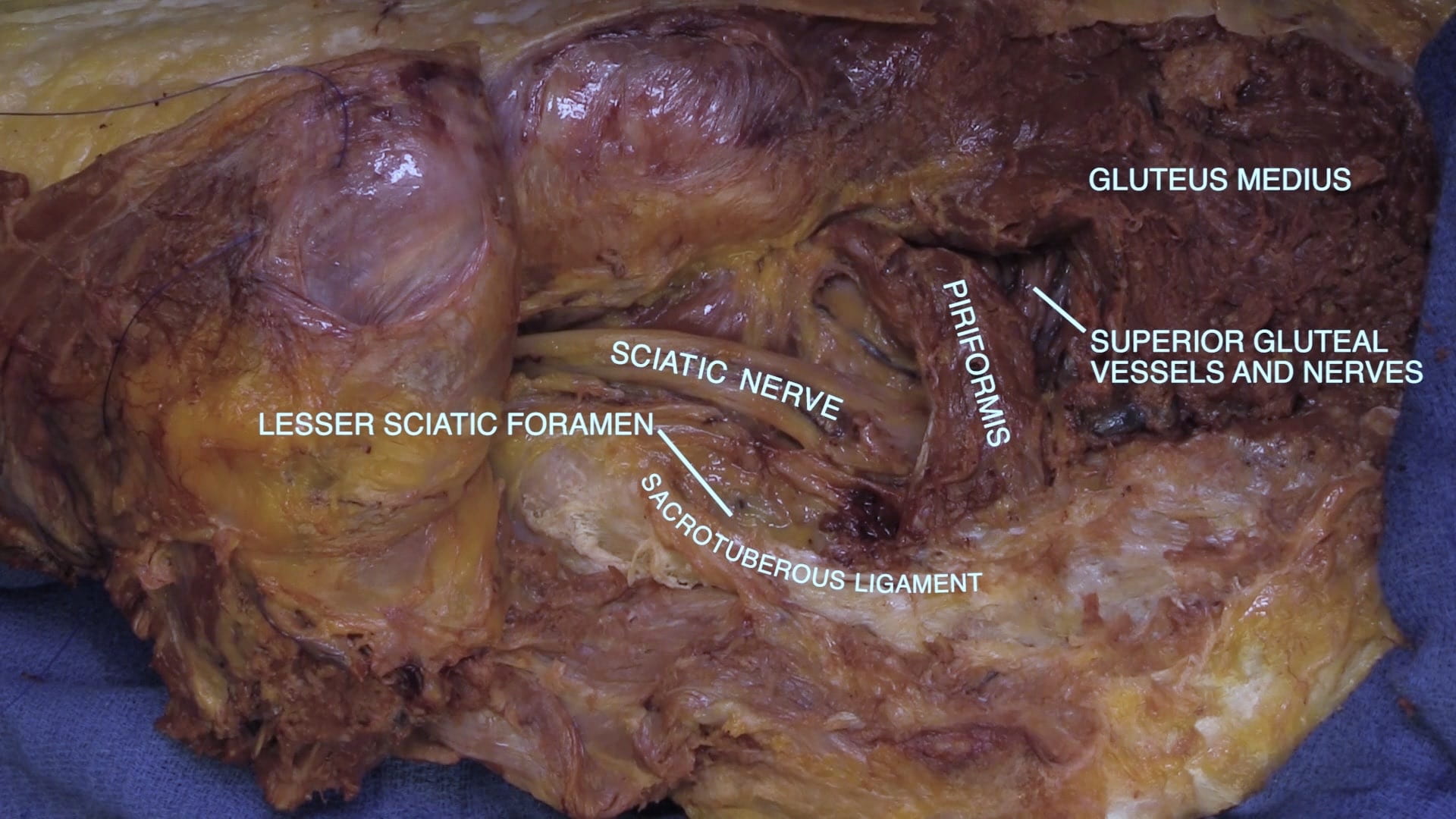
Further detach the gluteus maximus from its medial attachment on the sacrotuberous ligament. This ligament extends between the sacrum and ischial tuberosity medial to the sciatic nerve.

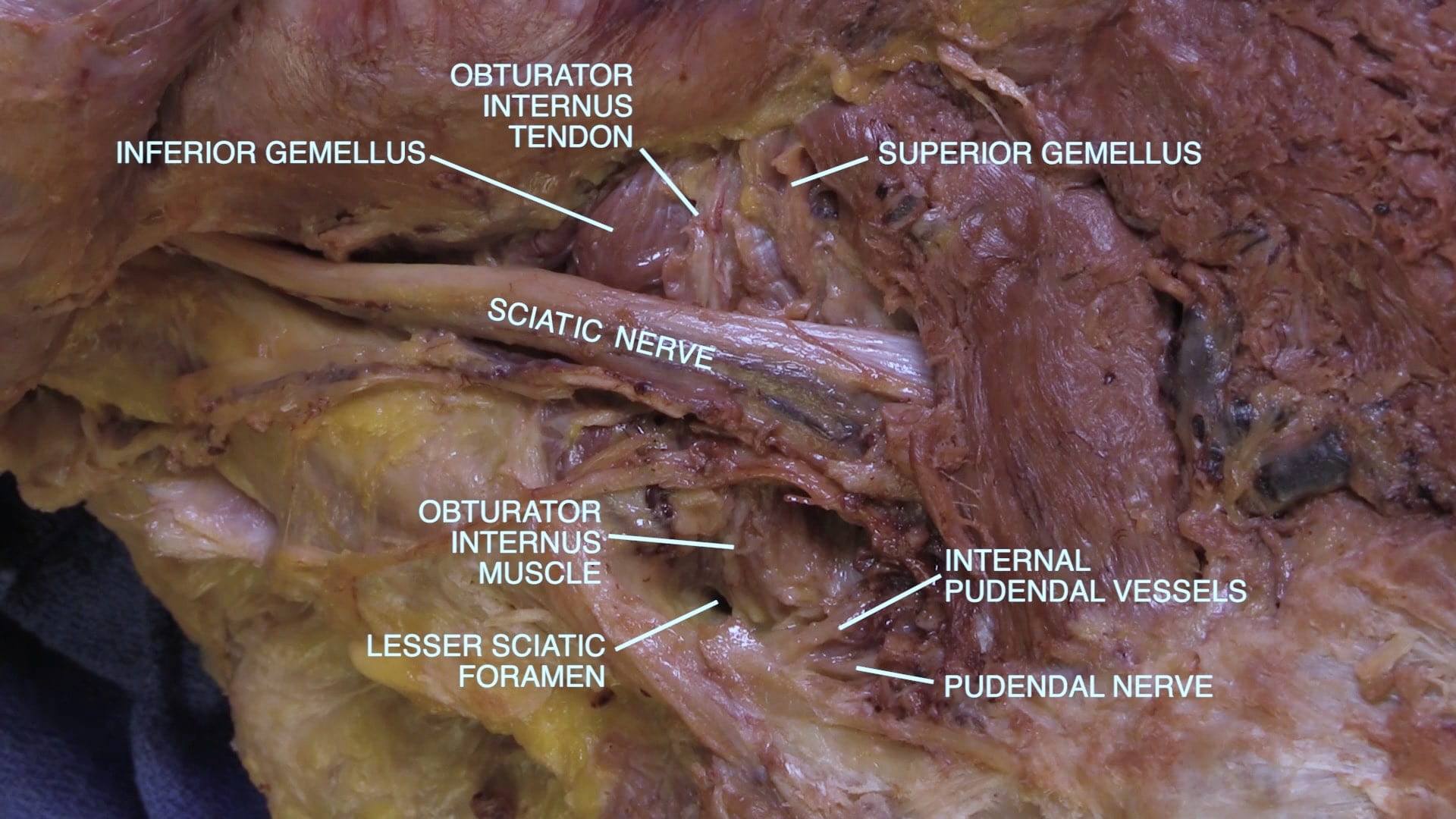
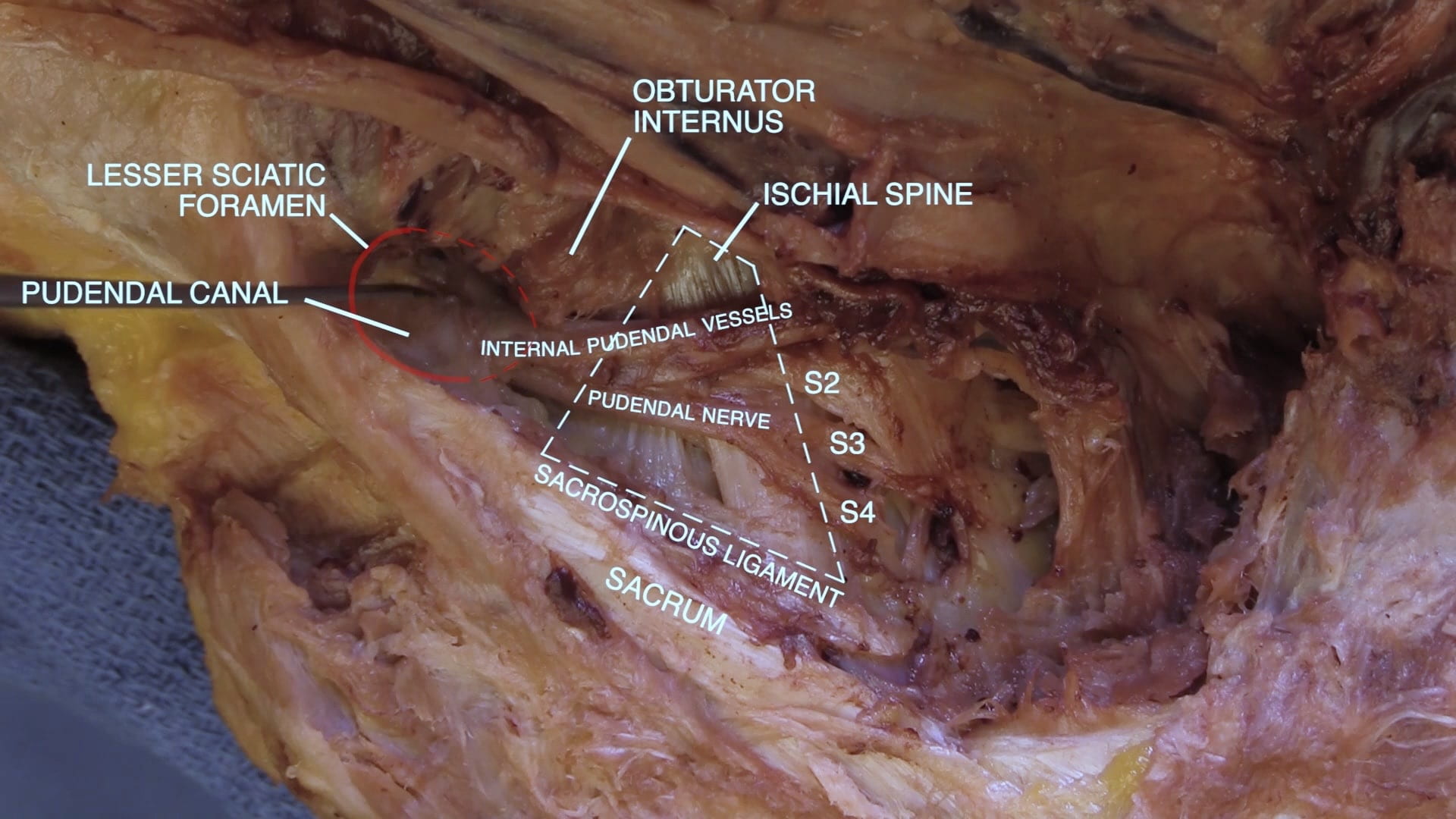
Locate the pudendal nerve and the internal pudendal vessels medial to the sciatic nerve and inferior to the piriformis. Follow the pudendal nerve to the sacrotuberous ligament.

Course of Pudendal Nerve

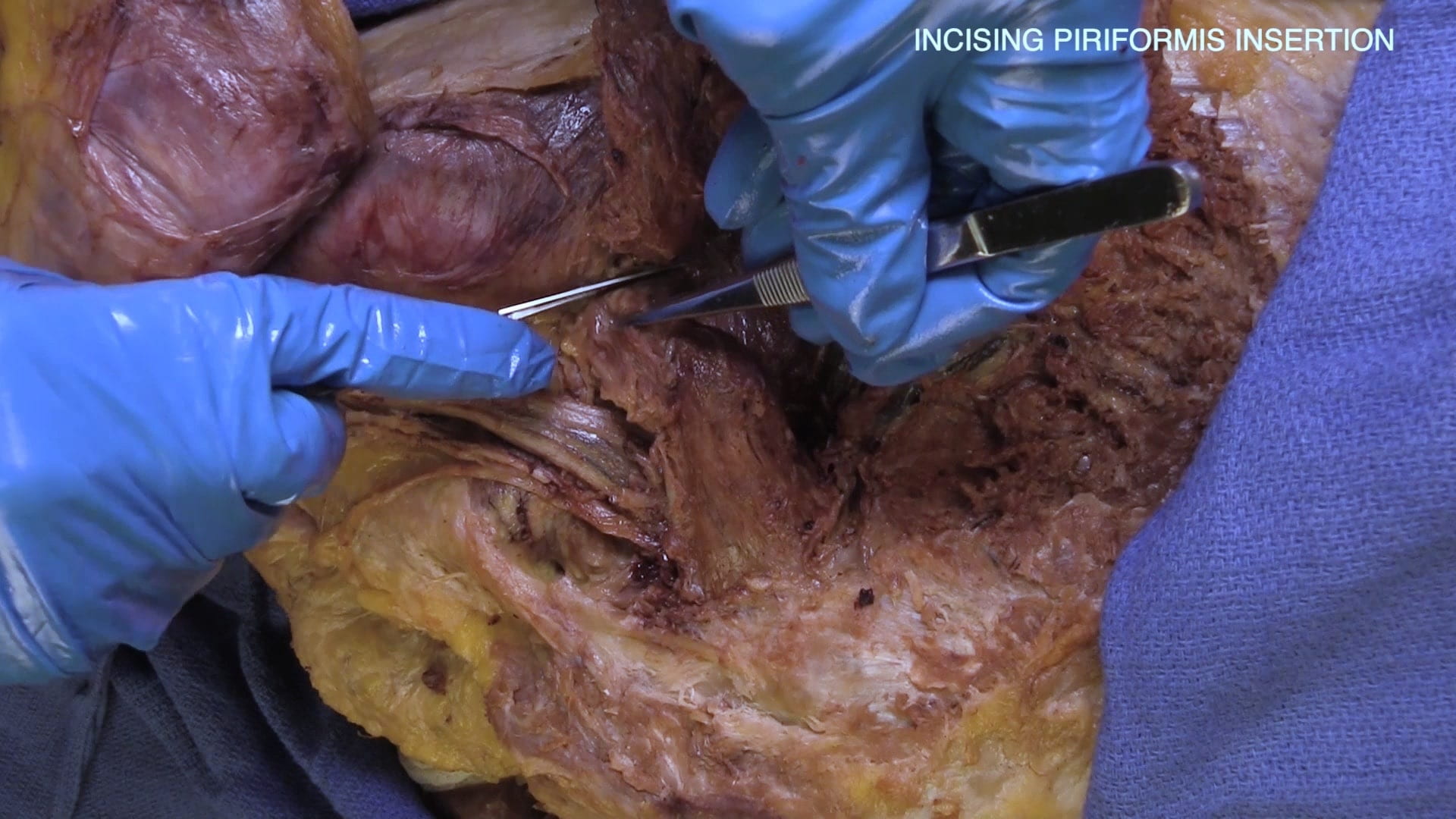
Divide the piriformis at its insertion on the greater trochanter and reflect it medially. Also, divide the piriformis medially at the level of the greater sciatic foramen.

Identify the S2, S3, S4 nerve roots as they combine to form the pudendal nerve. 

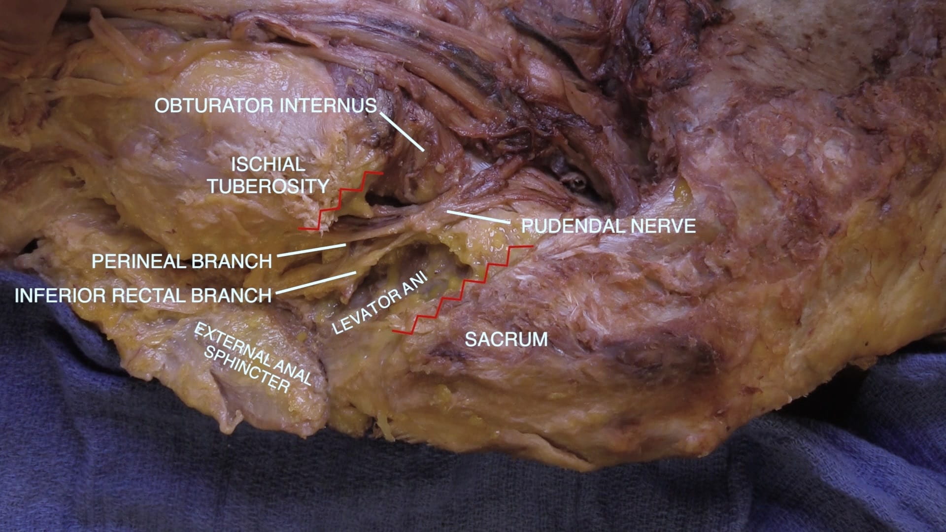
Divide the sacrotuberous ligament to follow the pudendal nerve through the lesser sciatic foramen to the ischiorectal fossa.

  • Pudendal nerve – Sensation to the perineum including external genitalia and motor innervation to urethral and anal sphincters

3D Leg

Review Quiz